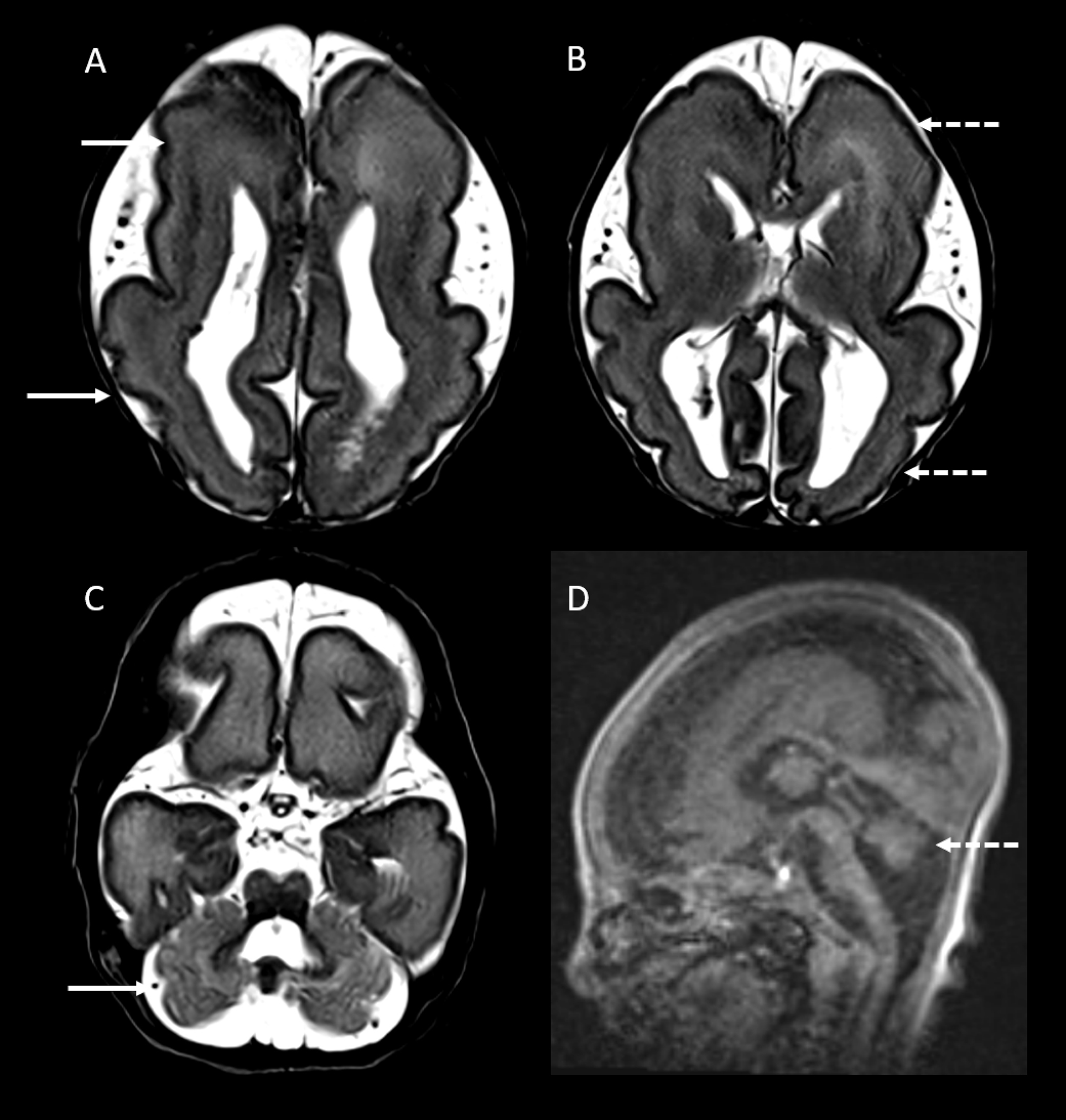
A male infant born preterm at 35+1 weeks (birth weight 2250 g), following a pregnancy complicated by polyhydramnios, had hypotonia, microcephaly and borderline cord gases at birth. Cranial ultrasound demonstrated a hypoplastic cerebellar vermis. Brain MRI at day 11 of life (Figure 1) demonstrated lissencephaly with cerebellar hypoplasia. Chromosomal microarray revealed an interstitial 5.7 Mb deletion spanning chromosome region 17p13.3–17p13.1. The deletion involved the critical 17p13.3 region associated with Miller–Dieker syndrome (MDS), including PAFAH1B1 (previously LIS1), while sparing YWHAE. He was discharged from the neonatal unit with pediatric neurology follow-up arranged. At 11 months of age, he presented with global developmental delay and new-onset bilateral tonic-clonic seizures followed by epileptic spasms. EEG showed hypsarrhythmia, confirming a diagnosis of infantile epileptic spasms syndrome.
MRI at day 11 of life. Axial T2-weighted images (A, B) demonstrate diffuse lissencephaly with cortical thickening and mixed areas of pachygyria (solid arrow) and agyria (dashed arrow), without a clear anterior–posterior or posterior–anterior gradient. Axial T2-weighted (C) and sagittal T1-weighted (D) images show cerebellar hypoplasia with an abnormal cerebellar foliation pattern (solid arrow) and a small cerebellar vermis (dashed arrow).

MRI at day 11 of life (Figure 1) demonstrated diffuse, severe paucity of cerebral sulcation with cortical thickening, consistent with lissencephaly exhibiting a mixed agyria–pachygyria pattern without a clear anterior–posterior gradient. There was also diffuse hypoplasia of the cerebellar hemispheres and vermis with abnormal foliation. Craniocaudal and anteroposterior vermian dimensions (13.2 and 9 mm, respectively) and transverse cerebellar diameter (42 mm) were </=3rd centile for age-established norms. Reference Tilea, Alberti and Adamsbaum1 Characteristic facial dysmorphic features of MDS were not present.
Deletions involving the short arm of chromosome 17 (17p) comprise a heterogeneous group of disorders. Reference Cardoso, Leventer and Ward2 Microdeletions of critical regions within this segment yield distinct imaging–genotype correlations. Deletion of the critical region at 17p13.3, leading to haploinsufficiency of PAFAH1B1 and YWHAE, causes MDS, characterized by severe lissencephaly with diffuse agyria. Reference Di Donato, Chiari and Mirzaa3,Reference Koenig, Dobyns and Donato4 MRI typically demonstrates the classic “figure-of-eight” brain configuration. Reference Cardoso, Leventer and Ward2 Microdeletions involving PAFAH1B1 but sparing YWHAE result in a less severe lissencephaly phenotype, usually a mixed agyria–pachygyria pattern without syndromic features, described as isolated lissencephaly sequence (ILS). Reference Di Donato, Chiari and Mirzaa3,Reference Koenig, Dobyns and Donato4 In contrast, microdeletions in 17p13.3 distal to and sparing PAFAH1B1 are not associated with lissencephaly. These include deletions involving YWHAE and CT10-regulated kinase (CRK), which give rise to a distinct syndrome characterized by impaired cognition, growth abnormalities and white-matter changes. Reference Schiff, Delahaye and Andrieux5 Smaller interstitial deletions between YWHAE and CRK have been linked to isolated, static leukoencephalopathy without cognitive impairment. Reference Emrick, Rosenfeld and Lalani6,Reference Wade, Williams and Labrum7
Cerebellar hypoplasia is the most frequent non-cortical malformation in lissencephaly but is not typical of 17p13.3 deletions. Reference Di Donato, Chiari and Mirzaa3,Reference Koenig, Dobyns and Donato4 The MRI pattern of lissencephaly with cerebellar hypoplasia (LCH) is linked to tubulinopathies (TUBA1A, TUBB2B) and to mutations in CDK5, RELN and VLDLR. Reference Di Donato, Chiari and Mirzaa3,Reference Koenig, Dobyns and Donato4
Only a few reports describe larger 17p13.3 deletions extending into contiguous segments (17p13.2–17p13.1), as in our case, Reference Emy Dorfman, Leite, Giugliani and Riegel8 and, to the best of our knowledge, none include detailed brain imaging on associated malformations. Although sparing of YWHAE predicts a milder phenotype, our patient’s course was severe and included congenital microcephaly, atypical for isolated PAFAH1B1-related lissencephaly, in which head circumference is usually normal at birth. More recently, a microdeletion syndrome involving the 17p13.1 segment has been described, characterized by intellectual disability, microcephaly and cerebellar hypoplasia in one of five cases. Reference Carvalho, Vasanth and Shinawi9 A recognizable distal 17p13.1 phenotype with microcephaly and distinctive facial features has also been delineated. Reference Zeesman, Kjaergaard, Hove, Kirchhoff, Stevens and Nowaczyk10 It is therefore plausible that the microcephaly and cerebellar hypoplasia in our case relate to involvement of an overlapping segment within 17p13.1.
Our case broadens the imaging phenotype associated with 17p13.3 deletions (MDS, ILS) to include lissencephaly with cerebellar hypoplasia (LCH), potentially due to involvement of an overlapping 17p13.1 segment.
Acknowledgments
The authors thank the patient and family for their participation.
Author contributions
MNN and PM conceived the case report. MNN and PM drafted the manuscript. PM prepared Figure 1 and interpreted the imaging. PM, SH, VS, MNN and MS acquired and interpreted the clinical and imaging data. MNN, SH, VS, MS and PM participated in revising the manuscript and approving the final version.
Funding statement
None.
Competing interests
The authors have no competing interests to declare.
Consent for publication
Informed consent for publication of the clinical details and neuroimaging was obtained from the parents/legal guardians of the patient.