Introduction
We present an interesting case of nasolacrimal duct obstruction secondary to a stone in the lacrimal sac (i.e. dacryolith). We discuss successful management via an endonasal endoscopic approach, enabling avoidance of an external approach, despite the presence of a dacryolith.
Case report
A 54-year-old man was referred to the multidisciplinary nasolacrimal clinic due to recurrent right eye infections and epiphora.
A clinical diagnosis of chronic dacryocystitis was made.
The patient had normal vision. An endonasal endoscopic examination was also normal.
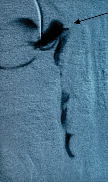
Dacryocystorhinography was conducted, which showed poor clearance of the dye and a filling defect in the right lacrimal sac and nasolacrimal duct (Figure 1).
Dacryocystogram showing filling defect (arrow) and poor clearance in the right nasolacrimal system.

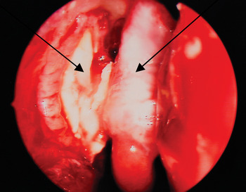
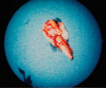
In view of the patient's persistent symptoms, elective, powered, endonasal dacryocystorhinostomy was performed using a 30° Hopkins rod. An incision was made 10 mm above the axilla of the middle turbinate, coming forward for 10 mm and then down to the root of the inferior turbinate. A D-shaped mucoperichondrial flap was elevated. A large bony osteotomy was created, using a Hajek–Koeffler punch below and a 15° curve, 2.9 mm rough diamond burr (Medtronic, Florida, USA) higher up, to expose the whole sac. The lacrimal sac was opened using an I-shaped incision created with a keratotome, which revealed a large stone filling the entire sac. The stone was removed (Figures 2 and 3) and the mucoperichondrial flap trimmed to surround the marsupialised sac. A Crawford stent was inserted and secured in the nose with a Ligaclip®.
Peri-operative endoscopic image of the dacryolith (left arrow) within the lacrimal sac, close to the middle turbinate (right arrow).

Dacryolith removed from the lacrimal sac.

The post-operative period was uneventful. The patient's epiphora improved immediately. After six months, the patient was asymptomatic and the dacryocystorhinostomy was patent; therefore, the stent was removed.
The patient had no further problems.
Discussion
Nasolacrimal duct obstruction has traditionally been managed by ophthalmologists. In recent years, various centres (including our own) have established a multidisciplinary team, comprising an otorhinolaryngologist and an ophthalmologist, in order to manage this condition.
Obstruction of the lacrimal sac or nasolacrimal duct can be acquired or congenital. Common cases of acquired obstruction are trauma and acute or chronic inflammation of the lacrimal sac. Patients with chronic dacryocystitis usually present with either recurrent dacryocystitis or epiphora, which is usually very uncomfortable.
Lacrimal sac calculi are an uncommon and often unrecognised cause of chronic epiphora. Their overall incidence is only 8.4 per cent.Reference Yazici, Hammad and Meyer1 Patients usually present with a short history of epiphora, with previous attacks of dacryocystitis.Reference Andreou and Rose2
Yazici et al. assessed the predictive factors and clinical characteristics of dacryoliths, and noted that, in patients with primary acquired nasolacrimal duct obstruction requiring dacryocystorhinostomy, dacryoliths were associated with male gender and sac distension.Reference Yazici, Hammad and Meyer1 Partial nasolacrimal duct obstruction and a history of cigarette smoking may also be relative risk factors for dacryolith formation.
Nasal endoscopic examination is essential in all cases of nasolacrimal duct obstruction. This will not only exclude any intranasal abnormalities, but also assess the state of the valve of Hasner. Piaton et al. have proposed an association between mucopurulent discharge at the valve of Hasner and lacrimal system patency, which has high specificity for dacryolithiasis.Reference Piaton, Keller, Sahel, Nguyen and Quesnot3 These authors have also hypothesised that dacryolithiasis may in many cases be caused by congenital malformation of the valve of Hasner.
Clinical suspicion of a dacryolith should arise when symptoms are intermittent or when nasolacrimal duct irrigation findings are inconsistent.Reference Wilkins and Pressly4 Dacryocystorhinography may occasionally detect a dacryolith, seen as a filling defect and poor clearance. Other imaging modalities (e.g. computed tomography) may also be useful in diagnosing both dacryolithiasis and dacryocystitis.Reference Asheim and Spickler5
In cases of dacryolithiasis, removal of stones and restoration of the nasolacrimal system is the treatment of choice.
Spontaneous expulsion of a dacryolith is rare but has been reported. However, following such expulsion, the formation of another dacryolith is likely, and the recurrence of symptoms in such cases may ultimately require a dacryocystorhinostomy.Reference Salam, Williams and Manners6
Non-surgical intervention (such as fluoroscopy-guided dacryolith removal during dacryocystoplasty) is a feasible alternative to dacryocystorhinostomy, with good clinical results.Reference Wilhelm, Hofer, Textor, Böker, Strunk and Schild7 However, this procedure requires an experienced interventional radiologist, expensive equipment and exposure to radiation.
The use of balloon dacryocystoplasty to treat obstructive epiphora is ideal for patients with circumscribed focal stenosis or nasolacrimal duct occlusion. The main contraindications to balloon dacryocystoplasty are active dacryocystitis, dacryocystolithiasis and post-traumatic lesions.Reference Berkefeld, Kirchner, Müller, Fries and Kollath8
Conventional external approach dacryocystorhinostomy is a good option, which gives direct access and good exposure. However, it has the disadvantages of invasiveness and facial scarring. The major causes of failure of the external approach are: the presence of granulation tissue, septal deviations or synechiae near the opening of the fistula; inadequate removal of the bony wall adjacent to the lacrimal sac; incorrect localisation of the lacrimal sac; and excessive peri-operative bleeding which impairs the surgical field. Most of these factors can be controlled by good surgical technique.
In contrast to external approaches, endoscopic dacryocystorhinostomy has the advantages of greater lacrimal sac visibility, quicker adaptation to anatomical variation and better results.Reference Ben Simon, Joseph, Lee, Schwarcz, McCann and Goldberg9
• Dacryoliths are an unusual cause of obstructive epiphora
• They are lacrimal sac concretions formed from cellular debris and proteins, which may calcify and cause further obstruction
• They are often involved in intermittent or chronic dacryocystitis
• Dacryocystitis is characterised by epiphora, pain, erythema, sac dilation and lacrimal punctum swelling
• Dacryoliths can be managed by a powered endonasal endoscopic approach, avoiding open surgery
Should revision be required, endoscopic dacryocystorhinostomy is safer than open approaches, with lower morbidity, and enables effective resolution of lacrimal obstruction.Reference Demarco, Strose, Araújo, Valera, Moribe and Anselmo-Lima10
Conclusion
The future of lacrimal surgery is certainly changing. Although external dacryocystorhinostomy remains the ‘gold standard’ against which other treatment methods are measured, endonasal dacryocystorhinostomy is becoming the treatment of choice for distal nasolacrimal system obstruction.Reference Shun-Shin and Thurairajan11 Similarly, we believe it is feasible to manage dacryoliths by endonasal endoscopic dacryocystorhinostomy, thus avoiding an external approach.