Introduction
Monochamus alternatus Hope (Coleoptera: Cerambycidae), as a serious wood-boring pest, is in the meanwhile a dominant vector of Bursaphelenchus xylophilus (Aphelenchida: Parasitaphelenchidae) causing destructive pine wilt disease (PWD). It follows that this pest has posed a double threat to pine forests in China and other East Asian countries (Mamiya & Enda, Reference Mamiya and Enda1979; Linit, Reference Linit1988; Hu & Wu, Reference Hu and Wu2018). In China, this beetle is currently distributed in the subtropical zone and tropics, such as Guangdong, Fujian, Hunan, and so on. Under the context of climate warming, extreme temperatures near 40°C are not uncommon in these regions (Xiong et al., Reference Xiong, Liu, Xiao, Tang, Liu, Lou, Wang and Jiang2019). For M. alternatus, the populations colonizing these regions are more vulnerable to high temperatures due to its higher per-capita energy consumption, and this detrimental effect is further aggravated by an energy trade-off during this process (González-Tokman et al., Reference González-Tokman, Córdoba-Aguilar, Dáttilo, Lira-Noriega, Sánchez-Guillén and Villalobos2020). As global warming worsens, the limitations of population dynamics and geographical distribution of M. alternatus largely arose from the frequent high temperatures in the subtropical and tropical regions (Garrad et al., Reference Garrad, Booth and Furlong2016). The development of heat tolerance, thus, is agreed to be a critical factor in determining the population fate of this specie.
M. alternatus showed strong heat tolerance in our previous study (Li et al., Reference Li, Zhao, Qiao, He, Tan and Hao2020). Only appropriately 50% of the M. alternatus population was deadly damaged when exposed to 42.5°C for 96 h and almost all individuals could endure 40°C for 96 h. This property, in M. alternatus, was achieved for a broad array of adaptive strategies against adverse high temperature, covering ecological, behavioral, and physiological aspects. In terms of individual physiology, RNA-seq data from the heat exposure revealed that genes involved in protein activity, immune responses, and signal transduction constituted the heat tolerance mechanism of M. alternatus (Li et al., Reference Li, Zhao, Qiao, He, Tan and Hao2020). Notably, the gene quantities, as well as the expression level of Heat Shock Proteins (HSPs) among the upregulated unigenes, were dominant in the physiological alterations (Li et al., Reference Li, Zhao, Qiao, He, Tan and Hao2020). Recently, it happened that there were similar cases confirming that HSPs commonly contribute to the physiological responses of insects to heat stress (Chen et al., Reference Chen, Xu, Li and Wu2014; Luo et al., Reference Luo, Ahola, Shu, Xu and Wang2015; Guo and Nian, Reference Guo and Nian2018).
HSPs can serve as molecular chaperones to conduct protein degradation, aggregation and folding under environmental stresses, such as extreme temperature, viral infection, pesticide exposure, starvation, and oxidative damage (Feder & Hofmann, Reference Feder and Hofmann1999; Crack et al., Reference Crack, Mansour, Sun and MacRae2002; Wang et al., Reference Wang, Li, Zhu, Fang and Ye2012; Zhang et al., Reference Zhang, Liu, Guo, Li, Gao, Guo and Xu2014). Moreover, certain HSPs can also participate in the biological processes of diapause, metamorphosis, fecundity, and immune responses in insects (Pockley, Reference Pockley2003; Sonoda et al., Reference Sonoda, Fukumoto, Izumi, Yoshida and Tsumuki2006; Huang et al., Reference Huang, Wang and Kang2009; Economou et al., Reference Economou, Kotsiliti and Mintzas A2017). Based on the molecular weight and function, HSP superfamily members can be generally divided into five families: HSP90, HSP70, HSP60, HSP40, and small heat shock protein (sHSP) (Feder & Hofmann, Reference Feder and Hofmann1999).
In the network of molecular chaperones, sHSPs are recruited as the first line of cell defense. Members of the HSP70 family, containing inducible HSP70 and heat shock cognate protein (HSC) 70 molecules, are capable of removing substrates from sHSPs and assisting in protein refolding and degradation in tandem with other HSPs or alone (King & Macrae, Reference King and Macrae2015). HSC70s are constitutively expressed under normal conditions, however, recent evidence indicates that their expressions can also vary in response to abiotic stresses (Prentice et al., Reference Prentice, Milton, Scheurle and Lutz2004; Wang et al., Reference Wang, Dong, Li, Hu and Ye2008; Sun et al., Reference Sun, Zhao, Sheng, Xiao, Zhang, Bai, Tan, Xiao and Xu2016). HSP70s possess two common features in most living organisms: a conserved N-terminus composed of an ATP binding domain and a variable C-terminus composed of a substrate-binding domain (Flaherty et al., Reference Flaherty, DeLuca-Flaherty and McKay1990; Lin et al., Reference Lin, Wang, Liu, Chen, Meyer, Barakat and Delseny2001; Matthias & Gierasch, Reference Matthias and Gierasch2019). Analyses of the ATPase sites of HSP70s have revealed that their activity relies on ATP supply (Mashaghi et al., Reference Mashaghi, Bezrukavnikov, Minde, Wentink, Kityk, Zachmann-Brand, Mayer, Kramer, Bukau and Tans2016). In subcellular locations, HSP70 proteins are ubiquitously detected, for instance, mitochondria, the endoplasmic reticulum (ER), the cytosol, lysosomes, and extracellular compartments (Stetler et al., Reference Stetler, Gan, Zhang, Liou, Gao, Cao and Chen2010; Wang et al., Reference Wang, Wang, Ban, Zhu, Liu and Wang2019b).
Since they were first discovered in Drosophila melanogaster larvae in 1962, HSP70 genes have been gradually identified and characterized in many insects, such as Tribolium castaneum, Bemisia tabaci, Locusta migratoria, Cydia pomonella, Lucilia sericata, and Megaphragma amalphitanum (Ritossa, Reference Ritossa1962; Qin et al., Reference Qin, Tyshenko, Wu, Walker and Robertson2003; Rizana et al., Reference Rizana, Yan and Bhadriraju2005; Tachibana et al., Reference Tachibana, Numata and Goto2005; Yin et al., Reference Yin, Wang, Tang, Hansen and Lurie2006; Chuvakova et al., Reference Chuvakova, Sharko, Nedoluzhko, Polilov, Prokhorchuk, Skryabin and Evgen'ev2017; Wang et al., Reference Wang, Zhang, Pan, Wang, Han, Niu, Shan, Hoffmann and Fang2019a). Both the transcript and protein levels of many HSP70s (PaptHAP70, AlucHSP70, and AovaHSP70) are significantly elevated after heat exposure, whereas the levels of HSC70 are only slightly increased (Vladimír et al., Reference Vladimír, Michaela and Marcelo2009; Cheng et al., Reference Cheng, Li, Wang, Liu and Keyan2016; Sun et al., Reference Sun, Zhao, Sheng, Xiao, Zhang, Bai, Tan, Xiao and Xu2016; Xin et al., Reference Xin, Lian, Li, Li, Shen, Liu and Xia2018). Moreover, the silencing of PaptHSP70 and BtabHSP70 via RNA interference significantly decreases the survival rates of Pyrrhocoris apterus and B. tabaci under high temperature (Vladimír et al., Reference Vladimír, Michaela and Marcelo2009; Wang et al., Reference Wang, Wang, Ban, Zhu, Liu and Wang2019b). Notably, in plants, further research in vitro has revealed that ATPase activity of HSP70 is stable under high temperature, thus corroborating the molecular chaperone function of HSP70s (Zhang et al., Reference Zhang, Zhou, Wang and Lin2018). However, little is known about the molecular chaperone activity of HSP70s in insects, especially in Coleopteran beetles.
Cai et al. (Reference Cai, Chen, Cheng and Lin2017) cloned one MaltHSP70 and analyzed its expression profile in M. alternatus larvae, and they revealed that the mRNA levels of this gene increased by 90-fold after exposure to 40°C for 1 h. However, the other HSP70s involved in the heat tolerance of M. alternatus remain to be further investigated. Here, six new HSP70 genes in M. alternatus were cloned and isolated from transcriptomic data. Quantitative polymerase chain reaction (qPCR) was performed to analyze the transcript levels of these genes in adults and larvae under different high-temperature treatments. Furthermore, we conducted recombinant expression and purification of the most sensitive HSP70 in M. alternatus to obtain the bioactive protein and then performed functional analyses. We aimed to provide molecular evidence for heat tolerance-related genes in M. alternatus and elucidate how this beetle utilizes HSPs to defend against extremely high temperature under global climate warming.
Materials and methods
Insects
The larvae of M. alternatus were obtained from infested host trees (Pinus massoniana) in Jiujiang, Jiangxi Province, China and reared individually on an artificial diet at a constant temperature of 25 ± 0.5°C under a relative humidity (RH) of 60 ± 5% as described by our previous experiment (Li et al., Reference Li, Zhao, Qiao, He, Tan and Hao2020). Once emergence, adults were individually provided with fresh 1- to 2-year-old P. massoniana twigs. Males and females (10:10) were mated in cages after receiving supplemental nutrition for 10 days, and the females spawned on P. massoniana trunks. The eggs were stripped from the bark carefully and hatched in opaque cell culture plates (25 ± 0.5°C, 60 ± 5% RH). The second-generation larvae were transferred into an artificial diet as described above and used for subsequent experiments.
Sample treatment and collection
To narrow the individual difference, larvae at the same stage and adults at sexual maturity were selected for the heat exposure treatment. In detail, 3-day-old fourth-instar larvae (3) and 10-day-old males (3) and females (3) were placed in environmental chambers (60 ± 5% RH) under a range of high temperatures (25, 35, 40, 45, and 50°C) and held at these temperatures for 1, 2, and 3 h.
The temperature of 40°C was close to the extreme temperature in the habitat of M. alternatus. Thus, the cuticles, haemolymph, fat bodies, guts, and Malpighian tubules were dissected from five 3-day-old molted fourth-instar larvae exposed to 40°C for 0, 1, 2, and 3 h. Three replicate groups were tested per treatment. All the samples were frozen in liquid nitrogen and stored at −80°C until utilization for qPCR.
RNA extraction, cDNA synthesis, and sequence cloning
The total RNA was isolated with RNA-easy Isolation Reagent and subsequently treated with DNase (Vazyme, Nanjing, China) following the manufacturer's instructions. The RNA quality was detected with a NanoDrop 2000c spectrophotometer (Thermo Fisher Scientific, West Palm Beach, FL, USA) and by gel electrophoresis. cDNA was synthesized from 1 μg of total RNA using a HiScript 1st Strand cDNA Synthesis Kit (Vazyme, Nanjing, China). This method was used to produce cDNA for both PCR and qPCR.
We obtained sequence fragments from the transcriptome database of M. alternatus (Li et al., Reference Li, Zhao, Qiao, He, Tan and Hao2020). Three HSP70 genes contained complete open reading frames (ORFs), while three others possessed sequence fragments as assessed with BLAST programs at the NCBI website (http://www.ncbi.nlm.gov/BLAST/) and ORF Finder (http://www.ncbi.nlm.nih.gov/gorf/gorf.html). Then, we performed PCR to correct the ORFs of the specific genes and used 5′ and 3′ rapid amplification of cDNA ends (RACE) to amplify the gene fragments (Takara, Dalian, China). The sequences of the primers used for gene cloning are listed in table S1. The PCR program included 3 min at 95°C; 30 cycles of 95°C for 30 s, 45°C for 30 s, and 72°C for 2 min; and 10 min at 72°C. Then, the PCR products were separated on a 1% agarose gel, purified with a DNA purification system (Tiangen, Beijing, China), cloned into a T/A blunt vector, and transformed into DH5α competent cells (Vazyme, Nanjing, China). Randomly selected positive clones for each construct were sequenced by JIE LI BIOLOGY (Shanghai, China).
Sequence analysis
Sequence alignment and splicing analysis were performed with the DNAMAN software package (Lynnon Corporation, Pointe-Claire, Quebec, Canada). The molecular weights and isoelectric points of the mature proteins were calculated using the ExPASy server program (http://www.expasy.ch/cgi-bin/pi_tool). The online protein structure prediction tool SWISS-MODEL (https://swissmodel.expasy.org) was used to predict the HSP70 tertiary structures. The prediction of subcellular localization was performed by CELLO v2.5 (http://cello.life.nctu.edu.tw/) (Yu et al., Reference Yu, Lin and Hwang2004).
Phylogenetic trees were constructed based on the deduced amino sequences of HSP70s from M. alternatus and insects of six other orders (Coleoptera, Lepidoptera, Hemiptera, Diptera, Orthoptera, and Hymenoptera) in the Unigene database of the NCBI. Maximum-likelihood phylogenetic trees were constructed using MEGA 6.0, with 1000 bootstrap replicates (Tamura et al., Reference Tamura, Peterson, Peterson, Stecher, Nei and Kumar2011).
qPCR
The total RNA of samples mentioned above was extracted, and 0.5 μg was reverse-transcribed into cDNA using a cDNA Synthesis Kit. The qPCR primers (table S1) were designed online (https://www.genscriptcom/tools/real-time-pcr-tagman-primer-design-tool). The Ribosomal Protein L10 (RPL10) gene was used as a reference gene (Li et al., Reference Li, He, Tao, Gong, Chen and Hao2018). Reactions were performed on an Applied Biosystem 7500 System (USA) using Hieff UNICON® qPCR SYBR Green Master Mix (YEASEN, Shanghai, China). The PCR procedure was as follows: 5 min at 95°C; 40 cycles of 10 s at 95°C, and 40 s at 60°C; and then a melting curve analysis for continuous fluorescence monitoring while the sample was slowly heated from 60 to 95°C. Ten-fold dilution series of the cDNA templates were utilized to calculate the efficiency of these primers for qPCR (table S1). The relative expression levels across samples were measured based on the 2−ΔΔCt method (Livak & Schmittgen, Reference Livak and Schmittgen2001).
Recombinant expression and purification of HSP70s
The PET-28a vector was digested with the restriction enzymes BamHI and NotI. The forward and reverse primers used for the amplification of the complete ORFs of the HSP70s contained homologous BamHI and NotI enzyme site sequences (table S1). The PCR products and linearized pET-28a (+) vector were ligated with a ClonExpress II One Step Cloning Kit (Vazyme, Nanjing, China). The recombinant plasmids (pET28-HSP70) were identified by sequencing and then transformed into Escherichia coli (E. coli) BL21 cells (Vazyme, Nanjing, China) for protein expression.
Single clones were selected and checked by PCR. The verified sample was transferred to 100 ml of fresh LB medium with kanamycin (30 μg ml−1) and then incubated until the optical density at 600 nm (OD600) approached 0.6. The samples were incubated for another 6 h at 37°C (with shaking at 200 rpm) with a final concentration of 1 mM isopropyl-β-D thiogalactopyranoside (IPTG; GENERAY, Shanghai, China). The cell pellets were suspended in balance buffer (50 mM NaH2PO4 and Na2HPO4, 300 mM NaCl, 20 mM imidazole, pH = 7.9), stirred and disrupted by sonication in ice-water mixtures. After centrifugation at 12,000 rpm for 15 min at 4°C, the supernatant protein was purified by His60 Ni Superflow Resin & Gravity Columns (Takara, Dalian, China). The purified recombinant proteins were analyzed by 7.5% SDS-PAGE and quantified using a total protein assay kit (with the standard BCA method) (Jian Cheng, Nanjing, China). Then, the obtained protein was stored at −80°C for the subsequent experiments.
Western blot
The antibody of the recombinant protein was generated and purified as previously described (Wang et al., Reference Wang, Fang, Bao, Jin, Zhu, Wang, Liu, Ji, Wang, Xu and Sima2014).
The total protein from three fourth-instar larvae, two adult females, and two adult males were individually extracted with phosphate-buffered saline (PBS). The protein concentration was measured by the method described above and diluted to 5 μg μl−1. Then, 20 μl extracts were separated on 7.5% SDS-PAGE gals and transformed to polyvinylidene difluoride membrane (PVDF). The membrane was blocked with 5% milk, incubated with purified recombinant protein antibody, and then incubated with horseradish peroxidase (HRP)-labelled anti-rabbit IgG (Beyotime, Shanghai, China). Proteins were visualized using the West Pico ECL (Biosharp, Wuhan, China).
HSP70 ATPase activity assay
ATPase activity was measured following the method described by Wang et al. (Reference Wang, Zhang, Pan, Wang, Han, Niu, Shan, Hoffmann and Fang2019a, Reference Wang, Wang, Ban, Zhu, Liu and Wang2019b) with slight modifications. Briefly, 100 μl reaction mixtures were created, with each containing 2.0 μg of purified HSP70/bovine serum albumin (BSA)/ddH2O, 1.0 mM Bis-Tris, 1.0 mM KCl, 0.1 mM MgCl2, and 0.01 mM ATP. The reaction mixtures were incubated at 25, 30, 35, and 40°C for 10 min. Then, 10 μl of 55% HClO4 was added to stop the reaction. The mixtures were cooled on ice for 10 min and centrifuged at 12,000 rpm for 3 min (Zhang et al., Reference Zhang, Zhou, Wang and Lin2018). The concentration of inorganic phosphorus was determined with a Micro Tissue Inorganic Phosphorus Content Assay Kit (Solarbio, Beijing, China). The ATPase activity was defined as the amount of inorganic phosphorus produced upon catalysis by 1 mg of MaltHSP70 in 1 min (nmol min mg−1).
Cell viability assay
The thermotolerance of E. coli BL21 expressing HSP70 was assessed following the method of Crack et al. (Reference Crack, Mansour, Sun and MacRae2002). After IPTG induction, as mentioned above, PET-28a-HSP70 cultures (0.5 mL) were diluted to a density of 1 × 10⁷ cells ml−1 and incubated at 50°C in a water bath. Ten-microliter samples were removed after 30 min of heat shock, diluted in 990 μl of cold LB, and maintained on ice. The solutions from the different heat shock treatments (100 μl each) were plated on LB agar containing 30 μg ml kanamycin. The colonies were counted to calculate the survival rates after 16 h at 37°C (the ratios of viable cell numbers per plate between the treated samples and untreated samples). All these experiments were conducted in triplicate. Cultures containing only PET-28a were used as control samples.
Statistical analyses
All statistical analyses were executed by SPSS 20.0 software (SPSS, Chicago, USA). Statistical differences in HSP70 gene expression profiles were determined by a one-way analysis of variance followed by Tukey's honestly significant difference (HSD) test. The survival rates of recombinant E. coli and the ATPase activity levels at different temperatures were both compared via t-tests. Differences were considered significant at P < 0.05.
Results
Sequence analysis of MaltHSP70s
Molecular characterization
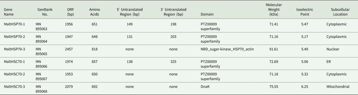
According to the transcriptomic database (Li et al., Reference Li, Zhao, Qiao, He, Tan and Hao2020), six HSP70s, MaltHSP70-1, MaltHSP70-2, MaltHSP70-3, MaltHSC70-1, MaltHSC70-2, and MaltHSC70-3, were identified and cloned (GenBank No.: MN895063-68). The ORFs of these genes encoded sequences of 648–692 amino acids with molecular weights of 71.16–75.55 kDa and isoelectric points of 5.00–6.25 except for the ORF of MaltHSP70-3 (table 1). A conserved domain search showed that MaltHSP70s and MaltHSC70s were characterized by the PTZ00009 superfamily domain (description: heat shock 70 kDa protein). Despite possessing a predicted molecular weight of 91.61 kDa, MaltHSP70-3 was screened to have an NBD_sugar-kinase_HSP70_actin domain (table 1). Thus, we assumed that MaltHSP70-3 was an isoform of MaltHSP70 given the information from the NCBI database.
Summary of HSP70 genes identified in M. alternatus

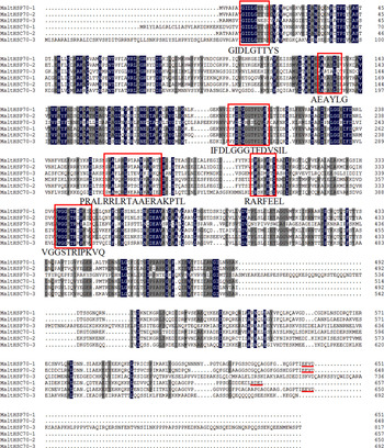
All three highly conserved regions (GIDLGTTYS, IFDLGGGTFDVSIL, and VGGSTRIPKVQ) defining HSP70 family signatures were found in the obtained sequences of MaltHSP70s and MaltHSC70s (fig. 1). Three other typical motifs, i.e., an ATP-GTP binding site (AEAYLGKS), a bipartite nuclear localization signal (PRALRRLRTAAERAKRTL), and a non-organellar consensus motif (RARFEEL), were also highly conserved in these genes (fig. 1) (Daugaard et al., Reference Daugaard, Rohde and Marja2007). The C-terminal domain was less conserved than the ATPase domain (fig. 1). Subcellular localization indicated that MaltHSP70-1, MaltHSP70-2, and MaltHSC70-2 were located in the cytoplasm, while MaltHSC70-1 was detected in the ER (table 1). The conserved motifs ‘EEVD’ and ‘RDEL’ in the C-terminus confirmed these results (fig. 1). MaltHSP70-3 and MaltHSC70-3 were predicted to localize to the nucleus and mitochondria, respectively (table 1).
Alignment of the deduced amino acid sequences of six HSP70s in M. alternatus. Amino acids conserved in all HSP70s and over 75% of HSP70s are shaded in dark blue and grey, respectively. HSP70 family signatures are indicated with red boxes. The cytoplasmic (EEVD) and ER (RDEL) C-terminal regions are emphasized with red underlines.

Structural analysis
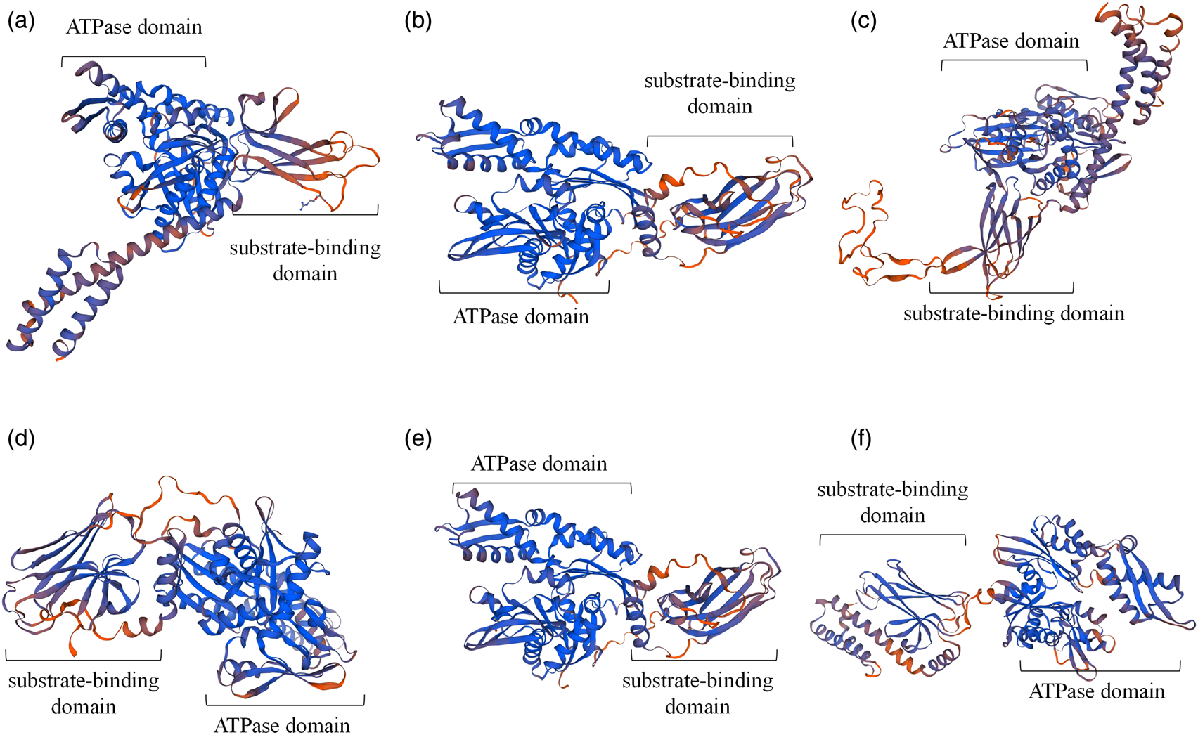
The three-dimensional structures of MaltHSP70s and MaltHSC70s were also predicted via the X-ray and NMR methods using the other HSPs and HSCs as the best templates, which shared 38.79%-88.77% sequence identity with MaltHSP70s and MaltHSC70s (table S2). As shown in figs 2a–f, all MaltHSP70s and MaltHSC70s were composed of an ATPase domain and substrate-binding domain enriched for β-strands.
Tertiary structures of six HSP70s in M. alternatus predicted by SWISS-MODEL. (a) MaltHSP70-1; (b) MaltHSP70-2; (c) MaltHSP70-3; (d) MaltHSC70-1; (e) MaltHSC70-2; and (f) MaltHSC70-3. The specific modelling information is shown in table S2.

Phylogenetic analysis
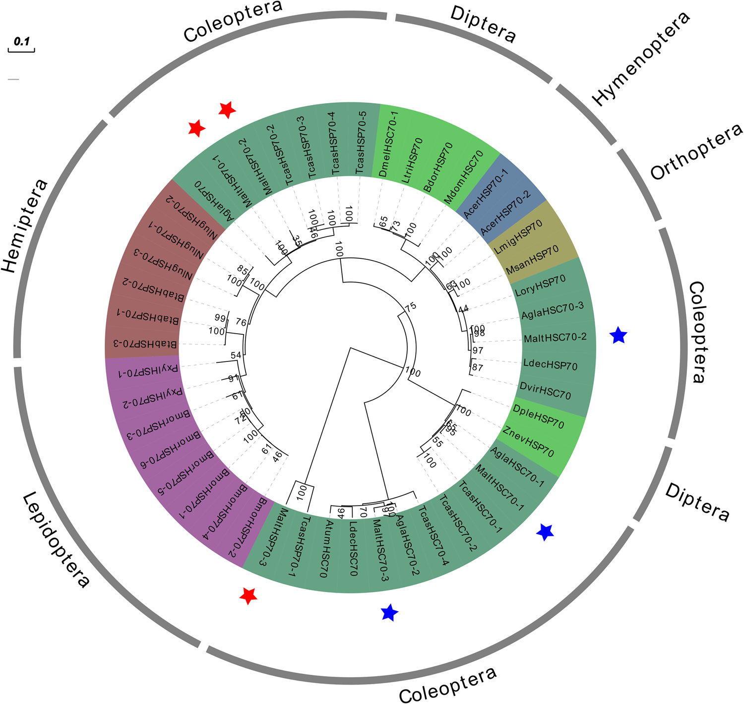
A phylogenetic tree of HSP70s from M. alternatus and insects in six other orders, Coleoptera, Lepidoptera, Hemiptera, Diptera, Orthoptera, and Hymenoptera, was constructed based on the amino acid sequences to investigate the evolutionary relationships (fig. 3). All MaltHSP70s and MaltHSC70s were classified into the order Coleoptera. Three MaltHSC70s clustered with HSCs from Anoplophora glabripennis, while three MaltHSP70s were on a branch with HSPs from
Phylogenetic analysis of amino acid sequences of HSP70s in M. alternatus and insects from six other orders conducted using the maximum-likelihood method with 1000 bootstrap replications. The sequence information used in the construction of this phylogenetic tree is listed in table S3. MaltHSP70s and of MaltHSC70s are marked with red and blue stars, respectively.

T. castaneum. In addition, HSP70s and HSC70s from the same orders of insects were located on nearby branches, which was consistent with their traditional taxonomic statuses.
Expression profiles of MaltHSP70s and MaltHSC70s
Real-time PCR was performed to examine the expression of MaltHSP70s and MaltHSC70s in fourth-instar larvae and 10-day-old adults subjected to heat stress. In addition, the transcript levels of these genes in the cuticles, haemolymph, fat bodies, guts, and Malpighian tubules of fourth-instar larvae after heat stress were determined.
Temporal expression patterns after heat stress in larvae
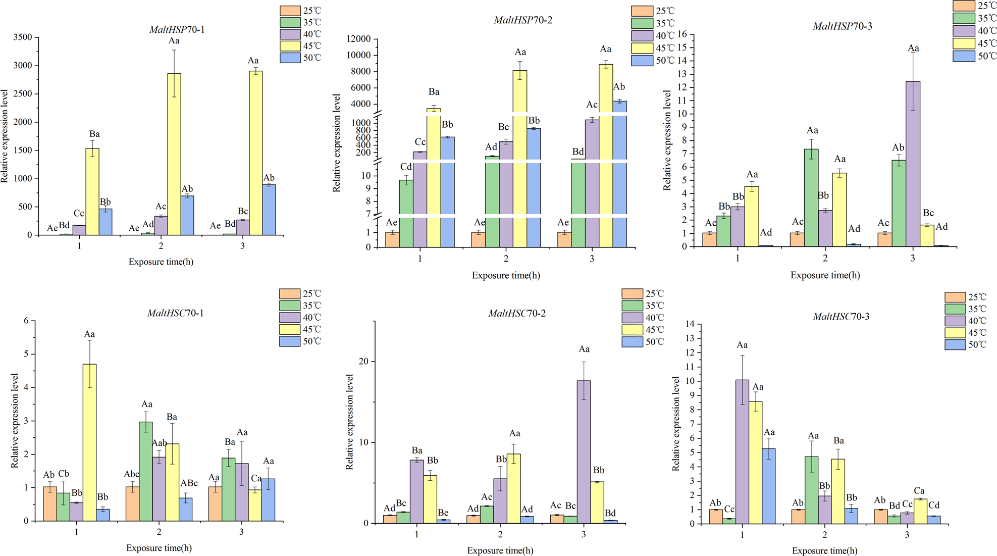
As shown in fig. 4, three MaltHSP70s were upregulated after exposure to 35, 40, 45, and 50°C for 1, 2, and 3 h. The relative expression levels gradually elevated with increasing treatment durations and temperatures except in the 50°C group. Notably, the transcript levels of MaltHSP70-1 and MaltHSP70-2 in the group under the 45°C treatment for 3 h were 2904- and 8898-fold higher than the levels in the control group, respectively.
Temporal expression patterns of six HSP70s after heat stress in M. alternatus larvae. Larvae reared at 25°C were used as controls. Different letters on error bars indicate significant differences among the different temperature treatments; and capital letters are used for different duration times at the same temperature. Significance was determined by analysis of variance (P < 0.05).

MaltHSC70s exhibited various expression patterns under high temperatures (fig. 4). For example, the relative mRNA levels of MaltHSC70-1, MaltHSC70-2 and MaltHSC70-3 peaked after exposure to 45°C for 2 h, 40°C for 3 h and 40°C for 1 h, respectively (5-, 17-, and 10-fold higher than the control levels, respectively).
Temporal expression patterns after heat stress in males and females
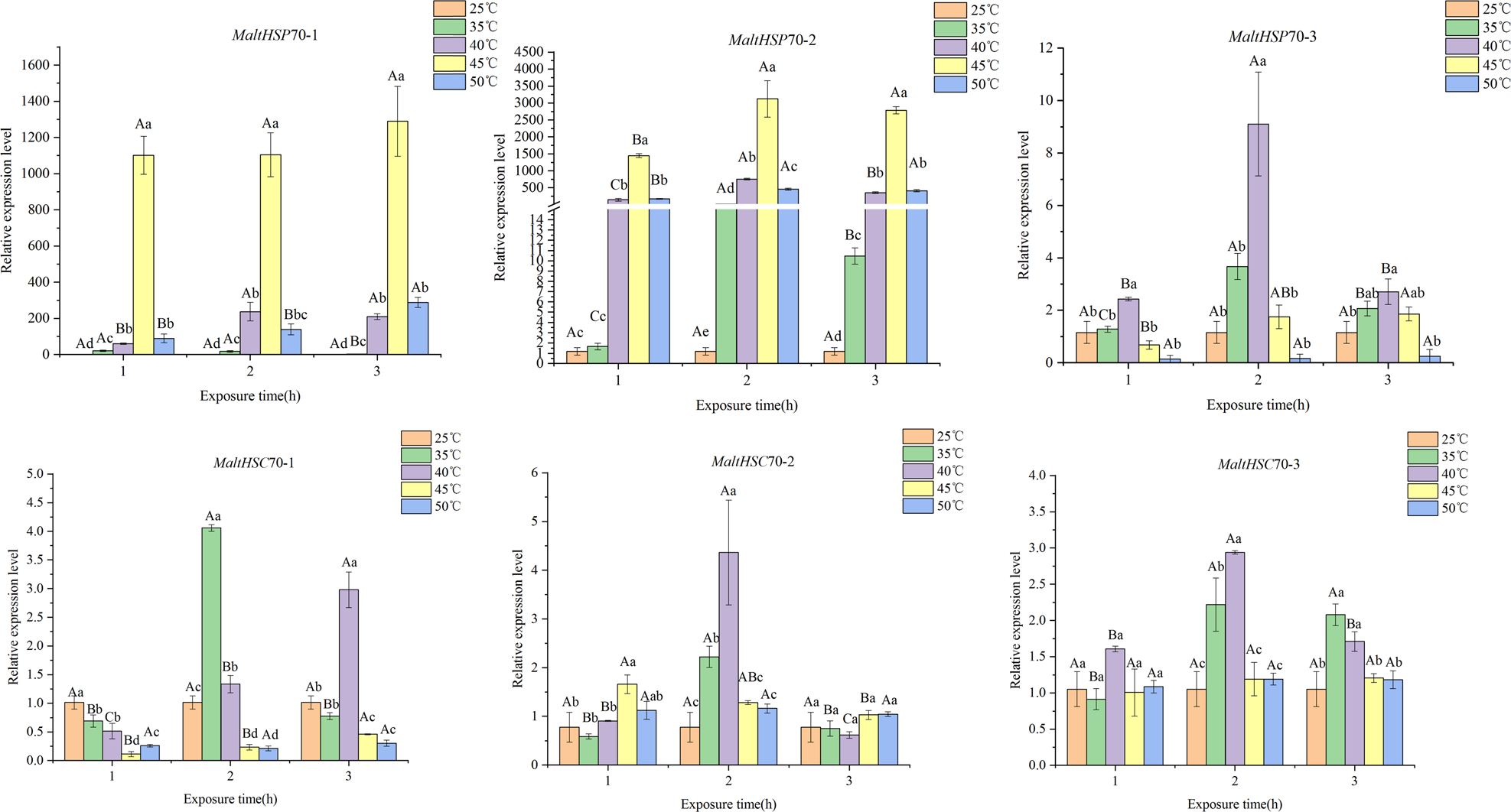
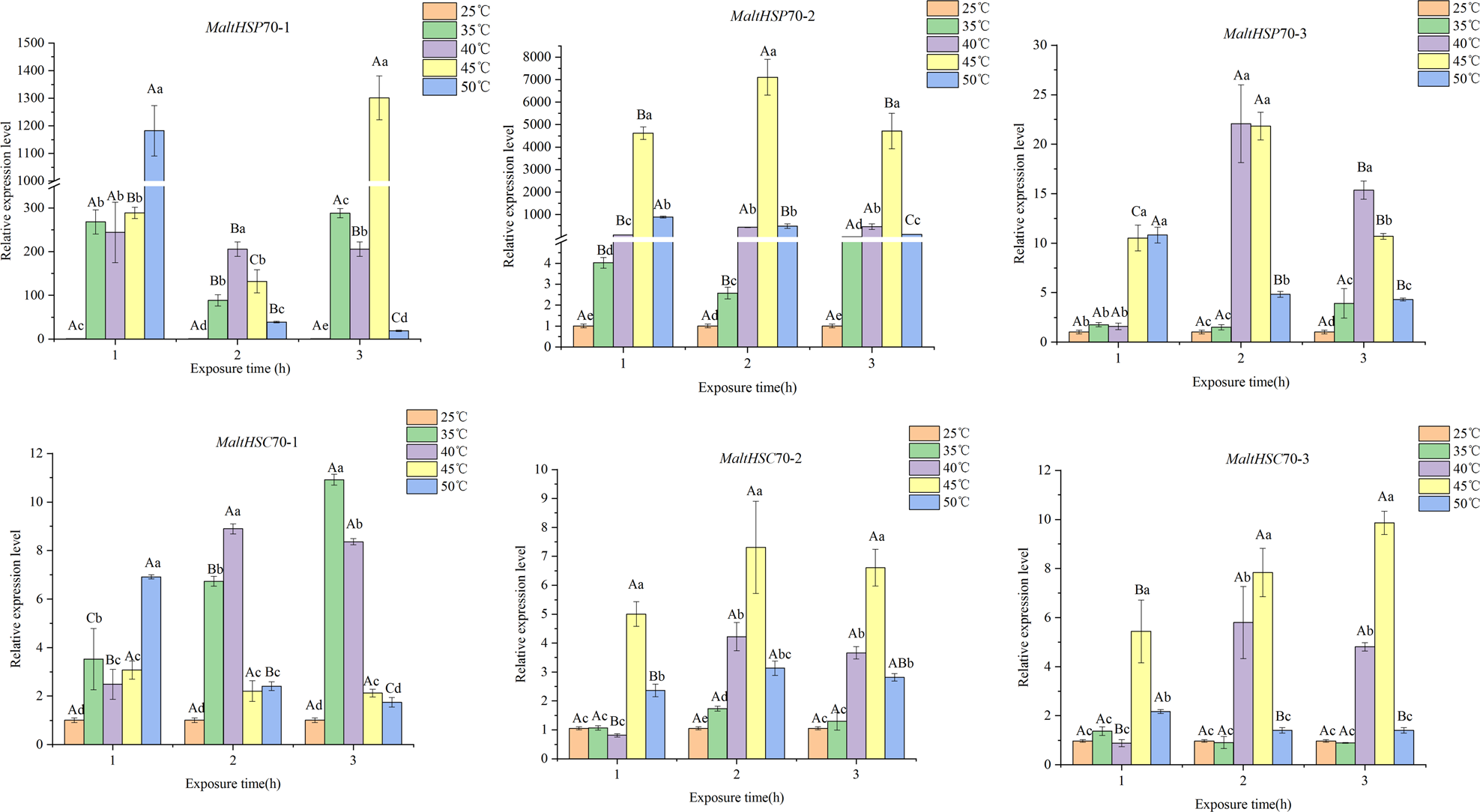
Similar to the case in larvae, MaltHSP70 expression in adults was markedly induced by high-temperature exposure (fig. 5). However, the transcript expression of MaltHSC70s in adults increased with the temperature only in a few treatment groups.
Temporal expression patterns of six HSP70s after heat stress in M. alternatus females. Females reared at 25°C were used as controls. Different letters on error bars indicate significant differences among the different temperature treatments; and capital letters are used for different duration times at the same temperature. Significance was determined by analysis of variance (P < 0.05).

In females, the expression of MaltHSP70-1 began to increase after exposure to 35°C for 1 h and peaked (1101-fold higher than control expression) after exposure to 45°C for 1 h, and did not show differences among the various time points. The transcript levels of MaltHSP70-2 increased with not only higher temperatures but also longer exposure times, and the highest relative expression level of this gene, which was 3121-fold higher than that under control conditions, occurred after exposure to 45°C for 2 h. The three MaltHSC70s were slightly upregulated after exposure to 35-40°C for 1 and 2 h.
The first two MaltHSP70s in males also showed upregulation under exposure to 35°C, and the highest expression levels, which were 1301- and 7109-fold higher than the control levels, were observed after exposure to 45°C for 3 and 2 h, respectively (fig. 6). The expression levels of the three MaltHSC70s in males were still slightly increased by approximately 10-fold.
Temporal expression patterns of six HSP70s after heat stress in M. alternatus males. Males reared at 25°C were used as controls. Different letters on error bars indicate significant differences among the different temperature treatments; and capital letters are used for different duration times at the same temperature. Significance was determined by analysis of variance (P < 0.05).

Tissue specific patterns
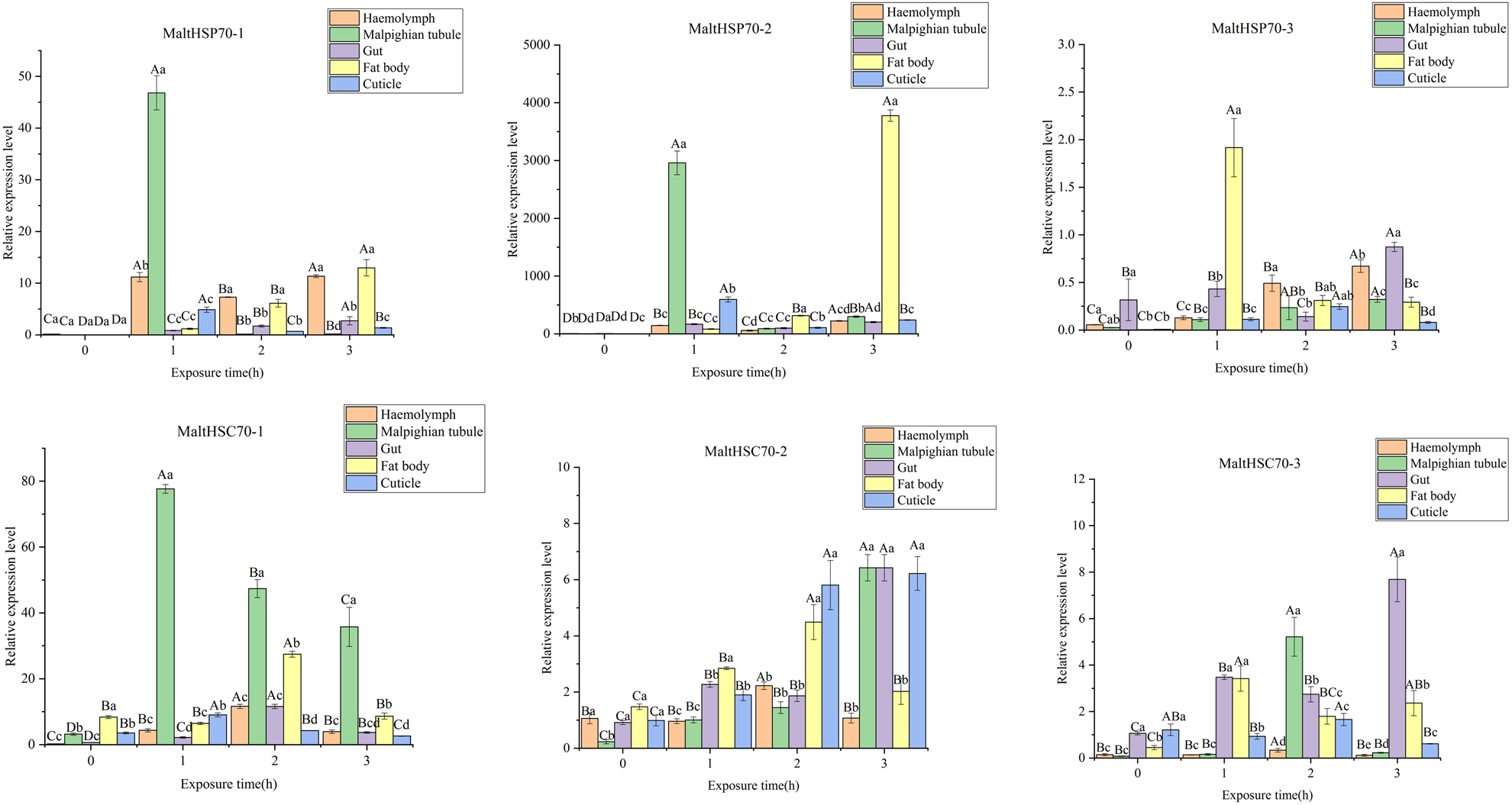
All MaltHSP70s and MaltHSC70s were commonly expressed in all tested tissues, and these genes had various levels of induction after exposure to 40°C (fig. 7). The upregulation of HSP70s mainly occurred in Malpighian tubules and fat bodies. For example, the highest expression levels of MaltHSP70-1, MaltHSP70-2, and MaltHSC70-1 were observed in Malpighian tubules after exposure to 40°C for 1 h. Both MaltHSP70-2 and MaltHSP70-3 exhibited abundant levels in fat bodies when the larvae were subjected to heat stress. Furthermore, the cognate MaltHSC70s showed moderate upregulation in the cuticle and gut in the process of heat shock.
Expression patterns of six HSP70s after exposure to 40°C for 0, 1, 2, and 3 h in M. alternatus larval tissues (haemolymph, Malpighian tubules, guts, fat bodies, and cuticles). Tissues from larvae reared at 25°C were used as controls. Different letters on error bars indicate significant differences among the different tissues; and capital letters are used for different duration times in the same tissue. Significance was determined by analysis of variance (P < 0.05).

Functional analysis of the MaltHSP70-2 protein
MaltHSP70-2 was the most upregulated protein not only in the M. alternatus larvae but also in the adults after heat stress; therefore, MaltHSP70-2 was overexpressed in E. coli BL21, and its function was investigated.
Recombination and purification of the MaltHSP70-2 protein
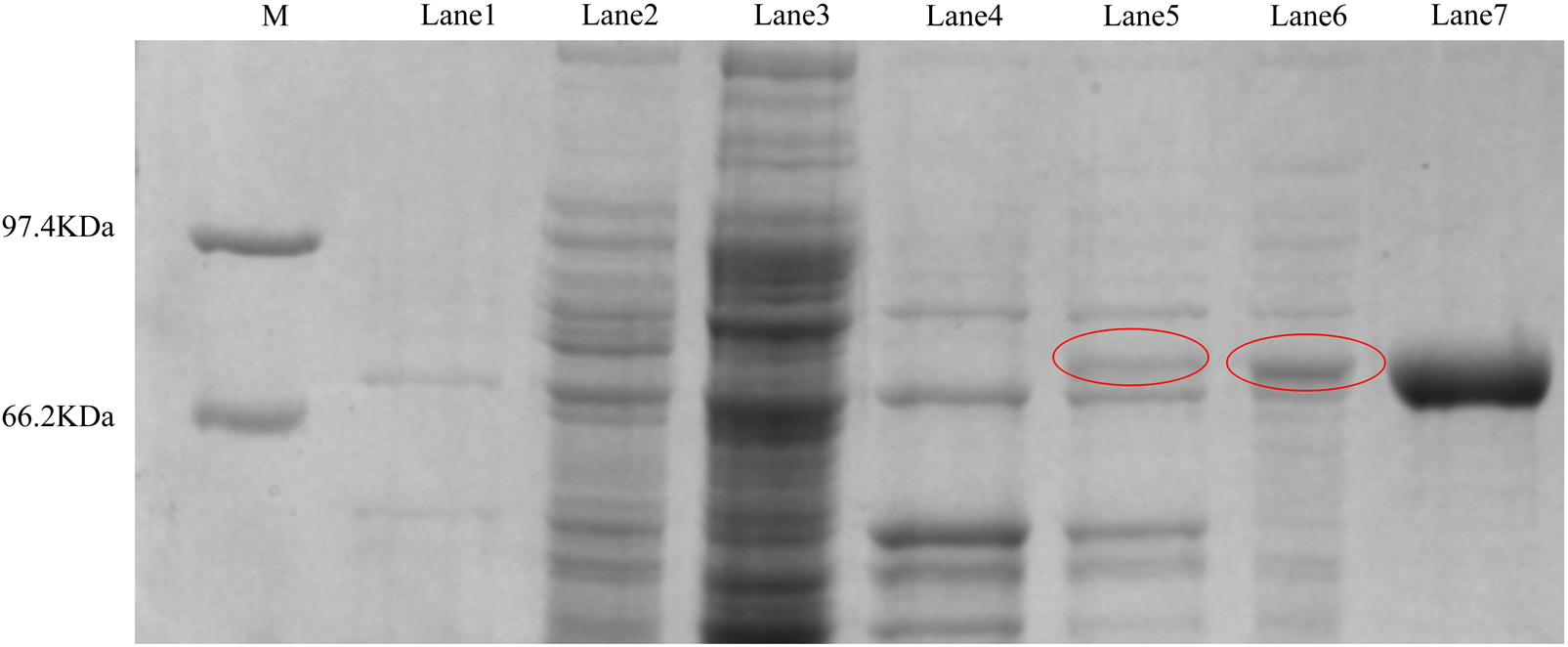
After 1 mM IPTG treatment, the supernatants and pellets of E. coli BL21 with expression plasmids were analyzed by SDS-PAGE. A distinct band with a molecular weight of ~71.16 kDa (fig. 8) was revealed, which was consistent with the predicted molecular mass. The soluble recombinant protein was further purified using a Ni2+-His column, and the concentration of the purified protein was 1.03 mg ml−1. The western blot showed that the antibody generated from the fusion protein could detect the target protein of approximately 70 kDa in the fourth instar larvae and females and males; therefore, the purified protein was identified as MaltHSP70-2.
SDS-PAGE analysis of MaltHSP70-2 after recombinant expression and purification. M: protein molecular standard; lane 1: supernatant of the sonicated pet-28a vector; lane 2: pellet of the sonicated pet-28a vector; lane 3: supernatant of sonicated MaltHSP70-2 (without induction); lane 4: pellet of the sonicated pet-28a vector (without induction); lane 5: supernatant of the sonicated MaltHSP70-2 after induction; lane 6: pellet of the sonicated MaltHSP70-2 after induction; and lane 7: purified MaltHSP70-2.

ATPase activity of the MaltHSP70-2 protein under different temperatures
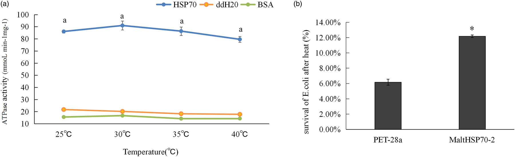
The ATPase activity levels of purified MaltHSP70 were investigated at temperatures of 25, 30, 35, and 40°C and determined to be 86.08, 90.99, 86.36, and 79.60 nmol min mg−1 higher than those of ddH2O and BSA, respectively (fig. 9a). Furthermore, there were no significant differences among the ATPase activity levels at different temperatures (P > 0.05).
(a) ATPase activity of MaltHSP70-2 at 25, 30, 35, and 40°C. Bars with different letters are significantly different (P < 0.05). (b) Survival of E. coli transformed with pet-28a containing MaltHSP70-2 after heat stress (50°C). Labelling of a column with an asterisk indicates significantly different survival between E. coli expressing MaltHSP70-2 and control E.coli as determined by t-test (P < 0.05).

Thermotolerance of E. coli after expression of MaltHSP70-2 cloned into pET-28a(+)
Bacteria transformed with PET-28a-HSP70 showed greater thermotolerance than cells transformed with PET-28a (+) (fig. 9b), and the survival of E. coli was increased by 6.17 to 12.17% after the overexpression of MaltHSP70-2. The results illustrated that the synthesis of MaltHSP70-2 proteins enhanced the thermotolerance of E. coli.
Discussion
HSP70s are found ubiquitously in all living organisms and serve as the dominant members in the HSP superfamily molecular network (Kim et al., Reference Kim, Hipp, Bracher, Hayer-Hartl and Hartl2013). HSP70s have already been identified in insects, and research has revealed that the accumulation of HSP70s is elicited by exposure to high temperatures, with peaks in HSP70 transcript levels showing significant positive correlations with heat tolerance (Sakano et al., Reference Sakano, Li, Xia, Yamamoto, Banno, Fujii and Aso2006; Sorensen et al., Reference Sorensen, Kristensen, Kristensen and Loeschcke2007; Wang et al., Reference Wang, Fang, Bao, Jin, Zhu, Wang, Liu, Ji, Wang, Xu and Sima2014).
In this study, three MaltHSP70s and three MaltHSC70s were found to constitute the HSP70 family of M. alternatus. The presence of a conserved 45 kDa ATPase pocket in the N-terminus indicated that MaltHSP70s and MaltHSC70s were characterized by rather weak ATPase activity (Jinwal et al. Reference Jinwal, Miyata, Koren, Jones, Trotter, Chang, O,Leary, Morgan, Lee, Shults, Rousaki, Weeber, Zuiderweg, Gestwicki and Dickey2009). In contrast, the diverse 25 kDa C-termini of these proteins contained substrate-binding sites that exhibited structures similar to that of HSP20 (Wang et al., Reference Wang, Fang, Bao, Jin, Zhu, Wang, Liu, Ji, Wang, Xu and Sima2014; Zhang et al., Reference Zhang, Liu, Guo, Li, Gao, Guo and Xu2014; Li et al., Reference Li, He, Tao, Gong, Chen and Hao2018). our results support the hypothesis that HSP70 can remove the outer sHSP shell and bind to the substrate core during protein degradation (Zwirowski et al., Reference Zwirowski, Klosowska, Obuchowski, Nillegoda, Piróg, Ziętkiewicz, Bukau, Mogk and Liberek2017). In many species, HSP70s are generally located in the cytoplasm and ER. The proteins in these locations are characterized by ‘EEVD’ and ‘RDEL’ motifs in the C-terminus, respectively (Chen et al., Reference Chen, Xu, Li and Wu2014; Giannetto et al., Reference Giannetto, Oliva, Mazza, Mondelo, Savastano, Mauceri and Fasulo2017; Wang et al., Reference Wang, Wang, Ban, Zhu, Liu and Wang2019b). In our research, interestingly, MaltHSP70s and MaltHSC70s were also predicted to exist in the cell nucleus and mitochondria. It is generally assumed that the nucleus and mitochondria are central hubs of cellular metabolism (Paine et al., Reference Paine, Moore and Horowitz1975). That said, MaltHSP70s and MaltHSC70s are likely to involve in cellular energy metabolism under normal or heat conditions. This assumption has the potential for further investigation. The phylogenetic analysis indicated that MaltHSP70 and MaltHSC70s shared the highest homology with sequences in Coleopteran species, such as A. glabripennis and T. castaneum, which was congruent with the close taxonomic relationships of these species. Lastly, high degrees of sequence conservation among insects belonging to the same orders suggested HSP70 has undergone order-specific duplication, supporting the point that HSP genes may have duplicated early in insect evolution (Huang et al., Reference Huang, Wang and Kang2008; Guo and Nian, Reference Guo and Nian2018).
Based on the verified sequences of MaltHSP70s and MaltHSC70s, we assessed the expression profiles of HSP70s in M. alternatus larvae and adults in response to heat stress. HSP70 is one of the critical predictors of insect tolerance to heat stress. Previous studies showed that higher induced levels of HSP corresponded to stronger heat tolerance in arthropods (Chang et al., Reference Chang, Zhang, Chen, Lu, Gong and Du2018; Chen et al., Reference Chen, Zhang, Jiang, Meng, Qiang and Wang2020). MaltHSP70s accumulated by over a thousand-fold after heat shock, which implied that MaltHSP70s were more sensitive than HSP70s in other insects (Li et al., Reference Li, Lu, Feng and He2009; Udaka et al., Reference Udaka, Ueda and Goto2010; Wang et al., Reference Wang, Yang, Zhao and Han2015; Quan et al., Reference Quan, Duan, Ladd and Krell2018). Furthermore, the maximum level of MaltHSP70 mRNA was observed after more severe heat exposure (i.e. higher temperature and longer exposure time) compared with that of the HSPs of other insects, such as P. apterus and Sitodiplosis mosellana (Vladimír et al., Reference Vladimír, Michaela and Marcelo2009; Cheng et al., Reference Cheng, Li, Wang, Liu and Keyan2016). These studies suggest that the extremely sensitive MaltHSP70s might be the intrinsic mediators of the strong heat tolerance of M. alternatus (Li et al., Reference Li, Zhao, Qiao, He, Tan and Hao2020).
The expression of MaltHSP70 as well as SmosHSP70, GmHSP70 were no longer increased when temperatures reached 50°C (Chen et al., Reference Chen, Xu, Li and Wu2014; Cheng et al., Reference Cheng, Li, Wang, Liu and Keyan2016). It is not surprising that the ability of HSPs to relieve damage and increase survival rates under severe temperature stresses might be limited (Feder & Hofmann, Reference Feder and Hofmann1999; Chen et al., Reference Chen, Xu, Li and Wu2014). An alternative explanation is a trade-off strategy of energy allocation will probably occur instead of the unrestricted synthesis of HSP70s or other HSPs requiring immense energetic costs.
This trade-off strategy is specifically reflected in exploiting heat tolerance mechanisms and fecundity (González-Tokman et al., Reference González-Tokman, Córdoba-Aguilar, Dáttilo, Lira-Noriega, Sánchez-Guillén and Villalobos2020). Sorensen et al. (Reference Sorensen, Kristensen, Kristensen and Loeschcke2007) found a significant difference between male and female fruit flies in the adaptability to high temperature, which was relevant to the fact redundant HSPs can impair female fertility (Sørensen et al., Reference Sørensen, Michalak, Justesen and Loeschcke1999; Chang et al., Reference Chang, Zhang, Chen, Lu, Gong and Du2018). The females of M. alternatus were more vulnerable to heat stress than males (data not shown), which might be caused by the lower induction of MaltHSP70-2 and MaltHSP70-3 in females. Moreover, M. alternatus likely exhibited a similar trade-off between heat resistance (in the form of HSP70 expression) and fecundity.
HSC70 family members are known to contribute to cellular homeostasis and immunity (Hartl & Hayer-Hartl, Reference Hartl and Hayer-Hartl2002; Luo et al., Reference Luo, Ahola, Shu, Xu and Wang2015; Cheng et al., Reference Cheng, Li, Wang, Liu and Keyan2016; Wang et al., Reference Wang, Wang, Ban, Zhu, Liu and Wang2019b). In our findings, the expression of the MaltHSC70 family was low but non-zero under some heat treatments, which demonstrated that the cognate HSP70s played a subtle role in the heat tolerance of M. alternatus.
In addition, the heat responses of HSP70s were investigated specifically in M. alternatus larval tissues. The diverse expression patterns of MaltHSP70s and MaltHSC70s suggested that individual HSP70 genes have unique roles in various tissues. Fat bodies and Malpighian tubules were found to be the tissues most sensitive to heat stress. Our previous study found that thermal stress damages energy reserves and metabolism in M. alternatus (Li et al., Reference Li, Zhao, Qiao, He, Tan and Hao2020). The fat body is a dynamic tissue involved in energy storage and release (Arrese and Soulages, Reference Arrese and Soulages2010; Chandrakanth et al., Reference Chandrakanth, Ponnuvel, Moorthy, Sasibhushan and Sivaprasad2015; Quan et al., Reference Quan, Duan, Ladd and Krell2018). Thus, HSP70s may confer protection to proteins related to metabolism in M. alternatus or even other insects. Heat stress can cause the production of toxic substances, and HSPs could be involved in the catabolism of toxic metabolites or the reabsorption of water in Malpighian tubules (Shen et al., Reference Shen, Gu, Huang, Zheng, Liu, Xu, Feng and Kang2011; Quan et al., Reference Quan, Duan, Ladd and Krell2018). Thus, the highly expressed MaltHSP70s in Malpighian tubules are hypothesized to participate in detoxification during heat exposure.
Our functional analysis based on prokaryotic expression focused on MaltHSP70-2, whose transcripts were at the highest levels upon heat exposure. As we have described above, HSP70s exist in an ATP-bound state under normal conditions. In the present study, MaltHSP70-2 had stable ATPase activity at 25–40°C. We hypothesize that MaltHSP70-2 sustains its refolding activity and remains efficient when M. alternatus undergoes heat shock and this characteristic of MaltHSP70-2 plays a vital role in the heat tolerance of M. alternatus. Additionally, MaltHSP70-2 expression increased heat resistance in recombinant E. coli, and similar phenomena have been observed in studies on Apis cerana cerana and Laodelphax striatellus (Zhang et al., Reference Zhang, Liu, Guo, Li, Gao, Guo and Xu2014; Wang et al., Reference Wang, Zhang, Pan, Wang, Han, Niu, Shan, Hoffmann and Fang2019a). These results indicate that MaltHSP70-2 expression could enhance cellular thermotolerance and support the hypothesis that MaltHSP70-2 is an important contributor to the mechanism of heat tolerance in M. alternatus.
Here, we have identified and characterized six HSP70s: three MaltHSP70s and three MaltHSC70s. These proteins possess conserved HSP70 family structures and are predicted to be located in diverse cellular components. We confirmed the sensitive HSP70s of M. alternatus was a potent weapon against heat stress, while the energy trade-off between heat tolerance and fertility restrained the induction threshold of HSP70s. In addition, the expression of HSP70s in larval fat bodies and Malpighian tubules might be related to metabolic reactions and detoxification under heat exposure. Recombinant MaltHSP70-2 was shown to have stable ATPase activity during heat treatment and to enhance the heat tolerance of E. coli in vitro, thus providing insights into the mechanism underlying the participation of MaltHSP70 in heat response. In conclusion, our results demonstrate that MaltHSP70s and MaltHSC70s are vital genes for the thermotolerance and widespread occurrence of M. alternatus. However, functional analyses of other HSP family numbers in M. alternatus should be performed and the mode of MaltHSP70-2 action in vivo remains to be further explored.
Supplementary material
The supplementary material for this article can be found at https://doi.org/10.1017/S0007485320000541
Acknowledgements
This work was supported by National Key R & D Program of China (2018YFC1200400), Postgraduate Research & Practice Innovation Program of Jiangsu Province (KYCX19-1083) and National Science Foundation of China under Grant (31470650 and 31170606).