Introduction
Assisted reproductive technologies (ARTs) can increase the risk of birth defects and other perinatal adverse events in the offspring.Reference Qin, Liu, Sheng, Wang and Gao1,Reference Lv, Diao and Du2 Furthermore, there is a positive association between ART, particularly in vitro fertilization (IVF), and a higher risk of imprinting disorders, possibly caused by epigenetic modifications in imprinted genes.Reference Cortessis, Azadian and Buxbaum3,Reference Henningsen, Gissler and Rasmussen4
Considering the Developmental Origins of Health and Disease (DOHaD), some features of ART, like culture media, incubation conditions and embryo manipulation, can impact in the embryo development and epigenome.Reference Gardner and Kelley5,Reference Feuer and Rinaudo6 This notion has raised concerns about health problems in IVF individuals during infancy or adulthood, such as low birth weight, diabetes, obesity and cancer.Reference Gardner and Kelley5,Reference Feuer and Rinaudo6 A yet controversial link between ART and pediatric cancer has been suggested. In 2005, a meta-analysis based on 11 cohort studies found no relation between increased risk of childhood cancer and ART patients.Reference Raimondi, Pedotti and Taioli7 In 2013, another meta-analysis that evaluated 25 cohort and case-control studies reported that ART and/or fertility treatment increased the risk for specific cancer types, namely leukemia, neuroblastoma, and retinoblastoma.Reference Hargreave, Jensen, Toender, Andersen and Kjaer8
More recently, two additional large meta-analyses have been published. Following the analysis of 327,884 children conceived after fertility treatment, in which 578 were diagnosed with cancer, Wang et al. Reference Wang, Chen and Yang9 found an increased risk of developing cancer, especially leukemia and hepatic tumors. The cancer risk is further increased when ART alone is considered, without the use of fertility drugs. Contradicting these findings, a meta-analysis based on 750,138 ART conceived children and 21,400,800 controls did not find an overall increase in risk of pediatric cancer in either ART or IVF.Reference Gilboa, Koren and Barer10
The investigation of possible epigenetic alterations caused by IVF may offer insights about its potential association with malignancies.Reference Wang, Chen and Yang9 Here, we investigated epimutations in the methylomes of a small cohort of eleven patients conceived by IVF who developed pediatric cancer.
Patients and methods
Samples
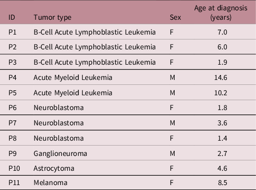
DNA was extracted from peripheral blood of eleven individuals conceived by IVF who developed pediatric cancer (Table 1). Individuals who underwent bone-marrow transplantation or who were diagnosed with hereditary cancer syndromes were excluded. Patients were referred from the ITACI - Childhood Cancer Treatment Institute (FMUSP), which is a reference pediatric cancer hospital in São Paulo, Brazil. Samples were provided after parents have signed the informed consent.
Clinical features of 11 individuals conceived by IVF who developed pediatric cancer

A control group was composed by peripheral blood samples collected from 12 children without cancer history matched by age and sex with patients (Control group 1 – C1) (Supplemental Table S1a).
A second group was added to this study in order to control the methylation status of children with similar diagnosis of cancer who were not conceived by IVF (Control group 2 – C2) (Supplemental Table S1b). This additional group was based on the recovery of germline Illumina 850K methylation data previously obtained from 16 children who developed pediatric cancer and were naturally conceived: five children with neuroblastoma (NB - peripheral blood methylomes provided by the ITACI center; data not published) and 11 children with acute myeloid leukemia (AML - bone marrow or peripheral blood methylomes public available on GEOReference Edgar, Domrachev and Lash11 accession GSE124413).
Infinium MethylationEPIC (850K) array hybridization
Genomic DNA samples were obtained from standard extraction procedures using the phenol/chloroform method. Evaluation of peripheral blood DNA methylation (DNAm) was performed using the Infinium MethylationEPIC (850K) array, according to the manufacturer’s instructions. A total of 500 ng of bisulfite-converted DNA samples (EZ DNA Methylation-Gold Kit; Zymo Research) were hybridized in the Infinium MethylationEPIC BeadChip array (Illumina). The raw image data with signal intensities were captured with the iScan SQ scanner (Illumina) and collected as IDAT files.
DNA methylation analysis
We applied the Chip Analysis Methylation Pipeline (ChAMP) package (version 2.20.1)Reference Tian, Morris and Webster12 in the R environment (version 4.0.4)13 for the methylation analysis. The quality filters removed 3,960 probes with a detection P-value above 0.01, 34,605 probes with a bead count <3 in at least 5% of samples, and non-CG sites (2,865). In addition, 94,529 SNP-related probes and 18 probes that aligned to multiple locations were removed. Lastly, 15,818 probes located on the X or Y chromosomes were excluded.Reference Aryee, Jaffe and Corrada-Bravo14,Reference Fortin, Triche and Hansen15
The beta-mixture quantile normalization (BMIQ) methodReference Teschendorff, Marabita and Lechner16 resulted in better Infinium I/II normalization compared to PBCReference Dedeurwaerder, Defrance, Calonne, Denis, Sotiriou and Fuks17 or SWAN.Reference Maksimovic, Gordon and Oshlack18 Singular value decomposition (SVD)Reference Teschendorff, Menon, Gentry-Maharaj and Aramayo19 reported the need for array and slide correction, made by ComBat.Reference Johnson, Li and Rabinovic20,Reference Leek, Johnson and Parker21 We adjusted the cell-type heterogeneity using the Refbase EWAS method.Reference Houseman, Accomando and Koestler22
Differential DNAm analysis
Methylation differences were identified by comparing patients and controls and using algorithms implemented by ChAMP.Reference Tian, Morris and Webster12 Differentially methylated positions (DMPs) analysis, i.e., methylation difference for a single CpG site, was performed using Limma.Reference Smyth23,Reference Wettenhall and Smyth24 Differentially methylated regions (DMRs) analysis was performed using the Bumphunter algorithm,Reference Jaffe, Murakami and Lee25 based on the detection of methylation differences in stretches of the genome in which there are several consecutive CpG sites exhibiting similar methylation alterations. Both analyses were performed considering P-value <0.05. In addition, DMRs should have a minimum of seven consecutive probes with changes in DNAm in the same direction (hypo or hypermethylated).
Results
Eleven patients with pediatric cancer who were conceived by IVF were evaluated (Table 1). Cancer types were hematological malignancies (three patients with B-cell acute lymphoblastic leukemia, and two with acute myeloid leukemia), and nervous system tumors (three patients with neuroblastoma, one with astrocytoma, and one with ganglioneuroma). One patient developed melanoma.
Following exclusion of probes during quality control steps, the methylation analysis proceeded with 714,443 probes for data correction and normalization. The differential methylation analysis compared the group of 11 patients conceived by IVF who developed childhood cancer (IVF/cancer) with 12 unrelated healthy controls (group C1); the goal of this analysis was to detect differences in the blood methylomes of IVF/cancer group possibly related to patient’s phenotypes. However, no significant differential methylated position - DMP (adjusted P-value <0.05) was found after Benjamini–Hochberg adjustment for multiple testing.Reference Benjamini and Hochberg26
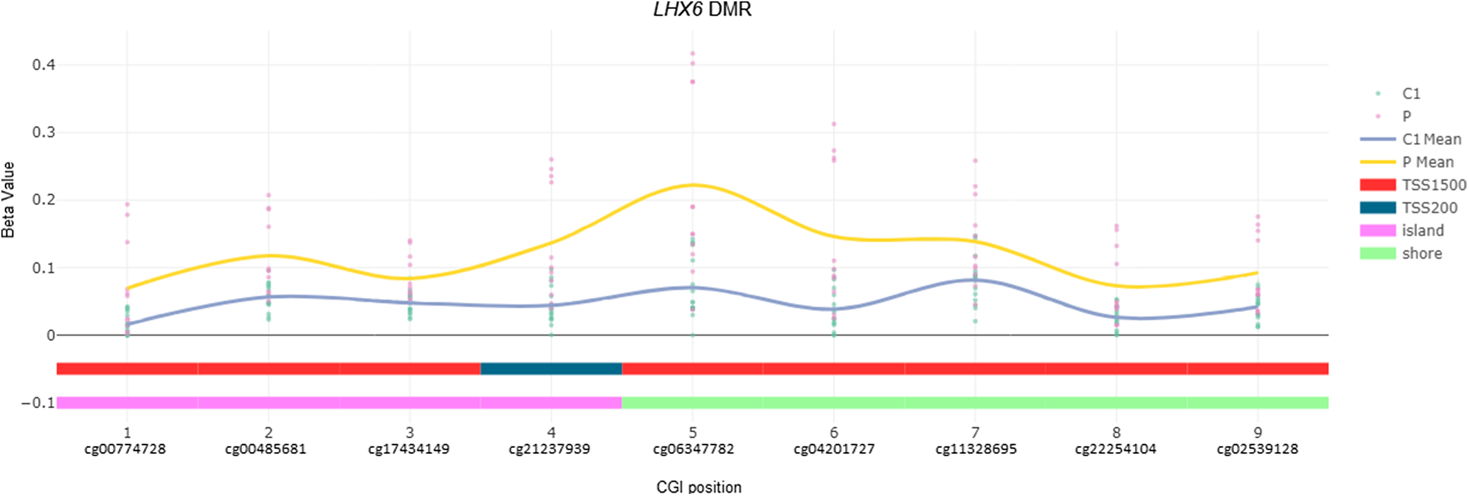
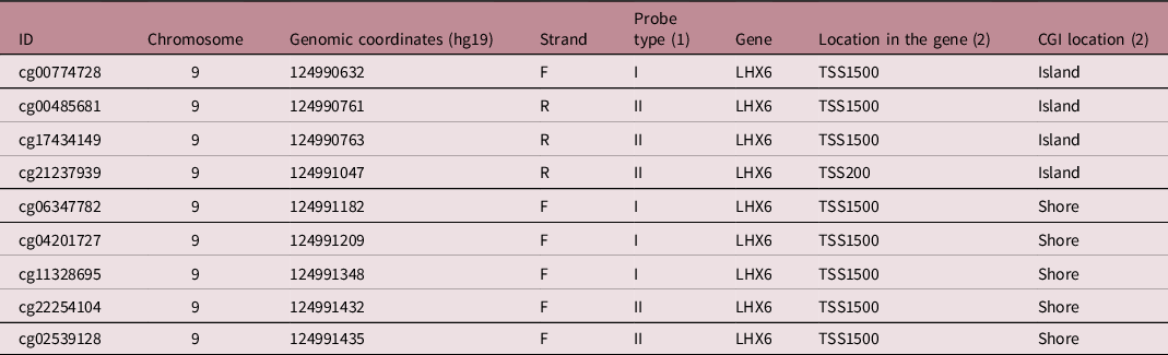
The methylation analysis detected 25 DMRs (Supplemental Table S2), 17 of which were hypermethylated and 8 were hypomethylated in patients. The most relevant DMR was hypermethylated in patients compared to healthy individuals from control C1 (Δβ = 0.07). This DMR is located in the promoter region of the LHX6 gene, mapped to 9q33.2. The LHX6 DMR encompassed nine CpG sites (Fig. 1), extending on a genomic segment of 803 bp. This genomic segment included only CpGs mapped to transcription start sites 1500 and 200 (TSS1500 and TSS200); four of these CpGs (cg00774728, cg00485681, cg17434149, cg21237939) map to a CpG island (chr9: CpG island 254, hg19), and five CpGs (cg06347782, cg04201727, cg11328695, cg22254104, cg02539128) to the shore (Table 2).
Hypermethylated DMR at the promoter region of the LHX6 gene. Plot (image extracted from ChAMP)Reference Tian, Morris and Webster12 showing the beta values of methylation of each CpG site mapped to the LHX6 gene. Green dots: individuals from the healthy control group (C1); Pink dots: individuals from the patient group (P); C1 mean: average beta value from the control group; P mean: average beta value from the patient group; TSS1500: transcription start site 1500; TSS200: transcription start site 200; island: CpG island; shore: CpG shore.

Promoter region of the LHX6 gene and its DMR genomic features

(1) Probe type according to different probe designs (Infinium I and Infinium II) in the Illumina Methylation chip
(2) Annotations for the CGIs genomic location in relation to gene sequence were done according to Illumina’s CpG loci database.
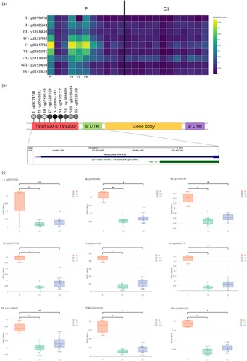
The heatmap using the beta values of the CpGs mapped to LHX6 of patients and controls C1 (Fig. 2a) 27 revealed that mostly four patients in the IVF group (P1, P4, P5, and P6) contributed to the identification of the DMR, although patients P3, P7 and P8 also contributed to this pattern of increased methylation. Considering this observation, we added a second control group to this study (control C2), based on the recovery of Illumina Infinium MethylationEPIC germline data previously obtained from 16 children who developed pediatric cancer and were naturally conceived: five children with neuroblastoma and 11 children with acute myeloid leukemia. Methylation levels of the nine CpG sites mapped to LHX6 were retrieved from all cases for comparison between the three groups: the four IVF patients, the control C1 (healthy children) and the cancer control C2 groups (Fig. 2b). Seven out of nine CpGs (cg00485681, cg17434149, cg21237939, cg06347782, cg04201727, cg22254104, and cg02539128) exhibited higher methylation levels in the subgroup of patients when compared to both control groups (Fig. 2c and Supplemental Table S3).
DNA methylation (DNAm) pattern from the CpGs located in the LHX6 DMR that was found in all analyses. a. Heatmap (image extracted from Plotly)27 showing the DNAm level of nine DMPs located in the original DMR revealed in the analysis (P and C1 groups separated by the black line). b. Regions of the LHX6 gene with the CpG sites from the DMR depicted as numbered lollipops. Promoter region image extracted from UCSC genome browser. c. Boxplots (images extracted from ChAMP)Reference Tian, Morris and Webster 12 of each CpG site with the respective methylation level for the three groups (orange: 4 patients - P, green: healthy controls - C1, blue: cancer controls - C2). n.s. are non-significant DMPs. *Statistically significant DMPs.

Discussion
Parental subfertility, parental age at conception, children sex, low birth weight, and other environmental factors, such as tobacco and alcohol consumption during pregnancy, can result in a biased risk of childhood cancer associated with ART.Reference Hargreave, Jensen, Toender, Andersen and Kjaer8,Reference Wang, Chen and Yang9,Reference Latino-Martel, Chan, Druesne-Pecollo, Barrandon, Hercberg and Norat28 Thus, a link between ART and pediatric cancer remains controversial. Considering the yet disputed association between pediatric cancer and ART,Reference Raimondi, Pedotti and Taioli7–Reference Gilboa, Koren and Barer10 in which ART is suggested to cause epigenetic modifications that could increase the risk of cancer, it is crucial to investigate DNA methylation in these patients.Reference Cortessis, Azadian and Buxbaum3,Reference Wang, Chen and Yang9 In previous studies, increased risk rates for specific types of cancer such as leukemia and neuroblastoma were detected among children born following fertility treatment.Reference Hargreave, Jensen, Toender, Andersen and Kjaer8,Reference Wang, Chen and Yang9 Likewise, our study IVF cohort included eight patients diagnosed with leukemias or neuroblastoma.
Current research regarding epigenetic alterations associated with ART are generally related to imprinting disorders.Reference Cortessis, Azadian and Buxbaum3,Reference Henningsen, Gissler and Rasmussen4 Previous studies that investigated the association between ART and pediatric cancer did not explore the presence of DNAm changes in patients conceived by IVF that developed pediatric cancer.
DMPs were not detected after multiple testing adjustments. A possible explanation for this situation can be the small number of patients. Small group analysis may not identify real changes because they do not reach statistical power when evaluating hundreds of thousands of sites simultaneously.Reference Mansell, Gorrie-Stone and Bao29 In addition, one could argue that robust DNAm changes were not detected because the methylation analysis was done in peripheral leukocytes obtained from patients and not on tumor tissues which presents a higher specificity.Reference Teschendorff, Menon, Gentry-Maharaj and Aramayo19,Reference Moore, Pfeiffer and Poscablo30 Even though DNAm changes in blood samples are generally small, most epigenetic alterations related to diseases occurring during development can be detected in surrogate tissues.Reference Woo, Kim and Christensen31 Recently, a large study evaluated the cord blood DNAm from 205 ART cases and 2,439 naturally conceived controls revealing two CpG sites associated with ART, as well as related to cancer, aging and HIV infection by EWAS studies.Reference Caramaschi, Jungius and Page32–Reference Xu, Bonder and Söderhäll35 Therefore, the use of peripheral blood samples seems to be a suitable strategy for searching for epigenetic variations, which may serve as good biomarkers for cancer risk.
The most significant DMR found in this study maps to the promoter region of the transcriptional factor LHX6, which is involved in embryogenesis and head development.Reference Liodis, Denaxa, Grigoriou, Akufo-Addo, Yanagawa and Pachnis36,Reference Grigoriou, Tucker, Sharpe and Pachnis37 Thus an additional control group was included to verify the LHX6 methylation status in children with similar cancers who were not conceived by ART. These analyses excluded the possibility that this DMR was driven by the occurrence of the cancer and not the IVF itself. The higher LHX6 methylation levels in IVF patients compared to both control groups, healthy children and children conceived naturally who developed similar pediatric cancers (neuroblastoma and leukemia), suggested that hypermethylation at the promoter of the LHX6 is likely due to the IVF process and not secondary to the cancer itself. During neurodevelopment, LHX6 is expressed in the ventral forebrain and in tangentially migrating GABAergic interneurons from the neocortex and hippocampus.Reference Liodis, Denaxa, Grigoriou, Akufo-Addo, Yanagawa and Pachnis36,Reference Yuan, Fang and Hong38 This gene is a potential tumor suppressor gene in glioma,Reference Yan, Cai, Sun, Gui and Liang39 pancreatic,Reference Abudurexiti, Gu and Chakma40 head and neck,Reference Estécio, Youssef and Rahal41 breast,Reference Hu and Xie42 lung,Reference Liu, Jiang and Han43 and cervicalReference Jung, Jeong and Kim44 cancer. Particularly, LHX6 may affect signaling pathways such as the Wnt/β-catenin in breast,Reference Hu and Xie42 lungReference Yang, Han and Liu45 and liver cancer,Reference Chen, Zhao and Li46 TP53 in hepatocarcinomaReference Chen, Zhao and Li46 and PI3K/Akt/mTOR in breast cancer.Reference Bi, Men, Han and Li47 LHX6 promoter hypermethylation has already been related to transcriptional silencing, and it is described as either hypermethylated or partially methylated in cervical, head and neck, pancreatic, lung and liver cancers.Reference Abudurexiti, Gu and Chakma40,Reference Estécio, Youssef and Rahal41,Reference Liu, Jiang and Han43,Reference Jung, Jeong and Kim44,Reference Chen, Zhao and Li46,Reference Nathalia, Theardy and Elvira48 Likewise, the DMR we found is hypermethylated in the promoter region.
Therefore, the LHX6 promoter hypermethylation previously associated with cancer and detected in this study, can be suggested as an epimutation that increases the risk of cancer in the patients herein reported. There are possible confounding factors such as low birth weight, parental age, type of infertility leading to IVF, use of fertility drugs, maternal smoking, and patient tumor, which can result in methylation alterations.Reference Wang, Chen and Yang9 Another aspect not investigated here was the genetic susceptibility to cancer of this group of patients. A recent study that analyzed the methylation profile of 23,116 individualsReference Garg, Jadhav and Rodriguez49 reported that 2/3 of the epivariations segregated according to underlying sequence variants, while the other 1/3 occurred post-zygotically. Therefore, the identified epimutations in these patients could also be attributed to genetic variants, driving both the methylation pattern and increased cancer risk.
In conclusion, we searched for possible variations in DNAm that could be linked to increased risk of childhood cancer in children conceived by IVF. A hypermethylated DMR in patients was detected in the promoter region of LHX6, a gene previously associated with cancer when its promoter region is hypermethylated. Therefore, this DMR can be an epimutation contributing to increased cancer risk in some children conceived by IVF and thus deserves additional investigation.
Supplementary materials
For supplementary material for this article, please visit https://doi.org/10.1017/S2040174422000526
Acknowledgments
We would like to thank the patients and their families for participating in this study.
Financial support
This research was carried out with financial support from Coordenação de Aperfeiçoamento de Pessoal de Nível Superior, CAPES, Brazilian Federal Agency for Support and Evaluation of Graduate Education (grant number 88887.606266/2021-00); and Fundação de Amparo à Pesquisa do Estado de São Paulo, FAPESP, São Paulo Research Foundation (A.C.B.T., grant numbers: 2018/21047-9, 2018/05961-2)
Conflict of interest
None
Ethical standards
The authors assert that all procedures contributing to this work comply with the ethical standards of the relevant national guidelines on human experimentation (Resolution 466/12) and with the Helsinki Declaration of 1975, as revised in 2008, and has been approved by the Ethics and Research Committee of ITACI (CAAE 47277115.0.0000.0068), and Institute of Biosciences (University of São Paulo, São Paulo, Brazil) (CAAE: 09163818.4.0000.5464).