Introduction
Kazakhstan is a large, landlocked country in the northern hemisphere, of greater than 2.4 million square kilometres. Favourable natural and climatic conditions of Kazakhstan and large areas of pastures (180 million hectares) have resulted in animal husbandry as a traditional occupation of the indigenous population. Presently there are approximately 6.5 million cattle, 17.5 million sheep, 2 million horses and 172,000 camels. This large animal population potentially favours the transmission of Echinococcus granulosus, and infection is now widespread in the animal population (Shaikenov et al., Reference Shaikenov, Torgerson, Usenbayev, Baitursynov, Rysmukhambetova, Abdybekova and Karamendin2003; Torgerson et al., Reference Torgerson, Burtisurnov, Shaikenov, Rysmukhambetova, Abdybekova and Ussenbayev2003a).
At the end of last decade of the 20th century it became apparent that an increasing number of cases of human cystic echinococcosis (CE) were being recorded in Kazakhstan. Official government statistics document an increase in cases of CE from about 200 cases per year until 1994, rising rapidly to approximately 1000 cases per year by the beginning of the 21st century. This epidemic emerged at a time of rapid economic decline and decreases in medical services (Torgerson et al., Reference Torgerson, Shaikenov, Baitursinov and Abdybekova2002), and hence was unlikely to be an artefact caused by improved diagnosis.
This review gives an overview of more recent data concerning the reported cases of human cystic echinococcosis in Kazakhstan. We also review the data available for E. granulosus infection in animal hosts. There is less evidence with regard to human alveolar echinococcosis (AE). However, analysis of data from hospital records of a referral centre in Almaty indicates that it could be an increasing problem. Likewise, cases recorded in Kazakhstani expatriates, now resident in Germany, also indicated that human AE in Kazakhstan is far from negligible. Finally, we also review data on infection with Echinococcus multilocularis in animal hosts.
Notified human cases of echinococcosis
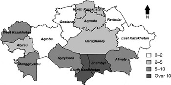
Echinococcosis is a notifiable disease in Kazakhstan and official government figures document a mean of 850 cases/year between 2007 and 2013. This varied between 917 cases notified in 2010 to 802 cases notified in 2013. The mean incidence of notified cases over this period was 5.1 cases per 100,000 per year. Most cases are seen in the southern regions of Kazakhstan, where the annual incidence is over 10 cases per 100,000 in Zhambyl and South Kazakhstan Oblasts (fig. 1). However, substantial numbers are reported in western regions (up to 7 cases per 100,000). Even in some areas which were thought of as low endemicity, such as in North Kazakhstan Oblast, an incidence of 3.2 cases per 100,000 is now reported (fig. 1). The database does not generally distinguish between the two types of echinococcosis (which could include CE and AE). However, four cases of ‘alveococcosis’, assumed to be AE, were notified in the database between 2007 and 2013.
The incidence of reported cases of echinococcosis (cases per 100,000 per year) reported for each Oblast (region) of Kazakhstan. The relative shadings indicate the mean annual incidence of the 7 years 2007–2013 inclusive. The data for the city administrations of Astana and Almaty have been merged with Aqmola and Almaty Oblasts, respectively.

Hospital records of echinococcosis and estimates of AE incidence
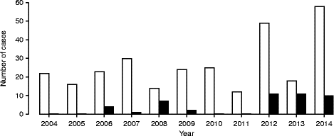
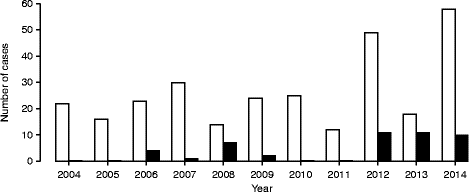
The notified data either do not differentiate between AE and CE, or AE is largely not reported. However, AE is an emergent disease in neighbouring Kyrgyzstan (Usubalieva et al., 2013). Furthermore, contact with dogs infected with E. multilocularis is believed to be an important risk factor for human AE, and high prevalences of E. multilocularis in dogs have been reported in some communities in south-eastern Kazakhstan (Torgerson et al., Reference Torgerson, Rosenheim, Tanner, Ziadinov, Grimm, Brunner, Shaiken, Shaikenov, Rysmukhambetova and Deplazes2009). Consequently, for one major referral centre in Almaty (The Sizganov National Scientific Surgical Centre), the hospital records for echinococcosis were examined to see if there was evidence of human AE. All cases of echinococcosis were examined between 2006 and 2014 (fig. 2). CE and AE were differentiated on clinical grounds (such as by imaging techniques) and AE cases confirmed by histology of resected lesions. Only original case numbers and not treatment episodes were included in the analysis. Between 12 and 58 cases (mean 26.5 cases per year) of CE were treated per year, with a total of 301 cases. Of these, 168 were female and 133 were male (P < 0.001). The mean age of cases was 33.8 years (SE = 0.94). Between 0 and 11 cases (mean of 4 cases per year) of AE were treated, with a total of 46 cases. The mean age of AE cases was 35.6 years (SE = 2.0) (range 14–58 years). This was not significantly different from the mean age of the CE cases (P= 0.24, t-test). Of the 46 cases, 21 were female and 25 male (not significantly different (NS)). Most of these cases (27), unsurprisingly, were from the Almaty region. However, a number of cases were referred from other regions, often a considerable distance away. In particular, eight cases were reported from East Kazakhstan.
Numbers of cases of cystic (CE; white bars) and alveolar (AE; black bars) echinococcosis cases treated in a single referral hospital in Almaty between 2004 and 2014.
Until cases of AE and CE are differentiated routinely in the reporting systems it is not possible to obtain an accurate picture of the incidence of AE. In the reported data, there were 5949 cases of echinococcosis reported between 2007 and 2013. But in the same database just four cases were definitively recorded as AE: three in Aqmola Oblast and one in Almaty Oblast. This is less than 10% of the total cases reported in one referral centre over the same time period, so, clearly, there is virtually no separate reporting of AE, despite it being a potential problem. However, estimates of the incidence of AE can be made. For example, if the ratio of CE to AE cases seen in the referral centre was representative of the country as a whole, then 46/301 = 15.3% of the total CE would be an estimate of total AE cases. Modelling uncertainty, assuming that AE cases and CE cases are presented as a Poisson process, results in a mean of 130 cases per year (95% confidence interval (CI) 93–174 cases). This is an incidence of 0.76 cases (CI 0.54–1.0) per 100,000 per year.
Estimates from data of AE in Kazakhstani emigrants
An alternative estimate can be made using data reported to the Robert Koch Institute (RKI) in Germany. AE reporting in Germany has been mandatory since 2001. Diagnosis of echinococcosis by serological testing and histopathological examination is reportable to RKI by microbiological laboratories and pathologists. The data are freely available (Robert Koch Institute, 2002–2014). The data also give the country of origin of each case. In the database, six cases in people of Kazakhstani origin were recorded between 2001 and 2013. Using a Poisson model, which assumes that the Kazakhstani cases in the RKI database are notified as a Poisson process, the six cases represent an incidence estimate with a 95% CI of between 2.2 cases and 13.1 cases over the 13-year period. These CIs were calculated using an internet Poisson CI calculator (see http://statpages.org/confint.html). Then, dividing by 13 results in a CI of 0.17–1 case per year. However, the data from RKI underestimate the incidence of AE in Germany and were calculated to have a sensitivity of just 33% (CI 25–40%) (Jorgensen et al., Reference Jorgensen, an der Heiden, Kern, Schöneberg, Krause and Alpers2008). So the mean annual number of cases and CI can be divided by the sensitivity of the RKI data, again modelling the uncertainty. This results in an estimate of between 0.049 and 0.30 cases per 100,000 annually (mean 0.14) in German residents of Kazakhstani origin, as there are 719,000 people resident in Germany with a recent migration history from Kazakhstan (State and Society, 2015). Applying this incidence from Kazakhstani expatriates to the population of Kazakhstan of 17 million results in an estimate of between 12 and 70 cases per year (mean 34 cases) for Kazakhstan.
Echinococcus granulosus in animals
Previous studies in dogs over 10 years ago indicated a high prevalence in rural dogs. In particular, based on arecoline purgation surveillance studies, village dogs had an estimated prevalence of 5.8% whereas shepherd dogs, more closely associated with livestock activities, had a prevalence of 23% (Torgerson et al., Reference Torgerson, Shaikenov, Rysmukhambetova, Ussenbayev, Abdybekova and Burtisurnov2003b). A study in the small community of Jalanash (located at latitude 46.47, longitude 80.48) in the Tien Shan mountains in southern Kazakhstan, described a total of 13 of 131 dogs infected with E. granulosus (Stefanić et al., Reference Stefanić, Shaikenov, Deplazes, Dinkel, Torgerson and Mathis2004). The G1 strain was initially isolated, and the G6/G7 strain was also shown to be present by sequencing of polymerase chain reaction (PCR) amplicons obtained from eggs isolated from faeces of two of these dogs (Trachsel et al., Reference Trachsel, Deplazes and Mathis2007). This is believed to be the first time the G6/G7 strain has been isolated in Kazakhstan. Further studies in the same community demonstrated that 85 of 632 dogs were infected with E. granulosus, with a mean abundance of 812 parasites per dog (Torgerson et al., Reference Torgerson, Rosenheim, Tanner, Ziadinov, Grimm, Brunner, Shaiken, Shaikenov, Rysmukhambetova and Deplazes2009).
There has also been a study of 41 wolves, necropsied between 2001 and 2008. These wolves were from the southern regions of Kazakhstan – South Kazakhstan, Zhambyl and Almaty Oblasts – which correspond to areas where most cases of human CE are described (fig. 1). Of these, eight were infected (c. 20%) with E. granulosus and the mean abundance was 1275 parasites (CI 405–2342) per wolf (Abdybekova & Torgerson, Reference Abdybekova and Torgerson2012).
In sheep the mean prevalence of infection has been estimated to be up to 48% in Almaty and 34% in Zhambyl and South Kazakhstan; the mean abundance of infection being 4.7 cysts per sheep and 2.54 cysts per sheep, respectively. Cattle appear to have a lower prevalence (7.2%) and lower abundance of infection (0.48 cysts per animal) (Torgerson et al., Reference Torgerson, Burtisurnov, Shaikenov, Rysmukhambetova, Abdybekova and Ussenbayev2003a). In this study the infection pressure was estimated through mathematical modelling as 1.98 cysts per sheep per year in Almaty Oblast, 1.24 cysts per year in Zhambyl and South Kazakhstan Oblasts, and 0.15 cysts per year for cattle.
Echinococcus multilocularis in animals
Echinococcus multilocularis has been described in two reports in dogs. In the study by Stefanić et al. (Reference Stefanić, Shaikenov, Deplazes, Dinkel, Torgerson and Mathis2004) six dogs from a sample of 131 (from Jalanash – see above) were proven to be infected with E. multilocularis. One of these dogs was co-infected with E. granulosus, and this is one of the first reports to confirm both species of parasite co-infecting the same dog. Further studies in the same district demonstrated that 29 dogs from 632 sampled (5%) were infected with E. multilocularis (Torgerson et al., Reference Torgerson, Rosenheim, Tanner, Ziadinov, Grimm, Brunner, Shaiken, Shaikenov, Rysmukhambetova and Deplazes2009). An extensive review (Shaikenov, Reference Shaikenov2006) describes the ecology of E. multilocularis in Kazakhstan. The parasite is distributed over much of Kazakhstan, but with a patchy distribution in arid areas. Rodents in such areas are more likely to be infected if they inhabit ecological niches where there is higher humidity, such as desert oases or river valleys. In more humid mountainous areas and northern steppes, the distribution appears to be more extensive. A large number of intermediate hosts from the genera Marmota, Lagurus, Microtus, Ondatra, Apodermus, Rhombomys, Meriones, Allactaga, Cricetulus and Clethrionomys are recorded as being infected. Two fox species – the red fox Vulpes vulpes and the corsac fox Vulpes corsac – are the main definitive hosts.
Conclusions
Echinococcus granulosus is widely distributed in Kazakhstan, with human cases of CE and animal infection, in both dogs and livestock, being widespread. The reported human incidence of echinococcosis appears to have stabilized at between 800 and 1000 cases per year following a rapid increase in incidence in the last decade of the 20th century. The situation with regard to AE is less clear. Infection in wild animal hosts has long been known; however, the increasing infection of dogs represents a potentially greater risk to humans, because of the closer contact between dogs and humans. The evidence in this review does suggest that there are significant numbers of cases of human AE in Kazakhstan. The estimate of 130 cases per year, based on hospital records and the ratio of AE:CE cases, could overestimate the number of cases, as the referral centre tends to deal with more complex cases of echinococcosis, and AE has more severe sequelae than CE. Likewise, the 34 cases per annum, estimated from an emigrant population, may not be representative because this population of emigrants is mainly of German descent, having been offered German citizenship after the dissolution of the Soviet Union. Furthermore, Germany is endemic for AE, so it is possible that some cases from the emigrant population were infected in Germany. However, because of the very long latent period of AE before clinical signs appear, clinical AE in recent immigrants is likely to be a result of infection outside of Germany. It is essential, therefore, that comprehensive monitoring for human AE is initiated, especially in light of the ongoing epidemic of human AE now being reported in Kyrgyzstan.
Financial support
The study was supported by the scientific research budget 2012–2014 of the Ministry of Agriculture of the Republic of Kazakhstan.
Conflict of interest
None.