Introduction
In mammalian oocytes, meiosis is initiated during fetal life and is subsequently arrested at the diplotene stage of the first meiotic division. Completion of the first meiotic division is triggered by the preovulatory gonadotropin surge and meiosis progresses to the metaphase II stage where it is arrested again until fertilization. Meiotically competent oocytes spontaneously resume meiotic maturation without hormonal stimulation when they are removed from follicles and cultured in suitable media. This situation suggests that the follicle provides an inhibitory factor(s) to prevent meiosis until the gonadotropin surge; however, mechanisms involved in meiotic arrest and induction are not fully understood.
Cyclic adenosine monophosphate (cAMP) levels within the oocyte apparently have an important role in the control of nuclear maturation, with high levels maintaining meiotic arrest and lower levels being permissive for meiotic maturation (Downs, Reference Downs, Grudzinskas and Yovich1995; Shimada et al., Reference Shimada and Tarada2002; Horner et al., 2003; Kalinowski et al., Reference Kalinowski, Berlot, Jones, Ross, Jaffe and Mehlmann2004; Thomas et al., Reference Thomas, Armstrong and Gilchrist2004). Cyclic AMP is produced by the enzyme adenylate cyclase in response to hormonal stimulation and its action is mediated by cAMP-dependent protein kinase (PKA). The amount of cAMP in cells is also controlled by the rate of its degradation by phosphodiesterase (PDE) enzymes, which hydrolyze the 3′ phosphoester bond of cAMP and cGMP to generate 5′AMP and 5′GMP respectively. Inhibitors of the oocyte-specific PDE can inhibit oocyte nuclear maturation in vitro (Tsafriri et al., Reference Tsafriri, Chun, Zhang, Hsueh and Conti1996; Mayes & Sirard, Reference Mayes and Sirard2002; Bilodeau-Goeseels, Reference Bilodeau-Goeseels2003) and also in ovulated oocytes in vivo (Wiersma et al., Reference Wiersma, Hirsch, Tsafriri, Hanssen, Van de Kant, Kloosterboer, Conti and Hsueh1998).
It was recently suggested that AMP, the product of degradation of cAMP by PDE, could have an active role in the control of oocyte meiotic maturation. The nucleotide AMP is a potent regulator of AMP-activated protein kinase (AMPK), a key regulatory enzyme that controls the activity of enzymes of carbohydrate and fat metabolism in response to changes in cellular energy levels (Hardie & Carling, Reference Hardie and Carling1997) or environmental stresses (Corton et al., Reference Corton, Gillespie and Hardie1994). The enzyme consists of a catalytic subunit α (two isoforms, PRKAA1 and PRKAA2) and two regulatory subunits: β and γ, with two and three isoforms, respectively (Hardie, Reference Hardie2004). The activity of AMPK is controlled by the intracellular AMP/ATP ratio, such that small increases in AMP or large decreases in ATP increase the ratio and lead to activation of the enzyme. Phosphorylation of the Thr172 residue of the α subunit is required for enzyme activation (Hardie, Reference Hardie2004). It was previously suggested that oocyte PDE activity could generate enough AMP from cAMP degradation to activate AMPK, which in turn would affect oocyte maturation (Downs et al., Reference Downs, Hudson and Hardie2002). Indeed, pharmacological activation of AMPK with 5-aminoimidazole-4-carboxamide 1-β-d-ribofuranoside (AICAR) induced oocyte maturation in arrested cumulus-enclosed and denuded mouse oocytes (Downs et al., Reference Downs, Hudson and Hardie2002; Chen et al., Reference Chen, Hudson, Chi, Chang, Moley, Hardie and Downs2006). Additionally, active AMPK was demonstrated in both FSH- and the EGF-like peptide amphiregulin (AR)-treated GV-stage oocytes within 3 h, supporting a role for AMPK in FSH- and AR-induced maturation (Chen & Downs, Reference Chen and Downs2008). In contrast, AICAR and metformin (MET), another activator of AMPK, inhibited meiosis in bovine and porcine oocytes (Bilodeau-Goeseels et al., Reference Bilodeau-Goeseels, Sasseville, Guillemette and Richard2007; Mayes et al., Reference Mayes, Laforest, Guillemette, Gilchrist and Richard2007). Although the presence of AMPK subunits was demonstrated in these studies and the effect of the activators did not appear to be through generation of purine nucleotides, whether AMPK was in fact activated in response to the AICAR and MET was not examined, and an inhibitor of AMPK was not yet available for testing; therefore, involvement of AMPK was not demonstrated. The objectives of the present study were to determine the effects of AMPK inhibitors, with and without AMPK activators, on bovine oocyte nuclear maturation, and to determine whether AICAR and metformin treatment lead to phosphorylation of bovine oocyte and cumulus cell AMPK.
Materials and Methods
Chemicals and antibodies
The AMPK inhibitor compound C was purchased from Calbiochem. A 12.5 mM-stock solution was prepared in DMSO; it was further diluted 1/10 and 1/100 in culture medium before being added to the culture wells. AICAR was purchased from Toronto Research Chemicals, Inc. A stock solution was prepared in water at 100 mM Metformin (1,1-dimethylbiguanide hydrochloride, Sigma-Aldrich) was prepared in water as 200 mM and 1 M solutions (5 μl was added to each well). Adenine 9-β-D-arabinofuranoside (AraA) and adenine 9-β-d-arabinofuranoside 5′-monophosphate (AraAMP) were purchased from Sigma-Aldrich Canada and prepared in water as 200 mM solutions. Okadaic acid (Biomol International) was prepared as a 500 μM stock solution in ethanol; it was used at 1 μM in the culture medium. Oligomycin A was purchased from Sigma-Aldrich, prepared as a 10 mM solution in ethanol, and 1 μl was added per well. Final concentrations of ethanol, DMSO, or additional water never exceeded 1% in the culture medium. These concentrations were tested and had no adverse effect on oocyte nuclear maturation (results not shown). Rabbit polyclonal antibodies against phospho-AMPKα (Thr172) and AMPK α-pan were obtained from Upstate Biotechnology Inc. Polyclonal antibodies against phospho-acetyl-CoA carboxylase (Ser79) and acetyl-CoA carboxylase were obtained from Cell Signaling Technology, Inc. Extracts from C2C12 cells treated with AICAR were from Cell Signaling Technology, Inc. All other reagents were from Sigma-Aldrich, unless otherwise indicated.
Oocyte recovery and culture
Heifer ovaries were collected at an abattoir approximately 30 min after slaughter and transported to the laboratory in saline solution (35°C), supplemented with 100 U/ml penicillin G, 100 μg/ml streptomycin sulfate, and 250 ng/ml amphotericin B. Cumulus-enclosed oocytes (CEO) were recovered from the ovaries by aspirating antral follicles >2 mm with a 10-ml syringe and an18-gauge needle. Oocytes with an unexpanded mass of cumulus cells and homogeneous cytoplasm were identified under a stereomicroscope (magnification, ×4) and recovered. When denuded oocytes (DO) were required for experiments, CEO were vortex-agitated for 1 min in the presence of trypsin (2.5 mg/ml in phosphate-buffered saline; PBS) to remove cumulus cells. Denuded oocytes or CEO were then washed twice in Medium 199-HEPES-BSA (Medium 199 with Earle's salts and l-glutamine, supplemented with 20 mM HEPES, 0.4% BSA, and 100 U/ml penicillin, 100 μg/ml streptomycin (Gibco)) and once in culture medium. For all experiments, CEO and DO were cultured in groups of 10–20 in 500 μl of culture medium. The control culture medium was Medium 199, supplemented with 10% fetal bovine serum (FBS; characterized, HyClone Laboratories, Inc.), 100 U/ml penicillin, and 100 μg/ml streptomycin. Oocytes were cultured at 39°C in a moisture-saturated atmosphere of 5% CO2 in air.
Fixation of oocytes and evaluation of nuclear maturation
At the end of the culture period, CEO were transferred to 1.5-ml tubes containing 400 μl of trypsin solution (as above) and vortex-agitated for 1 min to remove cumulus cells (DO were not vortexed before fixing). The oocytes were then recovered under a stereomicroscope and transferred to glass slides. A mixture of Vaseline and paraffin was used to maintain a coverslip in contact with the oocytes. Coverslips were secured with epoxy glue and the slides immersed in a fixative solution (ethanol:acetic acid, 3:1, v:v) for at least 24 h, stained with 1% aceto-orcein, and examined at ×320 magnification. Oocytes were classified as being at one of the following stages: germinal vesicle (GV), germinal vesicle breakdown (GVBD), condensed chromosomes, metaphase I (MI), anaphase I, telophase I, or metaphase II (MII). Oocytes with an abnormal chromatin configuration were classified as degenerated.
Western blot analysis
Oocytes and cumulus cells were frozen directly in lysis buffer (10 mM Tris pH 7.4, 150 mM NaCl, 1 mM EDTA, 1 mM EGTA, 0.5% Igepal; ICN Biochemicals Inc.), supplemented with protease inhibitors (2 mM phenylmethyl sulfonylfluoride, 10 mg/ml leupeptin, 10 mg/ml aprotinin) and phosphatase inhibitors (100 mM NaF, 10 mM sodium pyrophosphate, and 2 mM Na orthovanadate). Cell extracts were electrophoresed in 10% (w/v) polyacrylamide gels (BioRad) under reducing conditions. Proteins were transferred onto polyvinylidene fluoride (PVDF) membranes (Immobilon-P, Millipore) over a 4-h interval at 400 mAmp. Membranes were incubated at room temperature for 1.5 to 2.5 h in PBS supplemented with 5% non-fat dried milk powder and 0.1% Tween-20 to saturate non-specific sites. Membranes were then incubated overnight at 4°C with the appropriate antibody (final dilutions were 1:2000 for anti-phospho-PRKAA (Thr172), anti-phospho-acetyl-CoA carboxylase (Ser79) and anti-acetyl-CoA carboxylase and 1:7500 for anti PRKAA1/2) in PBS (or Tris-buffered saline (TBS) for acetyl-CoA carboxylase antibodies) supplemented with 0.1% Tween-20 and 5% BSA. Membranes were washed in PBS–Tween or TBS–Tween (0.1%) and incubated for 1 h at room temperature with a horseradish peroxidase-conjugated anti-rabbit immunoglobulin G (IgG; final dilution, 1:10,000). After washing, the signal was detected by enhanced chemiluminescence (ECL Plus, GE Healthcare). The films were scanned and the signals were quantified with the Quantity One software (BioRad). The membranes were stripped before being re-hybridized with another primary antibody.
Experimental design
Experiment 1: Effects of the AMPK inhibitor compound C on bovine oocyte nuclear maturation
Bovine CEO and DO were incubated for 7 or 21 h in control culture medium alone or supplemented with the AMPK inhibitor compound C (CC; 0.50, 1, 5, and 10 μM), AICAR (1 mM), AICAR (1 mM) + CC (1 and 5 μM), metformin (MET, 2 mM), and MET (2 mM) + CC (1 and 5 μM), and were then fixed for evaluation of nuclear maturation. To determine if the inhibitory effect of CC was reversible, CEO were cultured with CC at 5 μM for 7 h, washed twice and cultured in control medium for 16 h, and then fixed for evaluation of nuclear maturation.
Experiment 2: Effect of another inhibitor and another activator of AMPK on nuclear maturation
Bovine CEO were incubated for 7 h in control culture medium, alone or supplemented with the AMPK activator AraAMP (0.5 and 2 mM), or the AMPK inhibitor AraA (0.5 and 2 mM), and then fixed for evaluation of nuclear maturation. AraA (0.5 and 2 mM) was also tested in DO cultured for 7 h. AraAMP was not tested in DO, as it was no longer available for purchase.
Experiment 3: Effect of okadaic acid on the inhibitory effect of AICAR, MET and CC
Okadaic acid is an inhibitor of protein phosphatase-1 (PP1) and PP2A. It accelerates GVBD in cattle and pig oocytes (Kalous et al., Reference Kalous, Kubelka, Rimkevicova, Guerrier and Motlik1993). As activators (AICAR, MET, and AraAMP) and inhibitors (CC and AraA) of AMPK had inhibitory effects on oocyte nuclear maturation and these agents probably act through phosphorylation/dephosphorylation of target proteins, we tested whether the effect of AICAR, MET and CC would be reversed by okadaic acid to determine if they acted by different mechanisms. Bovine CEO and DO were incubated for 7 h in control culture medium alone or supplemented with okadaic acid (OA, 1 μM), AICAR (1 mM), AICAR + OA, MET (2 mM), MET + OA, CC (5 μM) and CC + OA and were then fixed for evaluation of nuclear maturation.
Experiment 4: Effects of AICAR, MET and CC on Thr172 phosphorylation
Cumulus-enclosed oocytes and DO were cultured for 0, 3 or 6 h in control medium alone, or supplemented with AICAR (1 mM), MET (2 mM) or CC (5 μM). Following culture, CEO were mechanically separated into oocytes and cumulus cells. Cells were lysed and protein extracts were subjected to western blotting with an antibody against phospho-PRKAA, followed by hybridization with a secondary antibody and detection. The membranes were stripped and re-hybridized with an antibody against PRKAA1/2 and a secondary antibody followed by detection. The signals were quantified and for each sample, the phosphorylated protein/total protein ratio was calculated.
Experiment 5: Effects of higher concentrations of AICAR and MET on AMPK Thr172 and acetyl-CoA carboxylase (Ser79) phosphorylation
Cumulus-enclosed oocytes were cultured for 3 h in control medium alone, or supplemented with AICAR (2 mM instead of 1 mM), MET (10 mM instead of 2 mM) or oligomycin (20 μM). Following culture, CEO were mechanically separated into oocytes and cumulus cells and protein extracts were subjected to western blotting with antibodies against phospho-PRKAA, PRKAA1/2, phospho-acetyl-CoA carboxylase (Ser79) and acetyl-CoA carboxylase (sequentially), followed by hybridization with a secondary antibody and detection. Signals were quantified, and the phosphorylated protein/total protein ratio was calculated for each sample.
Statistical analysis
At least three replicates were performed for each treatment for each oocyte maturation experiment. Data for oocyte maturation are expressed as mean ± SEM. They were arcsin-transformed and were analysed with a General Linear Model (GLM) of SAS (SAS Institute, Inc.). When GLM indicated a significant effect of treatment, differences were located with Duncan's multiple range test. Western blot analysis ratios are presented as mean ± SEM of 2 to 4 determinations and the effects of treatments were analysed by GLM, followed by the Duncan's multiple range test if significant differences were detected. For all analyses, p < 0.05 was considered significant.
Results
Experiment 1: Effects of the AMPK inhibitor compound C on nuclear maturation of bovine denuded and cumulus-enclosed oocytes
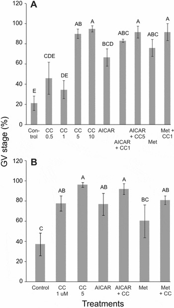
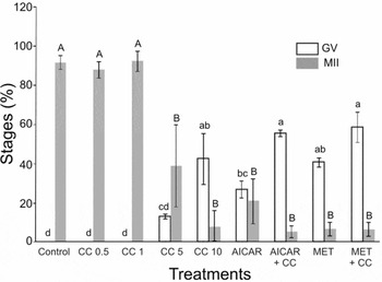
The majority of CEO cultured in control medium had resumed meiosis after 7 h of culture; only 21.1 ± 7.2% were still at the GV stage (Fig. 1a). The AMPK activators AICAR and MET increased (p < 0.05) the proportion of CEO remaining at the GV stage after 7 h of culture to 66.6 ± 8.6 and 75.8 ± 8.4%, respectively (Fig. 1a). The AMPK inhibitor CC at 5 and 10 μM also increased (p < 0.05) the proportion of CEO remaining at the GV stage compared with control, although lower concentrations had no significant effect (Fig. 1a). Additionally, when CEO were cultured in the presence of AICAR in combination with CC at 5 μM, the percentage of CEO remaining at the GV stage was higher (p < 0.05) than in the presence of AICAR alone (Fig. 1a). The AMPK activator AICAR and AMPK inhibitor CC also increased (p < 0.05) the percentage of DO remaining at the GV stage, compared to control, after 7 h of culture (Fig. 1b). When CEO were cultured for 21 h, CC 10 μM, AICAR and MET increased (p < 0.05) the proportion of CEO remaining at the GV stage and decreased (p < 0.05) the proportion of CEO reaching the MII stage compared with control (Fig. 2). When CEO cultured with CC at 5 μM for 7 h were washed and transferred to control medium for an additional culture period of 16 h and then fixed, 95.6 ± 1.6 of CEO (n = 46) and 77.6 ± 2.7% of DO (n = 49) progressed beyond MI.
Effects of the AMPK inhibitor compound C (CC, 0.5, 1, 5, or 10 μM), alone or in combination with AICAR (1 mM) or metformin (MET, 2 mM), on the percentage of bovine CEO (Panel A) and DO (Panel B) remaining at the GV stage after 7 h of culture. Values represent mean ± SEM of at least three replicates, with totals of 53 to 61 and 43 to 49 CEO and DO per treatment, respectively. A–ETreatments without a common superscript differed (p < 0.05).

Effects of the AMPK inhibitor Compound C (CC, 0.5, 1, 5, or 10 μM), alone or in combination with AICAR (1 mM) or metformin (MET, 2 mM), on the percentages of bovine CEO at the GV stage (solid bars) and at the metaphase II stage (open bars) after 21 h of culture. Values represent mean ± SEM of at least three replicates with totals of 51 to 60 CEO per treatment. Different lowercase letters (GV stage) and uppercase letters (MII stage) indicate significant differences (p < 0.05).

Experiment 2: Effect of another activator and another inhibitor of AMPK on nuclear maturation of bovine denuded and cumulus-enclosed oocytes
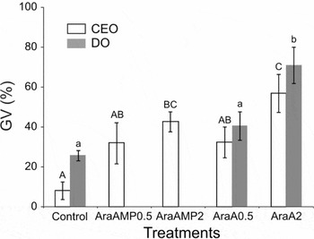
As the effect of the AMPK inhibitor CC was similar to the effect of the AMPK activators AICAR and MET, we tested another AMPK inhibitor, AraA, to rule out a non-specific effect of CC. We also tested the AMPK activator AraAMP. Both AraAMP and AraA at 2 mM increased (p < 0.05) the percentage of CEO remaining at the GV stage compared with control (Fig. 3). Furthermore, AraA at 2 mM increased (p < 0.05) the percentage of DO remaining at the GV stage after 7 h of culture (Fig. 3).
Effects of the AMPK activator AraAMP (0.5 and 2 mM) and the AMPK inhibitor AraA (0.5 and 2 mM) on the percentage of bovine CEO remaining at the GV stage after 7 h of culture. Values represent mean ± SEM of at least three replicates with totals of 48 to 67 CEO per treatment. Different uppercase letters indicate significant differences (p < 0.05).

Experiment 3: Effect of okadaic acid on the inhibitory effect of AICAR, MET and CC on bovine oocyte nuclear maturation in vitro
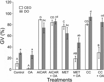
As previously observed, AICAR, MET and CC inhibited GVBD in CEO and DO (Fig. 4). Okadaic acid increased the percentage of CEO reaching the MI stage after 7 h of culture (62.3 ± 11.2 and 90.6 ± 4.1 for control and OA respectively, p < 0.05) and reversed the inhibitory effect of MET 2 mM and CC 5 μM (p < 0.05) but not of AICAR 1 mM in CEO. Okadaic acid did not reverse the effect of AICAR, MET and CC in DO (Fig. 4).
Effects of okadaic acid (1 μM) alone or in combination with AICAR (1 mM) or metformin (MET, 2 mM), on the percentage of bovine CEO and DO at the GV stage after 7 h of culture. Values represent mean ± SEM of at least three replicates with totals of 55 to 76 CEO per treatment. Different uppercase letters indicate significant differences (p < 0.05) according to Duncan's multiple range test.

Experiment 4: Effects of AICAR, MET and CC on AMPK phosphorylation level in bovine denuded oocytes, oocytes from complexes, and cumulus cells after IVM
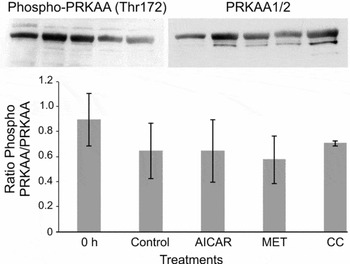
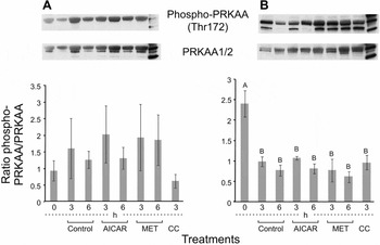
To determine whether AICAR, MET or CC inhibited nuclear maturation by changing AMPK activity level, we measured the levels of PRKAA phosphorylation in denuded oocytes, oocytes from complexes, and cumulus cells cultured in the presence or absence of AICAR (1 mM), MET (2 mM) and CC (5 μM) for 3 or 6 h. The level of PRKAA phosphorylation on Thr172 did not change in DO (Fig. 5) and in oocytes from complexes (Fig. 6a) in response to culture in the presence of AICAR, MET or CC (p > 0.05). In cumulus cells, PRKAA phosphorylation decreased (p < 0.05) after 3 or 6 h of culture compared to 0 h, regardless of the presence of AICAR, MET or CC in the culture medium (Fig. 6b).
Effects of culture in control conditions or with AICAR (1 mM), metformin (2 mM) or compound C (CC, 5 μM) for 6 h on the phosphorylation level of PRKAA in DO. After culture, DO were lysed and subjected to western blotting with antibodies against phospho-PRKAA (Thr172) and PRKAA1/2. A representative blot is shown. Bands were quantified by densitometry and the phosphorylated protein/total protein ratios are shown. The results are presented as mean ± SE of three determinations with 35–40 DO per group per experiment. There were no significant differences in the phosphorylation levels between treatments.

Effects of culture in control conditions or with AICAR (1 mM), metformin (MET, 2 mM) or compound C (CC, 5 μM) for 3 or 6 h on the phosphorylation level of PRKAA in oocytes (Panel A) and cumulus cells (Panel B) from COC. After culture, COC were mechanically separated into oocytes and cumulus cells. Oocytes and cumulus cells were then lysed and subjected to western blotting with antibodies against phospho-PRKAA (Thr172) and PRKAA1/2. The results are presented as mean ± SE of three determinations with 25–30 oocytes or cumulus cells from 12–15 COC per group per experiment.

Experiment 5: Effects of higher concentrations of AICAR and MET on AMPK Thr172 phosphorylation
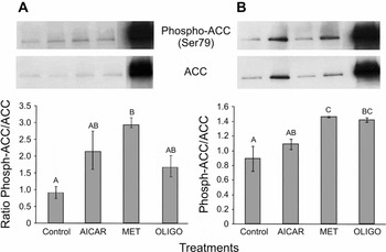
To determine whether the lack of effect of AICAR and MET in the previous experiment was due to the concentrations of AICAR and MET being too low to affect AMPK phosphorylation, we cultured COC in the presence of AICAR at 2 mM, MET at 10 mM, or oligomycin (20 μM). We tested oligomycin because this mitochondrial oxidative phosphorylation inhibitor is known to activate AMPK (Stefanelli et al., Reference Stefanelli, Stanic, Bonavita, Flamigni, Pignatti, Guarnieri and Caldarera1998). The level of Thr172 phosphorylation did not change in response to AICAR and MET at higher concentrations and oligomycin compared to control (results not shown). To confirm these results, we hybridized the membranes with phospho-acetyl-CoA carboxylase (Ser79) and acetyl-CoA carboxylase antibodies. Acetyl-CoA carboxylase is a substrate of AMPK and AMPK activation can be monitored by phosphorylation of Ser79 of acetyl-CoA carboxylase (Davies et al., 1999). Metformin 10 mM increased (p < 0.05) phosphorylation of Ser79 of acetyl-CoA carboxylase in oocytes and cumulus cells, whereas oligomycin increased (p < 0.05) Ser79 phosphorylation in cumulus cells only (Fig. 7).
Effects of culture in control conditions or with AICAR (2 mM), metformin (MET, 10 mM) or oligomycin (oligo, 20 μM) for 3 h on the phosphorylation level of acetyl-CoA carboxylase (Ser69) in oocytes (Panel A) and cumulus cells (Panel B) from COC. After culture, COC were mechanically separated into oocytes and cumulus cells. Oocytes and cumulus cells were then lysed and subjected to western blotting with antibodies against phospho-acetyl-CoA carboxylase (Ser69) and acetyl-CoA carboxylase. The strongly hybridizing band is from AICAR-treated C2C12 cell extract. The results are presented as mean ± SE of three determinations with 25–30 oocytes or cumulus cells from 12–15 COC per group per experiment.

Discussion
In a previous study, the AMPK activator AICAR inhibited bovine cumulus-enclosed and denuded oocyte nuclear maturation in vitro (Bilodeau-Goeseels et al., Reference Bilodeau-Goeseels, Sasseville, Guillemette and Richard2007). The effect of AICAR appeared to be through activation of AMPK rather than the generation of purine nucleotides, as inhibition of adenyl and guanyl nucleotide synthesis did not reverse the effect of AICAR and metformin, another AMPK activator, which also inhibited GVBD (Bilodeau-Goeseels et al., Reference Bilodeau-Goeseels, Sasseville, Guillemette and Richard2007). The AMPK activators AICAR and MET also delayed GVBD in porcine cumulus-enclosed, but not denuded, oocytes (Mayes et al., Reference Mayes, Laforest, Guillemette, Gilchrist and Richard2007). These results contrasted with murine oocytes, where AICAR and AMP stimulated GVDB in oocytes maintained in meiotic arrest with a cAMP analogue, a phosphodiesterase inhibitor or hypoxanthine (Downs et al., Reference Downs, Hudson and Hardie2002). Moreover, stimulation of oocyte meiotic maturation by AICAR was accompanied by increased AMPK activity in murine oocytes (Downs et al., Reference Downs, Hudson and Hardie2002). However, in studies with bovine and porcine oocytes mentioned above, AMPK activation in response to AICAR and MET was not examined.
In the present study, the inhibitory effect of AICAR and MET on bovine denuded and cumulus-enclosed oocyte meiosis was confirmed. However, the AMPK inhibitor compound C did not reverse the inhibitory effect of AICAR and MET on bovine oocyte nuclear maturation. Additionally, compound C and another AMPK inhibitor (AraA) had an inhibitory effect themselves on meiosis, suggesting that the inhibitory effect of AICAR and MET may not be through AMPK activation. In denuded murine oocytes, compound C on its own had a slight inhibitory effect on meiosis, prevented AICAR-induced maturation and decreased AMPK activity, as measured by the phosphorylation level of acetyl-CoA carboxylase (Chen et al., Reference Chen, Hudson, Chi, Chang, Moley, Hardie and Downs2006), an important substrate for AMPK, whose phosphorylation serves as an indirect assay for AMPK activation (Davies et al., Reference Davies, Carling and Hardie1989). We interpret these as differences between bovine and murine oocytes in the control of nuclear maturation, as AMPK activators were stimulatory in murine oocytes and inhibitory in bovine oocytes, and the AMPK inhibitor was inhibitory in murine and bovine oocytes, but to different degrees.
Based on western blot analysis, no changes in Thr172 phosphorylation were detected in cumulus cells, oocytes from complexes, or denuded oocytes, in response to AICAR or MET at concentrations that inhibited meiosis. The only significant change in Thr172 phosphorylation level was a decrease in cumulus cells after 3 or 6 h of culture, with or without AICAR or MET, compared with the level immediately after collection (0 h). Based on these findings, we inferred that the inhibitory effect of AICAR and MET on bovine oocyte meiosis may not be due to AMPK activation. Some of our results contrasted with those obtained by Tosca et al. (Reference Tosca, Uzbekova, Chabrolle and Dupont2007) who reported that MET inhibited meiosis in CEO, but not in DO, and meiotic arrest in CEO by MET (5 and 10 mM) was accompanied by increased PRKAA Thr172 phosphorylation in oocytes and cumulus cells from complexes, but not in DO as in our study. Moreover, culture with CC (10 μM) accelerated GVBD in CEO (Tosca et al., Reference Tosca, Uzbekova, Chabrolle and Dupont2007). These different results could be due to the culture media used: Tosca et al. (Reference Tosca, Uzbekova, Chabrolle and Dupont2007) used TCM with EGF whereas TCM with serum was used in the present study. Additionally, the higher metformin concentration used by Tosca et al. (Reference Tosca, Uzbekova, Chabrolle and Dupont2007) could account for the Thr172 phosphorylation detected in cumulus cells and oocytes from complexes. We chose a lower metformin concentration (2 mM) because this concentration inhibited meiosis in this study as well as in our previous study (Bilodeau-Goeseels et al., Reference Bilodeau-Goeseels, Sasseville, Guillemette and Richard2007). Our culture conditions could have resulted in a very transient increase of AMPK phosphorylation by metformin at 10 mM not detected after 3 h of culture, since increased phosphorylation of acetyl-CoA carboxylase, a substrate of AMPK, was observed in oocytes and cumulus cells in response to metformin, but not AICAR. However, this is probably not the mechanism responsible for the inhibitory effect of metformin on oocyte meiosis.
Several studies have shown that both AICAR and MET can have AMPK-independent effects. For example, AICAR induced apoptosis in Jurkat cells, but did not activate AMPK in these cells (López et al., Reference López, Santidrián, Campàs and Gil2003). In isolated rat hepatocytes, AICAR inhibited mitochondrial oxidative phosphorylation; this also occured in hepatocytes from mice lacking both α1 and α2 AMPK catalytic subunits and thus was not mediated by AMPK (Guigas et al., Reference Guigas, Taleux, Foretz, Detaille, Andreelli and Viollet2007). Metformin and AICAR inhibited the glucose-induced translocation of glucokinase from the nucleus to the cytosol in hepatocytes by a mechanism independent of AMPK (Guigas et al., Reference Guigas, Bertrand, Taleux, Foretz, Wiernsperger, Vertommen, Andreelli, Viollet and Hue2006). Also, it is well established that metformin inhibits the enzymatic activity of complex I of the respiratory chain (El-Mir et al., Reference El-Mir, Nogueira, Fontaine, Avéret, Rigoulet and Leverve2000; Brunmair et al., Reference Brunmair, Staniek, Gras, Scharf, Althaym, Clara, Roden, Gnaiger, Nohl, Waldhausl and Furnsinn2003) and this reduces the ratio of ATP to AMP and activates AMPK. Perhaps inhibition of meiosis by metformin is due to inhibition of complex I as rotenone, another inhibitor of complex I, inhibits meiosis in porcine cumulus-enclosed oocytes (Bilodeau-Goeseels, unpublished observation). This inhibition of meiosis may be independent of AMPK activation as an AMPK inhibitor did not reverse the effect of metformin.
What are the putative mechanisms of action of AICAR and MET if not through activation of AMPK? The pharmacological agents probably act by different mechanisms, as okadaic acid did not reverse the inhibitory effect of AICAR in CEO and DO; however, that it reversed the inhibitory effect of MET and CC in CEO, but not in DO, we inferred that OA-sensitive phosphatases are involved in MET- and CC-mediated inhibition of meiosis. Alternatively, inhibition of phosphatases (1 or 2A) in cumulus cells may result in a positive signal for maturation that can overcome the inhibitory effect of MET and CC in oocytes, but not the inhibitory effect of AICAR. It would be interesting to determine whether this positive signal depends on gap junctions. The effect of AICAR and metformin could also be due to their ability to inhibit inducible nitric-oxide synthase (Pilon et al., Reference Pilon, Dallaire and Marette2004). Inhibition of iNOS with aminoguanidine prevented GVBD in bovine and porcine CEO (Bilodeau-Goeseels, Reference Bilodeau-Goeseels2007; Tao et al., Reference Tao, Xie, Hong, Chen, Jang and Xia2005); however, in murine oocytes, NOS inhibitors suppressed the formation of the first polar body, but did not have any effect on GVBD (Bu et al., Reference Bu, Xia, Tao, Lei and Zhou2003). This could explain the opposite effect of AICAR and MET and bovine/porcine and murine oocyte maturation. Another mechanism by which AMPK activators could inhibit meiosis is through inhibition of protein synthesis (Horman et al., 2002). Protein synthesis is required for GVBD in bovine oocytes (Sirard et al., Reference Sirard, Florman, Leibfried-Ruthledge, Barnes, Sims and First1989) and metformin abolished the increase in ribosomal protein (RPS6) phosphorylation and the decrease in EEF2 elongation factor phosphorylation that occurred after 1 h of IVM in bovine oocytes, suggesting that metformin may inhibit protein synthesis in bovine oocytes (Tosca et al., Reference Tosca, Uzbekova, Chabrolle and Dupont2007). In rat hepatocytes, AMPK activation increased the phosphorylation state of eEF2, and in HEK293 cells, transfection of a dominant-negative AMPK construct abolished the oligomycin-induced inhibition of protein synthesis and eEF2 phosphorylation (Horman et al., 2002). Activation of AMPK by AICAR in rat skeletal muscle in vivo also inhibited protein synthesis (Bolster et al., Reference Bolster, Crozier, Kimball and Jefferson2002).
In conclusion, the inhibitory effect of AICAR and MET on bovine denuded and cumulus-enclosed oocytes was probably not mediated through activation of AMPK, since AMPK inhibitors also inhibited meiosis, and AMPK activation was not demonstrated after culture in the presence of AICAR and MET at inhibitory concentrations. The AMPK activators AICAR and MET, probably inhibited meiosis through different pathways.
Acknowledgements
The authors thank Sheila Torgunrud for graphics. Contribution no. 38709029 from Agriculture and Agri-Food Canada.