Subjects and methods:
We assessed 39 patients with PTSD and 37 sex and age matched healthy volunteers in two time points separated by 5.6 years (median; interquartile range: 5.4-6.3). Hormones were measured by radioimmunoassay and cytokines were determined by immunoenzyme assays. Immune function was assessed with the in vitro natural killer cell cytotoxicity (NKCC) toward 51Cr-labeled K562 target cells. Lymphocyte counts, immunophenotye and intracellular glucocorticoid receptor expression were determined by three-color flow cytometry.
Results:
In the first time point cortisol, prolactin, NKCC, lymphocyte count, total T cells and helper T cells were increased, while glucocorticoid receptor expression was decreased in all lymphocyte subpopulations in PTSD patients compared to controls. In the second time point only prolactin levels and lymphocyte counts remained elevated and no significant differences in other variables have been observed.
Conclusion:
Changes in the endocrine and immune systems are not static in persons with PTSD but rather depend on duration of alostatic load posed by the disorder and its impact on interactions involved in response to stress.
There is growing evidence that posttraumatic stress disorder (PTSD) is associated with increased medical morbidity, particularly from the autoimmune and cardiovascular diseases Reference Boscarino(1)). It has been implicated that the hypothalamic-pituitary-adrenal (HPA) axis disturbances and changes in the immune system are the key mediators in this process Reference Wong(2)).
Studies of the HPA axis activity Reference de Kloet, Vermetten, Geuze, Kavelaars, Heijnen and Westenberg(3)) in PTSD yielded controversial results. It is believed that PTSD is linked with low plasma cortisol levels and higher glucocorticoid receptor (GR) expression, suggesting enhanced feedback sensitivity to cortisol Reference de Kloet, Vermetten, Geuze, Kavelaars, Heijnen and Westenberg(3)). In contrast to these findings, we have found that Croatian combat veterans with PTSD had lower expression of the GCR in peripheral blood lymphocytes with higher serum cortisol concentration than healthy subjects Reference Gotovac, Sabioncello, Rabatic, Berki and Dekaris(4)). Shorter duration of allostatic load caused by adaptive response to stress in Croatian combat veterans compared to Vietnam veterans or Holocaust survivors could be the reason for this discrepancy.
The immune system interacts with the hypothalamic-pituitary-adrenal axis in a bidirectional fashion to maintain homeostasis. Studies of the leukocyte recirculation Reference Wilson, van der Kolk, Burbridge, Fisler and Kradin(5,Reference Boscarino and Chang6) and immune cells function Reference Laudenslager, Aasal, Adler, Berger, Montgomery, Sandberg, Wahlberg, Wilkins, Zweig and Reite(7)) in PTSD have also yielded various, often opposite, results. Overall findings support the hypothesis that immune activation in PTSD may be associated with Th2 cytokine shift and alterations in the proinflammatory cytokine system Reference Wong(2)). Considering the above mentioned discrepancies, we assumed that changes in endocrine and immune systems in PTSD are not static during the course of illness. To test this hypothesis we have measured hormones (cortisol and prolactin), proinflammatory cytokines (interleukin-6 (IL-6) and tumor necrosis factor-alpha (TNF-α)), components of HPA-axis (lymphocyte expression of glucocorticoid receptor (GR)), immune function (natural killer cell cytotoxicity) and peripheral blood percentages of various lymphocyte subpopulations (T cells, helper T cells, cytotoxic T cells, B cells and natural killer cells) in patients with posttraumatic stress disorder and healthy controls on two occasions separated by 5.6 years (median; interquartile range: 5.4-6.3).
Subjects and methods:
Subjects
Patients were 39 Croatian combat veterans (age: 38.46 ± 9.1 in the first and 44.10 ± 9.3 in the second time point), all men, recruited from the outpatient PTSD program at Vrapce Psychiatric Hospital, and control subjects were 37 sex matched healthy civilians (age: 34.2 ± 10.6 in the first time point), hospital and laboratory personnel with no combat experience. While all PTSD subjects were assessed on two occasions, we managed to follow only 25 healthy controls (age: 38.7 ± 8.3). The participants were tested in two time points separated by 5.6 years (median; interquartile range: 5.4-6.3) using exactly the same methods. The study was approved by the Ethic Committee of the Vrapce Hospital. All patients met the International Classification of Disorders (ICD)-10 PTSD criteria cit_bfcit_af ref_bf(World Health Organization, 1992 ref_num778)ref_af, the official classification in Croatian psychiatric practice. For purposes of this study, the diagnosis of PTSD, as well as the specificities of clinical picture was determined by using Diagnostic and Statistical Manual of Mental Disorders (DSM)-IV based Clinician-Administered PTSD Scale (CAPS) and all 39 patients met criteria for chronic PTSD. Related to DSM-IV A (traumatic event) criterion, each patient witnessed another person(s) death and experienced threat to individual's physical integrity and/or wounding. On the basis of intrusive thoughts content, the focal traumatic experience was identified and the time passed until assessment was recorded. The PTSD-group was relatively homogenous related to the severity of the illness. Patients with psychiatric premorbidity or comorbidity, including major depression, were excluded from this study.
Prior to blood drawing, healthy civilians were examined by experienced physician and relevant data for purposes of this study were recorded on the basis of patients' histories. They had negative history of any psychiatric disorder and no symptoms or signs of acute or chronic physical illness.
All participants were free of any psychotropic medication, drug or alcohol abuse for at least 1 month, and did not suffer from any infectious, allergic or endocrine disorder.
Samples
Heparinized and unheparinized blood samples (10 mL each) were collected by venipuncture between 8 and 9 AM in Vacutainer tubes (Becton Dickinson Vacutainer System Europe, Grenoble, France). The sera for determination of cortisol concentration were stored at -80°C.
Serum cortisol and prolactin determination
Serum cortisol and prolactin concentrations were determined by the radioimmunoassay kits (CORT-CT2 for cortisol and RIA-gnost PROL for prolactin, Schering S.A. Cis bio international, Gif-sur-Yvette, France). All samples were analyzed in duplicates following manufacturer's protocol. The sensitivity of the CORT-CT2 assay was 0.17 μg/100mL, and the intra-assay and inter-assay coefficients of variation were less than 6% and 8%, respectively. The sensitivity of the RIA-gnost PROL assay was 0.1 μg/mL, and the intra-assay and inter-assay coefficients of variation were 5.5-8.3% and 7.510.8%, respectively.
Cytokines
Serum concentrations of IL-6 i TNF-α were determined by ELISA (Enzyme-Linked Immunosorbent Assay) commercially available kits. In the first time point the concentrations were assessed with Milenia kits (Diagnostic Products Corporation, Los Angeles, USA) in duplicates following manufacturer's protocol. The sensitivity of IL-6 assay was 4 pg/mL, and for the TNF-α assay 6 pg/mL. In the second time point the concentrations were assessed with Quntikine kits (R&D Systems, Minneapolis, USA). The sensitivity of IL-6 assay was 1 pg/mL, and for the TNF-a assay 2 pg/mL.
Natural killer cell cytotoxicity
Peripheral blood mononuclear cells (PBMC) were separated by centrifugation on Ficoll-Hypaque (Pharmacia AB, Uppsala, Sweden) density gradient. The in vitro natural killer cell cytotoxicity (NKCC) toward 51Cr-labeled K562 target cells (185 MBq/mL, Amersham, UK) was performed according to previously described technique Reference Timonen, Saksela, Ranki and Hayry(8)) ccit_af ref_bf(Timonen, 1979 ref_num1078)ref_af.
Immunophenotyping and intracellular glucocorticoid receptor determination
Lymphocyte counts were determined by flow cytometry, using Flow-CountTM Fluorospheres (Coulter Corporation, Miami, USA) according to the manufacturer's protocol. Flow cytometric, three-color staining method was used for the simultaneous labeling of surface lymphocyte markers and intracellular lymphocyte glucocorticoid receptor expression. The method was previously described in detail 4. Briefly, surface staining was achieved by phycoerytrin (PE) conjugated anti-CD3, anti-CD16, 56, anti-IgG2a, and peridinin chlorophyll protein (PerCP) conjugated anti-CD4, anti-CD8, anti-CD20, anti-IgG1 monoclonal antibodies (MoAb) (Becton Dickinson, Heidelberg, Germany), 5 μL per 50 μL of whole blood each. After incubation in the dark for 15 minutes, cells were washed and fixed, followed by erythrocytes lysis. The cells were then washed in the permeabilization buffer, containing predetermined optimal concentration of fluorescein isothiocyanate (FITC) conjugated anti-glucocorticoid receptor MoAb Reference Berki, Kumáinovics, Kumáinovics, Falus, Ujhelyi and Németh(9)) or 5 μL of isotype control, left for 20 minutes in the dark, washed with permeabilization buffer again, and resuspended in 500 μL of fixation buffer. Prepared samples were run on FACSCalibur flow cytometer and analyzed by CELLQuest software (Becton Dickinson, Heidelberg, Germany). At least 5000 events in the light-scatter lymphocyte gate were acquired and the percentages of surface marker positive cells were determined in PE vs. PerCP dot plots. The relative quantity of lymphocyte glucocorticoid receptor (mean glucocorticoid receptor fluorescence), expressed as mean fluorescence intensity, was calculated as the difference between mean values of glucocorticoid receptor and isotype control. The instrument calibration was performed on a daily basis.
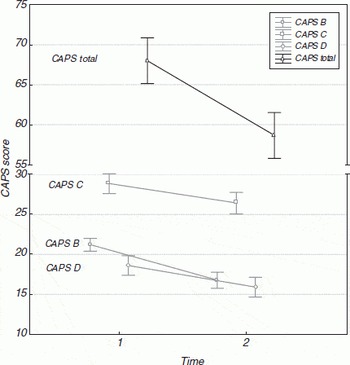
Changes in Clinician-Administered posttraumatic stress Disorder scale (CAPS) scores.

Statistics
Variables that did not follow Gaussian distribution were normalized using Box-Cox transformations. Since TNF-α could not be normalized we did not include it in overall model but tested the group difference in each time point using Mann-Whitney's U test.
Because of the presence of missing data, all other analyses were performed on 5 imputed datasets after multiple imputations. To assess group differences in measured variables in each time point, multivariate general linear model with age and smoking as covariates was performed. To test equality of covariance matrices Box's M-test was used followed by multivariate tests (Pillai's Trace, Wilks' Lambda, Hotelling's Trace, Roy's Largest Root) to test overall significance of the model. If the model was significant multiple univariate general linear model tests were performed to test the effect of group on single variable. To minimize possibility of type I error because of the multiple comparisons we used the false discovery rate method Reference Benjamini and Hochberg(10)). Statistical analyses were done with Statistica v6 (StatSoft, Inc., Tulsa, OK, USA).
Results:
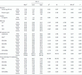
The total CAPS score, as well as all scores referring to all three clusters of symptoms have significantly declined over years in PTSD patients (Figure 1). The differences in biological variables between PTSD patients and controls in both time points are summarized in Tables 1 and 2. PTSD patients had higher cortisol and prolactin levels in the first assessment. Prolactin levels remained elevated in the second assessment. We did not observe the difference in IL-6 concentration between groups in both assessments while TNF-a was higher in the patients during first assessment (U = 309.0, P = 0.008). NKCC was elevated in PTSD patients and GR levels in lymphocytes, as well as all lymphocyte subpopulations were lower in PTSD patients in the first time point. These differences disappeared in the second time point. Lymphocyte count was higher in PTSD patients in both assessments. Percentages of circulating total T cells (CD3+) and helper T cells (CD3+CD4+) in particular, were higher in PTSD patients only in the first time point.
Discussion:
Our results indicate that changes in the endocrine and immune systems in PTSD are not static but change during the course of the illness in parallel with clinical symptoms of the disorder. To our knowledge this is the first prospective study of its kind.
Findings of low cortisol level with enhanced negative feedback sensitivity to cortisol, i.e. lower number of GR, are considered to be a distinctive biological feature of PTSD that separates this disorder from other psychiatric conditions such as depression Reference Yehuda(11)). Simultaneous determination of the cortisol and its receptor enables better characterization of the HPA axis than cortisol alone Reference Yehuda(11)). Using this approach, we have previously reported that Croatian combat veterans had higher cortisol concentration with lower lymphocyte GCR expression Reference Olff, Guzelcan, de Vries, Assies and Gersons(14)). This finding was confirmed in this study as well, but we also managed to show that over the course of about 6 years lymphocyte GR expression tends to be higher in all lymphocyte subpopulations assessed while cortisol levels decline. Although we didn't observe group differences in GR expression in the second time point, we assume that distinct profile of HPA axis shown in the other studies is probably achieved later in the course of PTSD, more than 10 years following trauma. This hypothesis is further supported by finding of positive correlation between lymphocyte GR expression and number of years passed since trauma (data not shown).
Along with cortisol, prolactin is another stress hormone that we have assessed in this study. Opposite to cortisol, prolactin is considered to be mainly immunostimulatory hormone Reference Webster Marketon and Glaser(12)). Prolactin has shown to be increased in civilians displaced by war Reference Sabioncello, Kocijan-Hercigonja, Rabatic, Tomasic, Jeren, Matijevic, Rijavec and Dekaris(13)), decreased in civilian PTSD Reference Olff, Guzelcan, de Vries, Assies and Gersons(14)), or unchanged in earthquake survivors with PTSD Reference Song, Zhou and Wang(15)). Serum levels of prolactin were elevated in PTSD patients in both time points in our study. We have also observed week-positive correlation (r = 0.452; P = 0.012) between NKCC (25:1) and prolactin level in PTSD patients in the first time point. Since NKCC was elevated in PTSD patients in the first time point, there is a possibility that prolactin contributed to this effect. This is further supported by in vitro findings of the effect of prolactin on human NK cells function Reference Sun, Li, Wei and Tian(16)). Since PTSD patients were taking medications (not systematically assessed) in the years between assessments there is also potential long term effect of drugs, especially commonly prescribed serotonin reuptake inhibitors (SSRI), on prolactin levels. Further studies are needed to elucidate this finding.
Elevated proinflammatory cytokines support the hypothesis for low-grade systemic proinflammatory activity in patients with PTSD Reference von Kanel, Hepp, Kraemer, Traber, Keel, Mica and Schnyder(17)). Consistent with previous findings Reference von Kanel, Hepp, Kraemer, Traber, Keel, Mica and Schnyder(17,Reference Sutherland, Alexander and Hutchison18) we found TNF-a to be higher in PTSD patients compared to controls, but only in the first time point. IL-6 was found both higher and unchanged in PTSD patients compared to controls Reference Miller, Sutherland, Hutchison and Alexander(19,Reference Maes, Lin, Delmeire, Van Gastel, Kenis, De Jongh and Bosmans20,Reference Baker, Ekhator, Kasckow, Hill, Zoumakis, Dashevsky, Chrousos and Geracioti21). We did not observe any differences in IL-6 between patients and controls in our study. The finding of higher cortisol levels in the first time point suggests active suppressive action of endogenous cortisol on proinflammatory cytokine production Reference Gander and Von Kanel(22)). Since this may be the case for IL-6, elevated TNF-α in the first time point does not support this notion. More likely, severity of symptoms which indicates activation of stress response systems could be associated with TNF-α elevation. The symptoms of the PTSD were more pronounced in the first time point in our study and others showed that TNF-a correlated with most of the PTSD symptoms determined by CAPS Reference von Kanel, Hepp, Kraemer, Traber, Keel, Mica and Schnyder(17)).
Research on the immune function in PTSD showed generally an increased delayed-type hypersensitivity Reference Watson, Muller, Jones and Bradley(23)), higher percentage of perforin positive natural killer cells Reference Skarpa, Rubesa, Moro, Manestar, Petrovecki and Rukavina(24)), and inconclusive natural killer cell cytotoxicity measurements Reference Laudenslager, Aasal, Adler, Berger, Montgomery, Sandberg, Wahlberg, Wilkins, Zweig and Reite(7,Reference Ironson, Wynings, Schneiderman, Baum, Rodriguez, Greenwood, Benight, Antoni, LaPerriere, Huang, Klimas and Fletcher25,Reference Mosnaim, Wolf, Maturana, Mosnaim, Puente, Kucuk and Gilman-Sachs26). According to the results from chronic stress studies of decreased NKCC Reference Segerstrom and Miller(27)), the same would be expected in the chronic PTSD. Nevertheless, it has already been shown that Croatian professional soldiers released from concentration camp had higher NKCC Reference Lauc G, Dumic and Flogel(28)). Our study showed that initially increased NKCC could not be confirmed 6 years after because of the elevation of NKCC in healthy individuals. Although our data indicate that NKCC in PTSD follows acute stress pattern Reference Segerstrom and Miller(27)) further studies are needed to elucidate the dynamics of NKCC changes during the course of the disease.
Group differences in biological variables in the second time point

CD, cluster of differentiation; FDR, false discovery rate; GR, glucocorticoid receptor; IL-6, interleukin-6; mfi, mean fluorescence intensity; NKCC, natural killer cell citotoxicity;
SE, standard error of the mean.
* Statistically significant (P < 0.05).
1 The time passed from traumatic experience was 11.4 years (median; interquartile range 11.1-2.4); 39 PTSD patients and 25 controls were assessed.
2 Estimated mean after 5 imputations.
3 Multivariate general linear model was significant (Pillai's Trace, Wilks' Lambda, Hotelling's Trace i Roy's Largest Root) in each of 5 imputed datasets (P < 0.001). β, SE, t and P are pooled values obtained after performing multiple univariate general linear model tests on 5 imputed datasets.
4 False discovery rate at α = 0.05 for total number of comparisons (P < 0.05 if P < d).
5 25:1, 50:1, 100:1 refer to effector to target cell ratio.
6 CD3+: T cells; CD3+CD4+: helper T cells; CD3+CD8+: cytotoxic T cells; CD20+: B cells; CD16+CD56+: natural killer cells.
SD, standard deviation.
Our results from the first assessment confirmed the previous findings of higher peripheral blood percentages of helper T cells (CD4) in Croatian PTSD patients Reference Skarpa, Rubesa, Moro, Manestar, Petrovecki and Rukavina(24)) or higher percentages of T cells (CD3) and CD4 in Vietnam veterans Reference Boscarino and Chang(6)). This is consistent with the hypothesis that immune activation in PTSD may be associated with Th2 cytokine shift and recruitment of CD4 cells Reference Wong(2)). However, in the second assessment we did not find any differences between patients and controls. Moreover, the results from other studies indicate the lack of differences in the peripheral blood lymphocyte phenotype in combat-related Reference Laudenslager, Aasal, Adler, Berger, Montgomery, Sandberg, Wahlberg, Wilkins, Zweig and Reite(7)), or childhood sexual abuse-related Reference Wilson, van der Kolk, Burbridge, Fisler and Kradin(5)) chronic PTSD. All of this suggests that immunopehnotype specific changes may be observable earlier in the course of the disease. It was shown that Vietnam veterans with PTSD had higher lymphocyte counts, consistent with the higher prevalence of autoimmune diseases Reference Boscarino(1)). Our results showed that PTSD patients had higher lymphocyte counts in both time points. This finding seems to be very stable in Croatian PTSD veterans since we have observed the same in other assessed groups as well (unpublished data). The risk for developing autoimmune disease according to lymphocyte counts should be systematically assessed.
According to the results of this study we can conclude that changes in the endocrine and immune systems are not static in persons under prolonged stress but rather depend on duration of alostatic load and its impact on interactions involved in response to stress. Long term prospective studies could further elucidate the dynamics of changes in endocrine and immune systems in the PTSD patients implicated by this study.
Acknowledgements:
This work was supported by grants from the Ministry of Science, Education and Sports of the Republic of Croatia (No. 0021003 to Dragan Dekaris). We gratefully acknowledge the assistance of Timea Berki (Department of Immunology and Biotechnology, University of Pecs, Faculty of Medicine, Pecs, Hungary) who provided us with anti-glucocorticoid receptor monoclonal antibody.