Introduction
Sperm cells from total asthenozoospermic specimens and testicular tissue lack or have poor motility. The use of an inducer of spermatozoa movement led to the production of increased numbers of motile sperm cells that may result in turning an invasive technique, intracytoplasmic sperm injection (ICSI), into in vitro fertilization (IVF), and IVF to intra-uterine insemination (IUI) (Salian et al., Reference Salian, Nayak, Kumari, Patel, Gowda, Shenoy, Sugunan, Managuli, Mutalik, Dahiya, Pal, Adiga and Kalthur2019). Pentoxifylline (PTX) is a phosphodiesterase inhibitor that leads to a surge in cyclic adenosine monophosphate (c-AMP) levels and, therefore, improves the outcomes of sperm motility as well as acrosome reactions (AR) (Chehab et al., Reference Chehab, Madala and Trussell2015). The AR includes a chain of action that sperm cells undergo to naturally achieve fertilization of the oocyte. Following the AR, there is structural modification of related proteins placed in the sperm plasma membrane and disclosure of the inner acrosomal membrane. These events have essential roles in the fusion of spermatozoa and oocytes (Tsirulnikov et al., Reference Tsirulnikov, Huta and Breitbart2019). PTX can improve the ability of spermatozoa to undergo AR in response to a calcium ionophore (CI) (Salian et al., Reference Salian, Nayak, Kumari, Patel, Gowda, Shenoy, Sugunan, Managuli, Mutalik, Dahiya, Pal, Adiga and Kalthur2019). CI A23187 is known to induce the AR of human spermatozoa, but has adverse effects on some biological parameters such as spermatozoa movement and mitochondrial integrity (Engel et al., Reference Engel, Springsguth and Grunewald2018).
As the ICSI technique bypasses the zona pellucida (ZP) and the oolemma, most injected sperm cells probably have intact acrosomes (Gianaroli et al., Reference Gianaroli, Magli, Ferraretti, Crippa, Lappi, Capitani and Baccetti2010). The injection of sperm cells without intact acrosomes resulted in increased clinical outcomes (Gianaroli et al., Reference Gianaroli, Magli, Ferraretti, Crippa, Lappi, Capitani and Baccetti2010). Furthermore, some results of motile sperm organelle morphology examination (MSOME) indicated that the presence of nuclear vacuoles on sperm heads is typically related to the presence of acrosomal material. Therefore, the induction of AR may enhance the percentage of vacuole-free sperm cells (Kacem et al., Reference Kacem, Sifer, Barraud-Lange, Ducot, De Ziegler, Poirot and Wolf2010). However, other studies have indicated that sperm head vacuoles are created during the early stages of spermatogenesis (Leahy and Gadella, Reference Leahy and Gadella2011) and they do not appear or disappear under different conditions (Neyer et al., Reference Neyer, Vanderzwalmen, Bach, Stecher, Spitzer and Zech2013). Throughout the process of oxidative phosphorylation, protons are pumped from inside mitochondria to the outside, generating an electrochemical gradient named the mitochondrial membrane potential (MMP). The MMP is associated with standard semen parameters such as motility and viability (Yeste et al., Reference Yeste, Recuero, Maside, Salas-Huetos, Bonet and Pinart2021). In infertile men, sperm mitochondria produce an excess of reactive oxygen species (ROS) that correlates with the decrease in MMP and increased DNA fragmentation (Kowalczyk, Reference Kowalczyk2022).
Previously, the co-effects of PTX and CI A12387 have been assessed in normozoospermic and asthenozoospermic ejaculate samples (Salian et al., Reference Salian, Nayak, Kumari, Patel, Gowda, Shenoy, Sugunan, Managuli, Mutalik, Dahiya, Pal, Adiga and Kalthur2019). Previous research has pointed to controversial results regarding PTX, and mentioned the necessity for more reassuring data (Fallahzadeh et al., Reference Fallahzadeh, Rezaei, Rahimi, Barmak, Sadeghi, Mehrabi, Rabani, Kashani, Barati and Mahmoudi2017; Salian et al., Reference Salian, Nayak, Kumari, Patel, Gowda, Shenoy, Sugunan, Managuli, Mutalik, Dahiya, Pal, Adiga and Kalthur2019). In addition, there have been limited human studies that evaluated the associations between the biological characteristics of spermatozoa (Zhang et al., Reference Zhang, Yang, Zou, Jiang, Zeng, Chen, Sun, Yang, Zhou, Wang, Liu, Cao, Zhou and Ao2019). The present study was designed to investigate the consequences of treatment with chemical agents PTX and CI on some biological characteristics of sperm cells including AR, vacuole status, MMP and DNA fragmentation in the most severely abnormal ejaculate samples from oligoasthenoteratozoospermia (OAT) patients.
Materials and methods
Patients
Among the OAT patients who were referred to the Yazd Reproductive Sciences Institute, 25 patients aged 20–40 years were entered into the study after signing informed consent. OAT is a condition that is described by oligozoospermia (sperm number < 15 million/ml), asthenozoospermia (total motility < 40%) and teratozoospermia (normal morphology < 4%) (WHO, 2010). Specimens were obtained by masturbation following 2–5 days of sexual abstinence. Sperm preparation was performed using the single-layer density gradient (DG) (Nabi et al., Reference Nabi, Khalili, Fesahat, Talebi and Ghasemi-Esmailabad2017). The exclusion criteria were smoking and a history of varicocele or varicocelectomy. This study was approved by the ethics committee of the institute (code: IR.SSU.RSI.REC.1400.007). All patients signed informed consent to participate in the research.
Study design
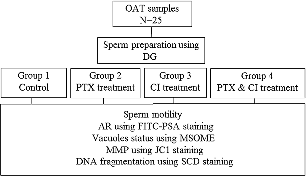
After preparation, each semen sample was divided into four groups including control (group 1), 3.6 mM PTX (group 2), 5 μM CI A12387 (group 3), and both PTX and CI A12387 (3.6 mM PTX and 5 μM CI A12387, simultaneously; group 4) (Nabi et al., Reference Nabi, Khalili, Fesahat, Talebi and Ghasemi-Esmailabad2017; Salian et al., Reference Salian, Nayak, Kumari, Patel, Gowda, Shenoy, Sugunan, Managuli, Mutalik, Dahiya, Pal, Adiga and Kalthur2019). As shown in Figure 1, AR was assessed using PSA-FITC staining; vacuole status using MSOME, MMP using JC-1 staining and DNA fragmentation via sperm chromatin dispersion (CSD) test.
Diagram of study design. AR, acrosome reaction; CI, calcium ionophore; DG, density gradient; JC1, unique fluorescent cationic dye, 5,5,6,6-tetrachloro-1,1,3,3-tetraethylbenzimidazolylcarbocyanine iodide; MMP, mitochondrial membrane potential; MSOME, motile sperm organelle morphology examination; OAT, oligoasthenoteratozoospermia; PTX, pentoxifylline; SCD, sperm chromatin dispersion.

Treatment with PTX and CI A12387
Washed sperm cells were incubated with 3.6 mM PTX (Sigma, USA) (Nabi et al., Reference Nabi, Khalili, Fesahat, Talebi and Ghasemi-Esmailabad2017) and 5 μm of CI A23187 (Sigma-Aldrich) (Salian et al., Reference Salian, Nayak, Kumari, Patel, Gowda, Shenoy, Sugunan, Managuli, Mutalik, Dahiya, Pal, Adiga and Kalthur2019) for 30 min at 37°C and in a 5% CO2 atmosphere. After incubation, sperm were washed with Ham’s F10 medium (Biochrome, Berlin, Germany) supplemented with 5 mg/ml human serum albumin (HAS; Vitrolife, Englewood, CO) and centrifuged at 400 g for 5 min. The supernatant was removed and the pellet was resuspended in 0.5 ml culture medium.
Assessment of sperm motility
Before and after sperm preparation, total motility (progressive and non-progressive) and progressive motility were evaluated manually according to the WHO criteria (WHO, 2010). A minimum of 100 sperm was assessed.
Assessment of AR
A smear was prepared using 25 µl from each sperm suspension and allowed to air dry. Cells were permeabilized using methanol (15 min at 4 C) and stained with 25 μg/ml FITC-conjugated Pisum sativum agglutinin (FITC-PSA; Sigma-Aldrich) (Salian et al., Reference Salian, Nayak, Kumari, Patel, Gowda, Shenoy, Sugunan, Managuli, Mutalik, Dahiya, Pal, Adiga and Kalthur2019). Slides were placed in an humidified chamber for 15 min in the dark and then washed in phosphate-buffered saline (PBS) (Kacem et al., Reference Kacem, Sifer, Barraud-Lange, Ducot, De Ziegler, Poirot and Wolf2010). Sperm cells in each group were assessed for AR using a fluorescence microscope (Imager A1, Carl Zeiss, Germany). Sperm cells with no green FITC-PSA fluorescence over the whole head or with a thin equatorial band were considered acrosome reacted and those with completely green acrosome caps were considered intact acrosome spermatozoa (Tsirulnikov et al., Reference Tsirulnikov, Huta and Breitbart2019).
Assessment of vacuole status
An aliquot of the sperm suspension was transferred to a microdroplet of polyvinyl pyrrolidone (PVP) medium (Irvine Scientific) in a sterile glass-bottomed culture dish (GWSt 1000; Will Co. Amsterdam, The Netherlands). Motile spermatozoa presented in PVP droplets were examined using an inverted microscope equipped with a heated stage and differential interference contrast (DIC) optics and a Nikon Digital Sight DS-Ri1 Camera at ×6000 magnification. For each sample, MSOME analysis was carried out on 100 randomly selected sperm cells. Classification of MSOME was done based on the presence of vacuoles in the sperm head, in which class 1 was defined as sperm cells having no nuclear vacuole and class 2 was defined as sperm cells that had vacuoles in the head (Kacem et al., Reference Kacem, Sifer, Barraud-Lange, Ducot, De Ziegler, Poirot and Wolf2010).
Assessment of MMP
Variations in the MMP of spermatozoa were assessed using a unique fluorescent cationic dye, 5,5,6,6-tetrachloro-1,1,3,3-tetraethylbenzimidazolylcarbocyanine iodide, commonly known as JC-1. In total, a 25-µl sperm suspension from each group was incubated with 25 µl of JC-1 solution for 30 min at 37°C. Next, the cell suspensions were centrifuged at 400 g in the dark for 5 min at room temperature. After removal of the supernatant, the pellet was resuspended in 0.5 ml culture medium and the sperm cells were observed using a fluorescence microscope (Olympus Co., Tokyo, Japan) at ×1000 magnification (Isachenko et al., Reference Isachenko, Isachenko, Weiss, Kreienberg, Katkov, Schulz, Lulat, Risopatrón and Sánchez2008). Based on the fluorescence colour intensity in the mid-piece region, the spermatozoa were categorized as intact (red fluorescence) or damaged mitochondria (green fluorescence) (Barbagallo et al., Reference Barbagallo, La Vignera, Cannarella, Aversa, Calogero and Condorelli2020).
Assessment of sperm DNA fragmentation
Next, a 20-µl sperm suspension from each group was added to 20 μl of 1% low-melting-point agarose gel and mixed. Here, 20 µl of this mixture was pipetted onto a glass slide pre-coated with 0.65% standard agarose and then covered with a coverslip. The slide sample was left to solidify at 4°C for 5 min. Coverslips were then removed and the slides were stained according to the manufacturer’s recommendation (SDFA kit, Tehran, Iran). Depending on the halo (dispersed DNA loops) size, sperm cells were categorized as no, small, medium, or large halos by observation under the microscope. Spermatozoa with no or small halos were considered to contain DNA damage and the results were expressed as percentages (Anbari et al., Reference Anbari, Khalili, Agha-Rahimi, Maleki, Nabi and Esfandiari2020).
Statistical analysis
The assessment of data distribution was performed using the Kolmogorov–Smirnov test. Based on this test, the difference between total motility and progressive motility before and after processing, as well as data from AR, MMP, DNA fragmentation, and vacuole status had a normal distribution in all groups (P > 0.05) were assessed. The study design considered dependencies between groups and was compared using repeated measurements analysis and the least significant difference (LSD) post-test. Mauchly’s sphericity test was used to validate a repeated measures analysis of variance. Statistical analysis was performed using Statistical Package for the Social Sciences 20 (SPSS Inc., Chicago, IL, USA) with a significance level of P < 0.05. Results were expressed as mean ± standard deviation (SD).
Results
Tables 1 and 2 show the mean difference between total motility and progressive motility before and after processing in all groups. Mauchly’s sphericity test for total motility and progressive motility was 0.624 (P = 0.057) and 0.648 (P = 0.080), respectively. Overall, the present data showed that CI A12387 decreased total sperm motility and progressive motility significantly. Moreover, the use of PTX along with CI, led to a significant enhancement in total motility and progressive motility compared with using CI alone (P < 0.05). In addition, treatment with PTX led to a significantly increased total sperm motility and progressive motility compared with the other groups (P < 0.05).
Mean difference of total motility before and after processing in all groups

* Repeated measures analysis and Mauchly’s sphericity test.
a – dAccording to the LSD post-test, significant differences were found between group 1 and groups 2 and 3; group 2 and groups 3 and 4; and also between group 3 and group 4 (P < 0.05).
Mean difference of progressive motility before and after sperm processing in all groups

* Repeated measures analysis and Mauchly’s sphericity test.
According to LSD post-test, significant difference was found between group 1 and groups 2, 3 and 4; group 2 and groups 3 and 4; and between group 3 and group 4 (P < 0.05).
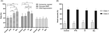
For AR analysis, Mauchly’s sphericity test was 0.843 (P = 0.568). As shown in Figure 2(A), the acrosome-reacted sperms were significantly lower in groups 1 and 2 compared with groups 3 and 4 and also in group 3 compared with group 4 (P < 0.05). Moreover there was no significant difference between group 1 and group 2 (P > 0.05) (Figure 2A). Therefore, PTX did not have a significant influence on the AR process, but the addition of CI led to an increase in the numbers of sperm cells that underwent AR. Moreover, the application of PTX along with CI significantly increased acrosome-reacted sperm cells.
Effect of inducers of sperm motility (PTX) and inducers of the AR (CI A23187) on biological characteristics of human sperm cells from OAT patients. (A) Based on the PSA-FITC staining and JC1 test, there were significant differences between groups, but SCD staining did not show any significant differences between all groups (P = 0.529). (B) There were not any significant differences in head vacuole distribution between all groups. Data are shown as bar graphs representing mean ± SD. Graphs were plotted using the software GraphPad Prism 8 (Graph Pad Software, Inc., La Jolla, CA, USA). Significance is shown as *P < 0.05, **P < 0.01, *** P < 0.001.

Conversely, Mauchly’s sphericity test in MSOME analysis was 0.785 (P = 0.358) and 0.875 (P = 0.711) in class 1 and class 2, respectively (Table 3). As shown in Figure 2(B), processing the semen samples either with PTX or CI A12387 did not have any significant effect on vacuole status (P > 0.05).
Mean of acrosome-reacted sperm cells, sperm occupied with vacuoles, sperm with disrupted MMP, and DNA fragmentation in each group

* Repeated measures analysis and Mauchly’s sphericity test.
The acrosome-reacted sperms were significantly lower in groups 1 and 2 compared with groups 3 and 4, and also in group 3 compared with group 4 (P < 0.05). The application of mixed PTX along with CI significantly increased acrosome-reacted sperm cells.
MSOME analysis showed that processing the semen samples with either PTX or CI A12387 did not have any significant effect on vacuole status (P > 0.05).
Disrupted MMP was significantly lower in groups 1 and 2 compared with groups 3 and 4, and also in group 4 compared with group 3 (P < 0.05). The present data showed that PTX can modify the adverse effects of CI on MMP.
There were no significant differences in DNA fragmentation between all groups (P < 0.05) (Figure 2A).
MMP is the main sign of the functional integrity of sperm cells. In our study, Mauchly’s sphericity test of MMP analysis gave a value of 0.828 (P = 0.508) (Table 3). The results indicated that sperm cells with disrupted MMP were significantly lower in groups 1 and 2 compared with groups 3 and 4, and also in group 4 compared with group 3 (P < 0.05). Moreover there was not any significant difference between group 1 and group 2 (P > 0.05) (Figure 2A). The present data showed that PTX can modify the detrimental effects of CI on MMP.
Finally, Mauchly’s sphericity test for DNA fragmentation analysis gave a value of 0.282 (P = 0.00) (Table 3) and there were no significant differences in DNA fragmentation among all groups (P < 0.05) (Figure 2A).
Discussion
Spermatozoa from OAT men were incubated with inducers of sperm motility (PTX) and AR (CI A12387). The results indicated that PTX increased total motility and progressive motility significantly, which was similar to findings from earlier investigations (Mahaldashtian et al., Reference Mahaldashtian, Khalili, Nottola, Woodward, Macchiarelli and Miglietta2021). In the different sperm anomalies, such as oligoasthenoteratozoospermia (Rezaie et al., Reference Rezaie, Allahveisi, Raoofi, Rezaei, Nikkhoo and Mousavi Khaneghah2021), asthenozoospermic samples (Saber et al., Reference Saber, Shafey, Alabedeen, Massoud and Elwan2022) it was also shown that PTX could improve sperm parameters and motility significantly. This effect was derived from the phosphodiesterase inhibitor (PDE) properties of PTX, which increased cyclic adenosine monophosphate (c-AMP) levels and, consequently, sperm motility (Mahaldashtian et al., Reference Mahaldashtian, Khalili, Nottola, Woodward, Macchiarelli and Miglietta2021).
In a previous study, it was shown that CI A23187 led to a significantly decreased total motility and progressive motility (Engel et al., Reference Engel, Springsguth and Grunewald2018). Also, in our study, the use of PTX along with CI led to an increased sperm motility, which showed that PTX could rectify the adverse effects of CI on sperm motility. To date, there have been very limited studies that have investigated in the effects of PTX and CI, in combination. In one study, it was shown that PTX could improve the ability of frozen–thawed spermatozoa to undergo AR in response to CI (Esteves et al., Reference Esteves, Spaine and Cedenho2007).
The results of AR indicated that PTX alone had no effect on the AR process, but the simultaneous use of PTX and CI resulted in an increased number of acrosome-reacted sperm cells. Other studies have also shown that, although PTX could increase sperm motility, it did not have a significant effect on the acrosomal reaction of the thawed spermatozoa. In this study, they reasoned that PTX could not preserve the sperm membrane integrity, which was needed for the initiation of the AR but, when mitoquinone was added, sperm membrane integrity improved significantly (Nazari et al., Reference Nazari, Daghigh-Kia, Mehdipour and Najafi2022).
In accordance with our study, a study by Salian et al. (Reference Salian, Nayak, Kumari, Patel, Gowda, Shenoy, Sugunan, Managuli, Mutalik, Dahiya, Pal, Adiga and Kalthur2019) showed that higher AR was achieved in the CI-induced acrosome reaction when PTX was used in sperm preparation medium. In this mechanism, AR was triggered by CI, which varied from the physiological AR induced by the ZP. Even though physiological AR is associated with the activation of numerous signal transduction pathways, the CI-induced AR comprised a chemical effect on calcium influx (Esteves et al., Reference Esteves, Spaine and Cedenho2007). Also, PTX could increase sperm sensitivity to the AR-inducing stimulus (Esteves et al., Reference Esteves, Spaine and Cedenho2007; Salian et al., Reference Salian, Nayak, Kumari, Patel, Gowda, Shenoy, Sugunan, Managuli, Mutalik, Dahiya, Pal, Adiga and Kalthur2019). PTX may improve the initiation of AR rather than direct stimulation (Tasdemir et al., Reference Tasdemir, Tasdemir, Kodama and Tanaka1993). Therefore, sensitization of PTX to the AR response led to it being used as a potential candidate for the treatment of AR deficiencies (Mahaldashtian et al., Reference Mahaldashtian, Khalili, Nottola, Woodward, Macchiarelli and Miglietta2021). The outcomes of polarized light microscopy affirmed that, for severe male factors, the injection of reacted spermatozoa may result in improved gravidity (Gianaroli et al., Reference Gianaroli, Magli, Ferraretti, Crippa, Lappi, Capitani and Baccetti2010). MSOME is a noninvasive technique for the precise evaluation of motile sperm cells (Mangoli et al., Reference Mangoli, Khalili, Talebi, Kalantar, Montazeri, Agharahimi and Woodward2020). In this study, sperm head vacuoles were detected in many sperm cells from all groups. The precise origin of the vacuoles in sperm cells is unknown, but different hypotheses have suggested they could be derived from the acrosome (Kacem et al., Reference Kacem, Sifer, Barraud-Lange, Ducot, De Ziegler, Poirot and Wolf2010) or early stages of spermatogenesis (Leahy and Gadella, Reference Leahy and Gadella2011). Moreover, Boitrelle et al. (Reference Boitrelle, Ferfouri, Petit, Segretain, Tourain, Bergere, Bailly, Vialard, Albert and Selva2011) suggested that vacuole formation in spermatozoa started during the incubation and capacitation period after ejaculation. The results related to the effect of CI on sperm head vacuoles are controversial, with some speculation that it is used to trigger the activation of the apoptosis cascade, leading to an enhanced percentage of spermatozoa with head vacuoles (Engel et al., Reference Engel, Springsguth and Grunewald2018). Kacem et al. (Reference Kacem, Sifer, Barraud-Lange, Ducot, De Ziegler, Poirot and Wolf2010) claimed that the incubation of sperm cells with CI significantly decreased sperm head vacuoles. They further proposed that most nuclear vacuoles had an acrosomal source. Also, Neyer and co-workers, announced that exposure to oxidative stress or the use of CI did not alter the percentage of spermatozoa with vacuoles or the disappearance of pre-existing vacuoles (Neyer et al., Reference Neyer, Vanderzwalmen, Bach, Stecher, Spitzer and Zech2013). Based on our knowledge, there have been no studies that assessed the effect of PTX treatment on spermatozoa vacuoles. We observed that PTX does not induce any changes in the vacuole’s numbers. It may be assumed that vacuoles are formed at former stages of spermiogenesis and they do not become visible or invisible under different conditions (Neyer et al., Reference Neyer, Vanderzwalmen, Bach, Stecher, Spitzer and Zech2013).
A former study indicated that MMP is associated with standard semen parameters such as motility and viability (Wang et al., Reference Wang, Sharma, Gupta, George, Thomas, Falcone and Agarwal2003). The outcome of this study also confirmed the previous result. As mentioned above, the use of CI A12387 resulted in significantly decreased sperm motility. Also, the percentage of spermatozoa with disrupted MMP was lower in the PTX group than the other groups, even though it was not meaningful between the control and PTX groups. The other study that investigated the effects of PTX on MMP showed that semen sample processing with PTX did not show any significant effect on MMP, however damage would increase over time (Salian et al., Reference Salian, Nayak, Kumari, Patel, Gowda, Shenoy, Sugunan, Managuli, Mutalik, Dahiya, Pal, Adiga and Kalthur2019). In addition, the percentage of spermatozoa with disrupted MMP was significantly higher in the CI group rather than in the other groups. Based on the Amaral study, myoinositol, as an inducer of sperm motility, resulted in an enhanced number of spermatozoa with high MMP (Amaral et al., Reference Amaral, Lourenço, Marques and Ramalho-Santos2013). Additionally, Salian and team workers (2019) demonstrated that the treatment of semen samples with PTX did not have any adverse effect on MMP. Also, Engel and associates noted that CI significantly increased the rate of sperm cells that reacted with the acrosome and disrupted MMP (Engel et al., Reference Engel, Springsguth and Grunewald2018). According to our results, DNA fragmentation was not different from any stimulators. This may be due to the high compartmentalization of sperm cells that led to a limited separation of proteins (Baker et al., Reference Baker, Naumovski, Hetherington, Weinberg, Velkov and Aitken2013). The former study indicated that the use of CI did not alter DNA fragmentation because DNA in sperm cells is mostly protamine bound (Engel et al., Reference Engel, Springsguth and Grunewald2018) and so packed more tightly than in somatic cells, which could avoid DNA fragmentation (Engel et al., Reference Engel, Springsguth and Grunewald2018). Some studies using sperm chromatin dispersion (SCD) staining have indicated that PTX has no adverse effect on sperm chromatin/DNA integrity (Nabi et al., Reference Nabi, Khalili, Fesahat, Talebi and Ghasemi-Esmailabad2017; Mahaldashtian et al., Reference Mahaldashtian, Khalili, Mangoli, Zavereh and Anbari2022).
According to the results of our study, it seems that PTX not only could increase sperm motility, but also may rectify the detrimental effects of CI on MMP, without any adverse effects on the formation of head vacuoles and/or DNA fragmentation.
Acknowledgements
We thank the staff of the andrology and research laboratories for their excessive contributions.
Funding information
There was no funding.
Conflict of interest
The authors declare none.
Ethical standards
The authors assert that all procedures contributing to this work comply with the ethical standards of the relevant national and institutional committees on human experimentation and with the Helsinki Declaration of 1975, as revised in 2008. The authors assert that all procedures contributing to this work comply with the ethical standards of the relevant national and institutional guides on the care and use of laboratory animals.