1. General considerations and overview
The work of Christian Anfinsen and his colleagues in the late 1950s established that a polypeptide chain contains sufficient information in its amino acid sequence to direct folding into the functionally active native state, lying at an energetic minimum (summarized by Anfinsen, Reference Anfinsen1973). Over the past decades, the traverse of polypeptides down energy landscapes to their native state has been intensively studied in vitro, with considerable understanding gained of the driving forces and pathways. The same forces and pathways are surely at work in the cell, but in this context, many polypeptides cannot reach the native state without assistance from a group of specialized proteins known as molecular chaperones, which collectively comprise a machinery that assists this final step of information transfer between DNA and functional protein.
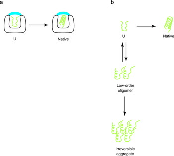
Chaperone machines provide kinetic assistance to the process of protein folding, essentially preventing or reversing steps of misfolding and aggregation that are disposed to by in vivo conditions that involve higher temperature and greater solute concentration than employed in vitro. In particular, the chaperones share in common the property of binding exposed hydrophobic surfaces that are presented by non-native proteins through their own variously exposed hydrophobic surfaces. Chaperones thus prevent polypeptide chains from being recruited to each other through such hydrophobic surfaces to form multimolecular aggregates. Thus, in effect, the chaperones prevent the exploration of an entirely different folding energy landscape, one that contains the possibility of multimolecular aggregation, which can present deep minima that are alternatives to the native state and from which recovery cannot occur on a physiologic timescale.
Typically, substrate proteins are released from the chaperones by the action of ATP binding, triggering a trial at correct folding, with failure to reach native form enabling rebinding by the chaperone machinery. Released proteins may also be subject, where appropriate signals are present, to engagement by trafficking machinery or by the degradation apparatus, enabling cellular compartmentation or removal of misfolded proteins, respectively. In general, it appears that proteins released from chaperones undergo a kinetic partitioning among various components, whose outcome is determined by the relative concentration of the cellular components and their respective affinities for any particular conformation of the substrate protein.
Many of the molecular chaperones are heat shock proteins, whose abundance in the cell is strongly induced by thermal as well as other stresses. This is consistent with the early recognition by Pelham and his co-workers in the mid-1980s that, under heat shock conditions, proteins are liable to misfolding and aggregation and that the heat shock proteins act to forestall or reverse these processes (Pelham, Reference Pelham1984, Reference Pelham1986). Yet subsequent studies made clear that the chaperones provide an essential action also under non-stress conditions to enable productive folding (Deshaies et al. Reference Deshaies, Koch, Werner-Washburne, Craig and Schekman1988; Fayet et al. Reference Fayet, Ziegelhoffer and Georgopoulos1989; Cheng et al. Reference Cheng, Hartl, Martin, Pollock, Kalousek, Neupert, Hallberg, Hallberg and Horwich1989; Reading et al. Reference Reading, Hallberg and Myers1989), further implying that kinetic errors leading to misfolding and aggregation can also occur in the cell under baseline physiologic conditions.
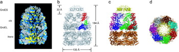
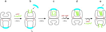
Perhaps the most remarkable of the chaperone machines are the ring assemblies known as the chaperonins, which release many of the substrates they bind into an encapsulated chamber. The best understood of the chaperonins, the bacterial GroEL, binds non-native protein in the hydrophobic central cavity of an open ring (Fig. 1a, trans; Braig et al. Reference Braig, Otwinowski, Hegde, Boisvert, Joachimiak, Horwich and Sigler1994) and then, upon binding ATP and a ‘lid’ component, the co-chaperonin GroES, to the same ring as non-native polypeptide, mediates productive folding to the native state inside the now-encapsulated and hydrophilic cavity (Fig. 1a, cis; Weissman et al. Reference Weissman, Hohl, Kovalenko, Chen, Braig, Saibil, Fenton and Horwich1995; Mayhew et al. Reference Mayhew, da Silva, Martin, Erdjument-Bromage, Tempst and Hartl1996; Weissman et al. Reference Weissman, Rye, Fenton, Beechem and Horwich1996; Xu et al. Reference Xu, Horwich and Sigler1997). Folding of a solitary polypeptide chain in this chamber is the longest-lived step of the reaction cycle, proceeding for a period of approximately 10 s (Fig. 2c; Rye et al. Reference Rye, Burston, Fenton, Beechem, Xu, Sigler and Horwich1997, Reference Rye, Roseman, Furtak, Fenton, Saibil and Horwich1999; Ranson et al. Reference Ranson, Burston and Clarke1997). This step is followed by ATP hydrolysis in the folding-active (cis) ring, gating subsequent binding of ATP in the opposite (trans) ring, which in turn sends an allosteric signal that ejects the GroES lid and substrate protein (Fig. 2d, e; Rye et al. Reference Rye, Burston, Fenton, Beechem, Xu, Sigler and Horwich1997, Reference Rye, Roseman, Furtak, Fenton, Saibil and Horwich1999), whether refolded or not (Todd et al. Reference Todd, Viitanen and Lorimer1994; Weissman et al. Reference Weissman, Kashi, Fenton and Horwich1994; Smith & Fisher, Reference Smith and Fisher1995; Taguchi & Yoshida, Reference Taguchi and Yoshida1995). Released non-native protein is then subject to partitioning, as just mentioned, but, as observed in vitro, it can be rapidly rebound by GroEL and subject to a further cycle of attempted folding (Weissman et al. Reference Weissman, Kashi, Fenton and Horwich1994). This overall reaction cycle, diagrammed in Fig. 2 and detailed in its legend, has been understood for some years, but the nature of how it acts on the conformation of non-native polypeptide at the various steps to facilitate productive folding has remained open to conjecture. This review focuses on current understanding of the fate of substrate polypeptide during the reaction: during the step of binding to an open GroEL ring, during ATP–GroES encapsulation, and during folding inside the GroEL–GroES cis chamber.
Architecture of the GroEL–GroES chaperonin complex, based on the GroEL–GroES–ADP7 crystallographic model (PDB 1AON; Xu et al. Reference Xu, Horwich and Sigler1997). (a) Cut-away view of a space-filling model of the GroEL–GroES complex, with hydrophobic side chains colored yellow and hydrophilic ones blue. Note the relative hydrophobicity of the trans, polypeptide-accepting ring, and hydrophilicity of the cis folding chamber formed by GroES binding to a GroEL ring. (b) Ribbon diagram of GroEL–GroES with the individual domains of one subunit in the upper, cis ring colored red (apical), green (intermediate), and blue (equatorial). (c) Same view as in (b), except each subunit in the cis ring is in a different color and GroES is colored yellow. (d) View of the complex from the top, looking down on GroEL, colored as in (c). Note that the apparent ‘hole’ down the long axis of the complex reflects that the flexible C-terminal tails of each GroEL subunit, lying in the equatorial regions of the central cavity of each ring, are not resolved in the crystal structure. Figure modified from Horwich et al. (Reference Horwich, Farr and Fenton2006), with permission. Copyright 2006, American Chemical Society.

The polypeptide folding cycle at GroEL. (a) The initial polypeptide acceptor state in vivo and in a cycling reaction in vitro is an asymmetric, ADP-containing complex, as shown in Fig. 1 (Rye et al. Reference Rye, Burston, Fenton, Beechem, Xu, Sigler and Horwich1997; Xu et al. Reference Xu, Horwich and Sigler1997). (b) ATP and non-native polypeptide (green) bind rapidly to the open ring of the ADP asymmetric complex, with ATP producing downward rigid body movement of the intermediate domain via contacts with the nucleotide pocket (Chaudhry et al. Reference Chaudhry, Farr, Todd, Rye, Brunger, Adams, Horwich and Sigler2003) and attendant small elevation and counterclockwise twist of the apical domains (Ranson et al. Reference Ranson, Farr, Roseman, Gowen, Fenton, Horwich and Saibil2001), the latter of which directly enable GroES binding. ATP binding also sends an allosteric signal to the opposite (trans) ring that ejects its ligands: GroES, substrate polypeptide, and ADP (Rye et al. Reference Rye, Burston, Fenton, Beechem, Xu, Sigler and Horwich1997). The binding of non-native polypeptide along with ATP accelerates such departure of GroES (Rye et al. Reference Rye, Roseman, Furtak, Fenton, Saibil and Horwich1999). (c) Initial GroES interaction with the ATP–polypeptide-bound ring, occurring by ~0·2 s (Cliff et al. Reference Cliff, Limpkin, Cameron, Burston and Clarke2006), is followed over ~0·8 s by large and forceful rigid body elevation and clockwise twisting movements (60° and 120°, respectively) that eject polypeptide into a GroES-encapsulated hydrophilic chamber where polypeptide attempts to fold (Weissman et al. Reference Weissman, Rye, Fenton, Beechem and Horwich1996; Xu et al. Reference Xu, Horwich and Sigler1997; Rye et al. Reference Rye, Burston, Fenton, Beechem, Xu, Sigler and Horwich1997). This folding-active cis complex is the longest-lived state in the folding cycle. (d) ATP hydrolysis, occurring with a half-time of ~10 s, primes the cis complex for discharge of its ligands by weakening affinity of GroEL for GroES (Rye et al. Reference Rye, Burston, Fenton, Beechem, Xu, Sigler and Horwich1997). (e) ATP hydrolysis gates the binding of ATP and non-native polypeptide to the trans ring (Rye et al. Reference Rye, Roseman, Furtak, Fenton, Saibil and Horwich1999), a function of allosteric behavior. In the case of ATP, such behavior is anti-cooperative between rings (sequential, KNF) and positive (concerted, MWC) within the rings, overall a ‘nested’ behavior (Yifrach & Horovitz, Reference Yifrach and Horovitz1995). ATP (and polypeptide) binding to the trans ring discharges GroES and polypeptide from the old cis ring and leads to a new round of cis encapsulation and folding in this (opposite) ring. Note that the 10 s or so of folding time in the cis complex is not sufficient for most molecules of stringent, GroEL–GroES-dependent substrate proteins to fold to native form; typically, only a few percent reach the native state. The other molecules are discharged at (e) in non-native states that, in vitro, are rebound by other GroEL complexes for another attempt at folding. In the cell, such non-native forms can also partition to other chaperones or the degradative apparatus, their fate depending on the relative concentrations and affinities of the various components. D indicates ADP, T indicates ATP.

Considering the findings that will be reviewed below, it seems increasingly likely that the GroEL–GroES machine functions essentially passively with respect to substrate polypeptide during all three steps, i.e. during binding, encapsulation, and cis folding. Concerning binding to an open ring, to which we devote the greatest attention here as this seems to be the least defined, it appears that GroEL simply uses its hydrophobic cavity-facing surface to capture collapsed non-native states. If it preferentially binds one or more of the most hydrophobic conformers in the rapidly dynamic ensemble of non-native conformers that is present in solution, it will shift the equilibrium in that direction, a thermodynamic partitioning. Where less folded states expose the greatest amount of hydrophobicity, this would effectively shift the equilibrium in the direction of less folded states, a passive unfolding action. But once a polypeptide is captured, during its dwell time of 1 s in an open ring (before ATP–GroES-driven release into the cavity), it may be subject to thermal motions that further optimize the binding of hydrophobic surfaces to multiple apical domains, effectively a passive ‘stretching’ action. In several cases, such action in association with binding appears to be able to rescue a kinetically trapped state of a substrate monomer, effectively pulling it up the energy landscape. Yet, surprisingly, there is little or no stable hydrogen-bonded secondary structure present in the non-native substrates examined to date, either free in solution or after binding to an open GroEL ring. This implies that there must be relatively low-energy barriers between the non-native states, which would seem in general to be surmountable by thermally induced motions, yet apparently where that fails, the passive stretching behavior of multivalent hydrophobic binding appears able to lift a number of substrates out of deeper kinetic traps.
During the subsequent step of ATP–GroES binding and encapsulation that triggers cis folding, there have been suggestions that an action of ‘forced unfolding’ of substrate polypeptide might be exerted by apical domain movements, before release of polypeptide into the GroES-encapsulated folding chamber. Most recently, this has taken the form of fluorescence studies observing, here also, ‘stretching’ when ATP is incubated with a chaperonin–substrate complex. We discuss these experiments, which do not reckon with what we find is an opposite order of addition in the reaction cycle, in which ATP binds rapidly to an open trans ring and has already driven apical movements by the time polypeptide substrate arrives. We also consider additional observations that seem to indicate that, regardless of the order of events, there is no requirement at this encapsulation stage for a stretching step in order to enable productive folding. Indeed, if there is any consistent behavior during the encapsulation step, it appears to be a compaction of the non-native protein (Lin & Rye, Reference Lin and Rye2004; Sharma et al. Reference Sharma, Chakraborty, Müller, Astola, Tang, Lamb, Hayer-Hartl and Hartl2008).
Finally, during the step of folding in the cis chamber, there is, based on recent studies, no evidence for any active assistance by the cavity or its walls to the folding process (Horst et al. Reference Horst, Fenton, Englander, Wüthrich and Horwich2007; Apetri & Horwich, Reference Apetri and Horwich2008). The chamber simply isolates non-native substrates as monomers and allows them to proceed down their energy landscape without the kinetic complication of multimolecular aggregation that supervenes if the same substrate proteins are placed free in solution.
Given such apparently passive action at these various steps, it appears that the GroEL–GroES machine uses ATP binding and hydrolysis largely to drive unidirectional progression through its reaction cycle. The only recognized point at which energy seems to be directly applied is during ATP–GroES binding, where forceful rigid body movements of the apical domains are needed to efficiently and concertedly eject substrate polypeptide into the cis chamber to trigger folding (Motojima et al. Reference Motojima, Chaudhry, Fenton, Farr and Horwich2004; Kipnis et al. Reference Kipnis, Papo, Haran and Horovitz2007; Papo et al. Reference Papo, Kipnis, Haran and Horovitz2008). We discuss below the underpinnings of the various conclusions as well as current uncertainties.
2. Polypeptide binding
2.1. Hydrophobic contacts as the major driving force
2.1.1 Site of bound polypeptide
Following initial biochemical observations of association of non-native polypeptide with both mitochondrial Hsp60 and bacterial GroEL chaperonin assemblies (Goloubinoff et al. Reference Goloubinoff, Christeller, Gatenby and Lorimer1989; Ostermann et al. Reference Ostermann, Horwich, Neupert and Hartl1989), microscopy studies localized bound protein in the central cavity of an open GroEL ring. Hartl, Baumeister, and their co-workers, using negative stain electron micrsoscopy (EM), observed ‘mass’ within the central cavity in end views of substrate-bound as compared with empty complexes (Langer et al. Reference Langer, Pfeifer, Martin, Baumeister and Hartl1992). Wall, Hainfeld, Horwich, and their collaborators, using scanning transmission electron micrsoscopy, observed localization of a nanogold particle attached to a substrate protein within the central cavity in end views and at a terminal end of the GroEL cylinder in side views of substrate-bound complexes (Braig et al. Reference Braig, Simon, Furuya, Hainfeld and Horwich1993). The localization of bound substrate was confirmed by Saibil and co-workers during a study using negative stain EM to resolve ATP-directed movements of GroEL. They consistently observed ‘masses’ in the terminal ends of the GroEL central cavities in side views, abutting the GroEL apical domains (Saibil et al. Reference Saibil, Zheng, Roseman, Hunter, Watson, Chen, Auf der Mauer, O'Hara, Wood, Mann, Barnett and Ellis1993). The masses very likely comprised an averaged collective of substrate proteins that remained bound by GroEL during purification. This study was followed by one from the same group using cryo-EM that similarly localized a substrate protein, malate dehydrogenase (MDH), to one end of the GroEL cylinder (Chen et al. Reference Chen, Roseman, Hunter, Wood, Burston, Ranson, Clarke and Saibil1994), consistent with biochemical studies indicating a stoichiometry of one non-native polypeptide bound per GroEL double ring particle (Goloubinoff et al. Reference Goloubinoff, Christeller, Gatenby and Lorimer1989), implying a negative cooperativity between rings with respect to polypeptide binding.
At the same time as the early EM studies, a study from Horowitz, Lorimer, and their co-workers inferred that hydrophobic interactions might be involved in polypeptide binding, observing that addition of the detergent lauryl maltoside could modestly improve the low recovery of native rhodanese enzyme following dilution from urea at 25°C in the absence of chaperonin (Mendoza et al. Reference Mendoza, Rogers, Lorimer and Horowitz1991). While the recovery was well below the near quantitative recovery achieved by GroEL–GroES–ATP, it suggested that the chaperonin, like the detergent, might be binding-exposed hydrophobic surfaces of the non-native rhodanese, preventing them from directing aggregation. An additional inference of the importance of hydrophobic interaction came from a thermodynamic study observing a negative heat capacity associated with binding of non-native subtilisin, an indication of hydrophobic interactions as a driving force in polypeptide binding (Lin et al. Reference Lin, Schwarz and Eisenstein1995).
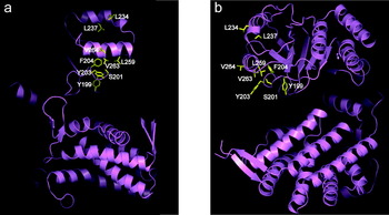
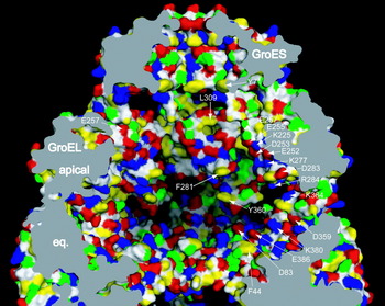
The X-ray structure of unliganded GroEL indeed revealed a hydrophobic cavity wall at the level of the apical domains at the terminal ends of the chaperonin cylinder, an apparent surface to which non-native polypeptides could become bound (Braig et al. Reference Braig, Otwinowski, Hegde, Boisvert, Joachimiak, Horwich and Sigler1994), apparently through their own hydrophobic surfaces that would be exposed specifically in the non-native state but that would be buried to the interior of native folded protein (for which GroEL does not have affinity). Consistently, mutagenesis studies showed that substitution of any one of eight different hydrophobic residues facing the GroEL central cavity at the level of the apical domains to hydrophilic or electrostatic character abolished polypeptide binding (Fig. 3; Fenton et al. Reference Fenton, Kashi, Furtak and Horwich1994). These residues were positioned on a tier of three different structures lining the cavity: helix H, helix I, and an underlying extended segment (see Fig. 3). Additional mutational studies used covalent rings that enforced a specific arrangement of subunits with wild-type and mutant apical domains (the mutant substituting a central residue in the binding site, Val 263, with Ser). It was observed that three or four consecutive intact wild-type apical domains were required in order to efficiently bind non-native substrate protein (Farr et al. Reference Farr, Furtak, Rowland, Ranson, Saibil, Kirchhausen and Horwich2000). Cross-linking studies validated this conclusion. Cysteines in a bound radiolabeled substrate protein, Rubisco, were assessed for ability to form disulfide bonds with multiple apical domains, each carrying a single cysteine substitution in the apical binding site. The radioactive cross-linked products were sized in an SDS gel to determine the number of GroEL subunits that had become cross-linked (Farr et al. Reference Farr, Furtak, Rowland, Ranson, Saibil, Kirchhausen and Horwich2000). This indicated that, indeed, bound substrate could simultaneously interact with three or four surrounding apical domains.
The apical residues that form the hydrophobic binding surface of a GroEL subunit. A ribbons model is shown, with side chains of residues whose mutation abolishes polypeptide binding shown as yellow sticks and labeled with residue numbers. The residues are grouped on three structures – helix H, helix I, and an underlying extended segment – from top to bottom, respectively. (a) View en face from inside the central cavity. (b) View from one side of a subunit, with the cavity to the left. From Horwich et al. (Reference Horwich, Farr and Fenton2006), with permission. Copyright 2006, American Chemical Society.

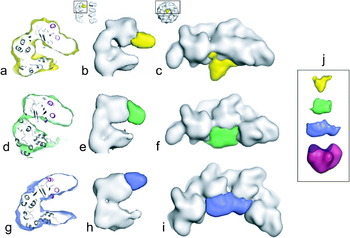
More recently, a multivalent binding topology has been directly observed by cryo-EM studies examining one of the well-studied substrate proteins, MDH, while bound to an open GroEL ring (Elad et al. Reference Elad, Farr, Clare, Orlova, Horwich and Saibil2007). Multiple image classes revealed MDH lodged against three or four apical domains of an open GroEL ring (Fig. 4). At the level of the individual domains, there were varying topologies observed. One image class, for example, showed electron density from MDH perched at the outside rim of the cavity (Fig. 4g–i), reflecting interaction with helices H and I, and another showed MDH density deeply buried in the cavity, abutting both helix I and the underlying extended segment (Fig. 4a–c). The GroEL apical domains themselves appeared to adjust to polypeptide binding, with those physically contacting substrate bunched together and those at the opposite aspect of the ring exhibiting a wider separation from each other (see also Falke et al. Reference Falke, Tama, Brooks, Gogol and Fisher2005).
Topologies of non-native MDH bound to GroEL. Three image classes from a cryo-EM reconstruction (Elad et al. Reference Elad, Farr, Clare, Orlova, Horwich and Saibil2007) are illustrated in side and end views. (a, d, g) Sections through the MDH-bound GroEL subunits in each class, showing the fitting of the GroEL apical domain secondary structural elements. Note the areas at the end of each domain without fitted elements, with density contributed by the bound non-native MDH. (b, e, h) Side views of a substrate-bound subunit (see thumbnail at top), with the GroEL rendered as a surface at 14 Å and the difference density corresponding to MDH colored yellow, green, or blue. (c, f, i) Top views of substrate-bound subunits (see thumbnail) rendered as in (b, e, h). Three GroEL subunits are shown in (c) and (f) and four in (i), corresponding to the number of subunits contacted by substrate in each of these classes. (j) The individual difference densities corresponding to MDH for each class, compared with the surface representation of an MDH subunit monomer (red) filtered to 20 Å; the exposed dimer interface of the MDH subunit in the bottom image is colored purple. Modified from Elad et al. (Reference Elad, Farr, Clare, Orlova, Horwich and Saibil2007), with permission. Copyright 2007, Elsevier.

An additional cryo-EM study of the large T4 phage-encoded capsid protein, gp23 (54 kDa), in complex with GroEL, has recently been reported, showing localization of density of the non-native capsid protein deep within the cavity, contacting five to seven surrounding GroEL subunits of an open ring (Clare et al. Reference Clare, Bakkes, van Heerikhuizen, van der Vies and Saibil2009). Bound substrates thus interact simultaneously with multiple surrounding hydrophobic apical domains and can interact at multiple points across the hydrophobic surface, although in general it appears that helix I represents a central shared position of association.
For polypeptides of size greater than 30–40 kDa, it seems clear that only a portion of the bound non-native chain can be contained within the central cavity, whereas other portions must reside in the bulk solution. Such portions have not been observable by EM but have been inferred to lie in solution immediately above the cavity opening. For example, the oblong shape of GroEL–rhodanese complexes as determined from neutron scattering studies suggested that the polypeptide occupied a topology like that of a ‘champagne cork’ (Thiyagarajan et al. Reference Thiyagarajan, Henderson and Joachimiak1996). Consistently, additional cysteine cross-linking studies employing cysteine scanning across the interior and exterior surfaces of GroEL do not observe GroEL-bound polypeptide forming cross-links with the top surfaces of the apical domains (Elad et al. Reference Elad, Farr, Clare, Orlova, Horwich and Saibil2007). Rather, cross-links are only formed within the cavity, occurring at all levels. That is, while the polypeptide chain is physically associated with the apical domains, it can occupy space throughout the cavity, including the equatorial zone. Thus, for example, the mass of C-terminal tails of the GroEL subunits (aa 523–547) bearing flexible GGM repeats, lying in the equatorial zone of the cavity and forming a mass (~20 kDa per ring) that is visible by EM but not crystallographically resolvable, can form contacts with the non-native polypeptide. The C-terminal tails presumably serve as a barrier that contains substrate within a ring, although their deletion does not produce any significant growth defect of Escherichia coli (Burnett et al. Reference Burnett, Horwich and Low1994; McLennan et al. Reference McLennan, McAteer and Masters1994). Notably, however, in Mycobacterium tuberculosis, there are two GroEL homologs, one of which, GroEL1, has C-terminal tails bearing a poly-histidine stretch. This segment might contribute to binding specific substrate proteins, such as KasA, involved in synthesis of mycolic acids that are required for biofilm formation, a property dependent on GroEL1 function (Ojha et al. Reference Ojha, Anand, Bhatt, Kremer, Jacobs and Hatfull2005).
Interestingly, the portion of bound polypeptide in solution outside the cavity appears in general to be also non-native in character, as opposed to occupying a folded state. For example, the large substrate protein, aconitase (82 kDa), was entirely susceptible to proteolysis and hydrogen exchange while bound by GroEL (Chaudhuri et al. Reference Chaudhuri, Farr, Fenton, Rospert and Horwich2001, unpublished observation). This, as discussed below, suggests that the chaperone binds various exposed hydrophobic surfaces in a spatially averaged as well as probably a time-averaged fashion.
2.1.2 Elements recognized in bound polypeptide
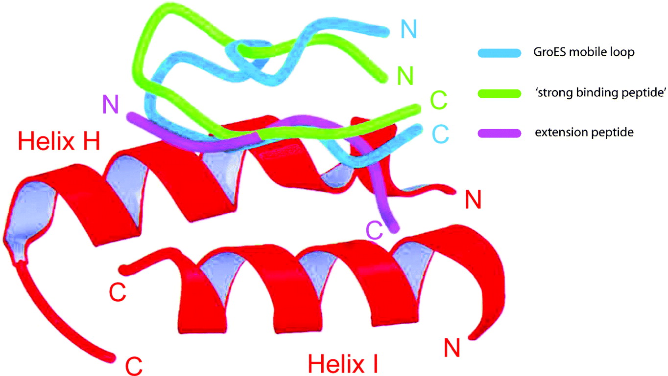
The proffering of hydrophobic side chains on the part of GroEL-bound non-native substrate proteins themselves has also been explored. For example, Itzhaki, Fersht, and their co-workers engineered single substitutions into a variety of residues in the protein CI2, observing the greatest reduction in binding with substitution of hydrophobic side chains with alanine or glycine (Itzhaki et al. Reference Itzhaki, Otzen and Fersht1995). In a further study from the Fersht group, the crystallization of an isolated apical domain led unexpectedly to the crystallographic resolution of seven residues, GLVPRGS, in a larger N-terminal His tag, bound in an extended conformation within a groove between helices H and I in a neighboring apical domain molecule in the asymmetric unit (Buckle et al. Reference Buckle, Zahn and Fersht1997). The leucine was positioned in a cavity between the two α-helices, while the C-terminal portion of the peptide aligned along helix I, forming numerous apolar contacts with the same hydrophobic side chains that had been identified by mutagenesis. In a more directed study, Chen & Sigler (Reference Chen and Sigler1999) used filamentous phage display to select a 12 residue peptide called SBP (strong binding peptide) that bound to isolated GroEL apical domain with ~10 μm affinity (compared with typically nanomolar affinity for polypeptides, bound multivalently). The SBP peptide, SWMTTPWGFLHP, was co-crystallized with isolated apical domain, binding as a β-hairpin with residues 7–12 associated with the cavity-facing surface of the apical domain via the groove between helices H and I, forming a number of hydrophobic contacts with residues that had been implicated in the earlier mutagenesis study (Fig. 5).
Topologies of three peptides bound to the apical domain of GroEL. The backbones of the GroES mobile loop segment (blue) (Xu et al. Reference Xu, Horwich and Sigler1997), the ‘strong binding peptide’ (green) of Chen & Sigler (Reference Chen and Sigler1999), and the N-terminal extension peptide from Buckle et al. Reference Buckle, Zahn and Fersht1997 (magenta) are shown overlaid on aligned Cα ribbon trace of apical domain helices H and I of GroEL (red) from the respective structures. Note that the bound regions of all three peptides appear to occupy nearly the same position in the groove between the two helices. From Fenton & Horwich (Reference Fenton and Horwich2003), with permission. Copyright 2003, Cambridge University Press.

These two studies of peptide–apical interactions provide the highest resolution to date of apical interactions. It has to be noted, however, that they resemble quite strongly the interaction formed by the hydrophobic mobile loop of GroES when it binds to the GroEL apical domains (Fig. 5; see Xu et al. Reference Xu, Horwich and Sigler1997; Shewmaker et al. Reference Shewmaker, Maskos, Simmerling and Landry2001). One might suggest that these interactions between peptide and the apical interhelical groove represent one physiologic mode of polypeptide binding, potentially a favored one, but, given the cryo-EM data, there are surely others that have yet to be resolved, not to mention that the ‘tracking’ of a non-native polypeptide across multiple apical domains has yet to be observed at high resolution. The problems of trapping such a state for X-ray studies remain daunting, as discussed earlier (Fenton & Horwich, Reference Fenton and Horwich2003).
One of the most informative studies concerning the role of substrate hydrophobicity in polypeptide binding has been carried out by the Gierasch group, systematically arranging hydrophobic side chains and secondary structure of 20 different synthetic peptides of 12–14 residues, assessing their overall hydrophobicity by retention time on reverse phase high-performance liquid chromatography and measuring affinity of GroEL for them by either line broadening or by the transferred NOE (trNOE) method of nuclear magnetic resonance (NMR) (Wang et al. Reference Wang, Hwa-Ping, Landry, Maxwell and Gierasch1999). First, reversal of chirality of an α-helical peptide using d-amino acids did not affect the extent of binding, and the trNOE pattern suggested that the helical character (now left-handed) was preserved. Interestingly, an l,d-mixed peptide was also recognized and could compete with the l-peptide but assumed a structure, as judged from the trNOE spectrum, that was not recognizable as either a right- or left-handed α-helix. Thus, the three peptides, exhibiting similar proffered hydrophobicity as judged from retention in reversed phase analysis, were bound with roughly similar affinity by GroEL, despite their substantially different secondary structures, pointing to hydrophobic side chains as a major recognized feature regardless of backbone context. Next, the binding of an amphiphilic α-helical peptide, placing hydrophobicity contiguously on one side, was compared with that of a non-amphiphilic one of the same amino acid composition. Here the non-amphiphilic peptide exhibited both less line broadening and smaller trNOEs, reflecting lower affinity of GroEL for this peptide. This would support the idea that clustering of hydrophobic side chains, here on one face of an α-helix, favors binding (see also Preuss et al. Reference Preuss, Hutchinson and Miller1999; Li et al. 2009). Finally, peptides whose trNOE spectra were consistent with extended structure while associated with GroEL (e.g. as in SBP) were also studied, and one with alternating hydrophobic/hydrophilic character was observed to be recognized by GroEL. Overall, when the retention time of the 20 peptides was compared with intensity of transfer NOEs as a measure of affinity, there was an excellent correlation, suggesting that extent of contiguous hydrophobic exposure determines affinity of GroEL for any given stretch of polypeptide chain, irrespective of structural context.
Indeed, GroEL has substrates that are all α-helical, like citrate synthase (Buchner et al. Reference Buchner, Schmidt, Fuchs, Jaenicke, Rudolph, Schmid and Kiefhaber1991), but also can bind all β-sheet proteins, like a partially disulfide-bonded Fab fragment (Schmidt & Buchner, Reference Schmidt and Buchner1992). Similarly, studies of different structural states of α-lactalbumin indicate that GroEL does not efficiently recognize the disulfide-bonded molten globule state (formed by chelating calcium ion; Okazaki et al. Reference Okazaki, Ikura, Nikaido and Kuwajima1994) but prefers a reduced, less structured state, which exposes substantial amounts of hydrophobic surface (Hayer-Hartl et al. Reference Hayer-Hartl, Ewbank, Creighton and Hartl1994). Parenthetically, disulfide-bonded species are not normally acted on by chaperonins, because they are, for the most part, secreted proteins that fold in the oxidizing environment of the ER, where such ring assemblies have not been identified. Chaperonins, rather, reside in reduced compartments of the cell (cytosol, mitochondrial matrix, chloroplast stroma), where disulfide bonds are not usually formed. However, disulfide bonded proteins, because of their ability to form relatively stabilized intermediate states, have been of use in vitro as ‘probes’ of GroEL function.
2.2 Binding an ensemble of non-native conformers – evidence for thermodynamic partitioning action
The foregoing collective of observations would indicate that, in general, collapsed non-native forms that expose substantial hydrophobic surface will become bound by GroEL. This goes a long way to explaining the early observation by Viitanen, Lorimer, and co-workers that, when a total E. coli extract solubilized in guanidine HCl was diluted into a mixture containing GroEL, greater than 50% of the proteins became stably bound to it as revealed by comigration in gel filtration (Viitanen et al. Reference Viitanen, Gatenby and Lorimer1992). The full range of sizes was recognized, including proteins greater than 60 kDa in molecular mass. Presumably they all share in common the exposure, after dilution from denaturant, of hydrophobic surfaces. This observation and one made more recently in vivo, observing wholesale aggregation in a cellular context of GroEL deficiency (Chapman et al. Reference Chapman, Farr, Usaite, Furtak, Fenton, Hondorp, Matthews, Wolf, Yates, Pypaert and Horwich2006), would indicate that the binding action of GroEL may have a broad role in the cell that extends beyond the set of a few dozen proteins whose proper folding must take place in the encapsulated GroEL–GroES chamber. That is, insofar as binding may forestall aggregation mediated by association of hydrophobic surfaces, and insofar as such binding may be able to reverse kinetically trapped states of at least some protein monomers (see below), this may already provide an assisting action to a broad collective of cellular proteins that do not require encapsulation for productive folding. Further proteomic work will be required to establish the breadth of such a role under basal growth conditions. It seems likely, however, that under heat shock conditions, where the level of GroEL is induced to 10–15% of total soluble protein (Herendeen et al. Reference Herendeen, van Bogelen and Neidhardt1979), the action of general binding of non-native proteins via exposed hydrophobic surfaces becomes a dominant and protective role.
One must presume that non-native states of any given chaperonin substrate protein in solution are present as an equilibrium ensemble, with relatively low-energy barriers separating the various states. If GroEL is simply binding those states that expose the greatest amount of hydrophobic surface with the greatest affinity, then this will effectively shift the equilibrium, by mass action, toward such states. This model of so-called thermodynamic partitioning was first proposed by Zahn & Plückthun (Reference Zahn and Plückthun1994). One study that has probably lent the greatest amount of direct support to the model was from Walter et al. (Reference Walter, Lorimer and Schmid1996). They employed a mutant form of RNase T1 and carboxyamidated its four thiols, producing two-state folding behavior between a near native conformation and an unfolded state that could be followed by tryptophan fluorescence. The addition of increasing concentrations of GroEL progressively shifted the equilibrium to the unfolded state but, as revealed by kinetic studies, addition of GroEL did not affect the microscopic rate of unfolding. This indicated that GroEL shifted the equilibrium by selectively binding the non-native form, as opposed to actively catalyzing unfolding (see also Bhutani & Udgaonkar, Reference Bhutani and Udgaonkar2000).
Such a mechanism seems likely to operate not only at the level of an ensemble of non-native monomeric states free in solution but also at the level of low-order oligomers that form a reversible equilibrium with binding-proficient monomers. Early work of the Clarke group measured rates of recovery and yield of native MDH in relation to relative concentration of GroEL, enabling kinetic modeling of a competition for non-native MDH free in solution between a slow unimolecular step that commits MDH to the native state and a reversible low-order oligomerization step that slowly gives rise to irreversible higher order aggregates (Ranson et al. Reference Ranson, Dunster, Burston and Clarke1995). Here, as with an ensemble of monomers, a kinetic partitioning by GroEL could be operative, in this case with a reversible equilibrium in solution between low-order aggregates and monomers, shifted toward the disassembled monomeric state by binding of monomers by GroEL. Such a shift would obviously reduce the population of irreversible higher order aggregates and increase the overall efficiency of MDH folding.
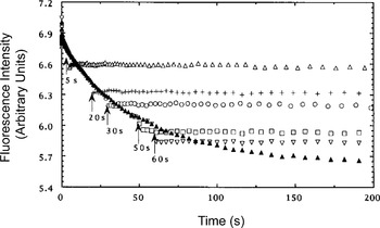
The thermodynamic partitioning model offers an explanation for a host of results observed over the years. For example, studies of DHFR (Goldberg et al. Reference Goldberg, Zhang, Sondek, Matthews, Fox and Horwich1997) refolding free in solution, under conditions where aggregation does not supervene, observed that addition of GroEL at a variety of times could entirely halt refolding, associated with efficient binding (Fig. 6). This would be consistent with the chaperonin accessing the entire ensemble of non-native states that have not as yet crossed a transition state to form the native state. This occurs quite rapidly, in the case of DHFR within a few seconds at most. Thus, within the lifetime of an open GroEL ring during a cycling reaction, 1–2 s (see Fig. 2), an ensemble of non-native states would be predicted to be efficiently captured. This appears to be the case, as evidenced by trap studies, in which various polypeptide substrates released from cycling reactions in non-native form are captured within seconds of the time of addition of a molar excess of a trap version of GroEL, able to bind non-native conformers but not release them (Weissman et al. Reference Weissman, Kashi, Fenton and Horwich1994; see also Brinker et al. Reference Brinker, Pfeifer, Kerner, Naylor, Hartl and Hayer-Hartl2001).
Tryptophan fluorescence of human DHFR refolding from urea. At the times indicated, an excess of GroEL was added. The solid triangles show the entire time-course of refolding in the absence of chaperonin. Note that addition of GroEL apparently stops the refolding of DHFR whenever it is added to the ongoing solution reaction, implying that GroEL can recruit the ensemble of non-native intermediates present throughout the course of the folding reaction. Redrawn from Goldberg et al. Reference Goldberg, Zhang, Sondek, Matthews, Fox and Horwich1997. Copyright 1997, National Academy of Sciences, USA.

2.3 Lack of secondary structure in substrate proteins
2.3.1 GroEL-bound substrates lack stable secondary structure
Exchange studies on binary complexes of cyclophilin (Zahn et al. Reference Zahn, Spitzfaden, Ottiger, Wüthrich and Plückthun1994), disulfide-reduced α-lactalbumin (Robinson et al. Reference Robinson, Gross, Eyles, Ewbank, Mayhew, Hartl, Dobson and Radford1994), human DHFR (Goldberg et al. Reference Goldberg, Zhang, Sondek, Matthews, Fox and Horwich1997), MDH (Chen et al. Reference Chen, Walter, Horwich and Smith2001), and Rubisco (Park et al. Reference Park, Fenton and Horwich2005) showed at best only low levels of protection in the bound proteins. In several cases, potentially weakly structured native regions were observed, but protection factors in these regions were below 100. The observation of a lack of any significant hydrogen bonded structure raised the question of whether secondary structure present in species free in solution might be getting disrupted as they became bound to the open GroEL ring.
Yet observations on two well-studied substrates while free in solution have failed to indicate any substantial secondary structure. When MDH was diluted from denaturant and pulse exchanged while free in solution 5 s later (for 1 s), it exhibited little or no protection (Chen & Walter, unpublished observation; see also Chen et al. Reference Chen, Walter, Horwich and Smith2001). This corresponds to the timescale on which non-native MDH would be fully bound by GroEL, because its bimolecular rate constant for binding to GroEL has been measured at 5×10⁶ M−1 s−1, and GroEL concentration is ~1 μm (Ranson et al. Reference Ranson, Burston and Clarke1997). Similarly, a metastable non-native intermediate of Rubisco, formed after dilution from denaturant under chloride-free conditions and able to be efficiently bound by GroEL during a lifetime of ~1 h, exhibited no protection from pulsed hydrogen–tritium exchange (Park et al. Reference Park, Fenton and Horwich2005).
2.3.2 Binary behavior of secondary structure during a cycling reaction – progressive vs. all-or-none folding
There is an implication from the observations above that there may really be only two principal states of secondary structure of a substrate protein in a chaperonin-mediated folding reaction, either non-native intermediates, an equilibrium ensemble of poorly structured states that are rapidly captured by GroEL via exposure of hydrophobic surfaces, or the end-product native state, no longer recognized by the chaperonin. This implies that partially folded non-native intermediates with substantial secondary structure do not accumulate during the reaction. Indeed, such states, that would manifest stable secondary structure in pulsed hydrogen–deuterium exchange, have not been observed. Rather, the evidence at hand would support that stable secondary structure formation seems to occur as all or none. Indeed, hydrogen exchange behavior does not substantially change if one compares the behavior of the substrate in a starting polypeptide–GroEL binary complex with a binary complex isolated after a number of ATP–GroES-driven cycles – the not-yet-native molecules of polypeptide remain exquisitely sensitive to protease and exhibit the same hydrogen exchange sensitivity from cycle to cycle (Weissman et al. Reference Weissman, Kashi, Fenton and Horwich1994; Groß et al. Reference Groß, Robinson, Mayhew, Hartl and Radford1996; Horst & Fenton, unpublished observations). In addition, topology studies by single-molecule FRET indicate the same distribution of topologies at both the beginning and after minutes of the cycling reaction (Sharma et al. Reference Sharma, Chakraborty, Müller, Astola, Tang, Lamb, Hayer-Hartl and Hartl2008).
2.3.3 Secondary structure unbroken by GroEL on the physiologic timescale in two bound secretory proteins bearing disulfides (that are not normally substrates for GroEL)
There are two examples to date of GroEL-bound proteins that exhibit substantial stable secondary structure while bound to an open chaperonin ring. Gervasoni et al. (Reference Gervasoni, Staudenmann, James, Gehrig and Plückthun1996) reported that E. coli RTEM β-lactamase heated to 48°C formed a stable complex with GroEL, in which it had lost its native state as evidenced by loss of enzymatic activity. When the binary complex was subjected to hydrogen–deuterium exchange, however, the β-lactamase was exchanged to an extent resembling that of native enzyme at 25°C, reflecting that a native-like degree of secondary structure was preserved in the bound protein. Further examination of the binary complex using partial proteolysis suggested a bound topology in which the central β-sheet was separated from a C-terminal α-helical region, exposing hydrophobic surfaces that were recruited to GroEL (1998). RTEM β-lactamase seems likely to be stabilized against further unfolding by a disulfide bond in its N-terminal α-helical domain. This disulfide would normally be acquired upon entry of the protein into the oxidizing environment of the bacterial periplasm. As mentioned earlier, the normal cytosolic substrates of GroEL lack such stabilizing bonds, residing in the relatively reducing environment of the cytosol. Thus, this may represent a situation where further unfolding associated with recruitment of additional hydrophobic surfaces of the bound non-native state to the GroEL apical domains cannot occur due to covalent stabilization by the disulfide bond, a type of stabilization that GroEL does not normally confront both due to the lack of such bonds in its natural substrates and the failure to form such bonds in the reducing environment of the cytosol.
In a second case, Park and co-workers have produced a partially disulfide-bonded intermediate of the secretory protein, trypsinogen, that exhibits substantial hydrogen bonded secondary structure while free in solution as indicated by exchange protection (Park et al. unpublished observations). This intermediate, produced by partial reduction of native trypsinogen, contains four out of the six native disulfides of trypsinogen. It behaves as a monomeric species in gel filtration, but nevertheless appears to expose substantial hydrophobic surface, because when mixed with GroEL, it becomes efficiently bound. In free solution, when subject to pulsed deuterium exchange (1 s), the four disulfide intermediate acquired 75 deuterons, whereas in the binary complex with GroEL, it acquired 142 deuterons, relatively close to the theoretical maximum incorporation of 183 deuterons. Yet the time employed for binary complex formation in this experiment was 30 min! In contrast, under physiologic conditions during a cycling reaction, a GroEL ring is open and available for binding for only 1 s before ATP and GroES arrive (see Fig. 2). Indeed, when binding was allowed for only 1 or 10 s before pulsed exchange, there was no exchange beyond 75 deuterons. Thus, on the short timescale, only the same surface-accessible amide protons that were exchanged in free solution were apparently subject to exchange in the binary complex. Indeed, only modest exchange beyond this level was observed even when binding was carried out for 5 min.
Thus, GroEL has only a weak ability to unfold the disulfide-bonded intermediates described and does so slowly. Because it appears to rely simply on passive hydrophobic binding, it does not have the potency of, for example, the AAA+ pulling machines such as ClpX or ClpA, which use forceful ATP-driven axial-directed movement of loops lying in their central channels to pull bound segments of polypeptide through the channels, mediating an unraveling action (Sauer et al. Reference Sauer, Bolon, Burton, Burton, Flynn, Grant, Hersch, Joshi, Kenniston, Levchenko, Neher, Oakes, Siddiqui, Wah and Baker2004; Hinnerwisch et al. Reference Hinnerwisch, Fenton, Farr, Furtak and Horwich2005).
2.3.4 Direct NMR examination of stably bound substrate proteins – lack of secondary and tertiary structure and evidence for conformational exchange
Structure in bound polypeptide has also been directly probed by recent NMR studies using transverse relaxation optimized spectroscopy (TROSY) techniques, directly observing isotope-labeled human DHFR (Horst et al. Reference Horst, Bertelsen, Fiaux, Wider, Horwich and Wüthrich2005) and rhodanese (Koculi, submitted) while stably bound to GroEL. In both cases, the uniformly 15N-labeled and deuterated proteins in complex with 15N-depleted GroEL produced resonances in the random coil region of two-dimensional 15N, proton-correlated spectra, reflecting a lack of regular secondary or tertiary structure. Line widths were broad, reflecting either the slow tumbling of the large complex or internal motion of the bound substrate, or its presence in multiple bound conformations. With both uniform labeling and, in the case of 15N-leucine specific labeling of DHFR, the number of detected residues of any specific amino acid type appeared to be underrepresented. For example, in the case of the leucines of DHFR, only a small number of overlapped cross-peaks were observed, out of 19 that would be predicted (Horst et al. Reference Horst, Bertelsen, Fiaux, Wider, Horwich and Wüthrich2005). This suggests that parts of the bound polypeptide chain are NMR invisible. These could conceivably be parts of the chain that are stably associated with the cavity wall. An attempt to resolve this with site-specific assignment using mutagenesis of the glycine residues in rhodanese, observing spectra of various alanine-substituted mutant forms while in complex with GroEL, has not so far resolved which out of 20 glycines are producing the three to four broad cross-peaks observed in this region of the spectrum.
The dynamics of SR1-bound DHFR was also examined by comparing two different types of magnetization transfer, CRIPT and INEPT, measuring 1H–15N-correlated cross-peak intensity in relation to different transfer times (Horst et al. Reference Horst, Bertelsen, Fiaux, Wider, Horwich and Wüthrich2005). The several-fold greater signal intensity with INEPT and persisting anti-phase character by both mechanisms of transfer indicated that DHFR is not rigidly bound to SR1 but is internally mobile. Because it is unknown whether bound portions of the chain are NMR visible, it is unclear whether such associated regions also undergo conformational exchange, e.g. with one region of the chain bound to an apical domain at one time but replaced by a different region of the chain at a subsequent time. This issue of whether apical bound segments are stably bound remains a fundamental question. Further NMR studies employing magnetization transfer between polypeptide and the apical domains might ultimately be able to address this. Such studies may become feasible in the near future considering both recent backbone assignment of the GroEL apical domains in the setting of an intact thermophilic GroEL assembly (Horst et al. unpublished observations) and recent successes in observing methyl side chains in large assemblies using methyl-TROSY techniques (e.g. Sprangers & Kay, Reference Sprangers and Kay2007). Meanwhile, fluorescence studies, discussed below, have provided additional measurements of chain behavior.
2.4 Measurements of long-range topology of substrates during binding using fluorescence resonance energy transfer
2.4.1 ‘Stretching’ action exerted on polypeptide topology in association with binding to an open ring – rescue of kinetically trapped monomers
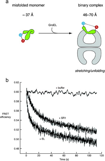
Lin & Rye (Reference Lin and Rye2004) placed fluorophores near the N- and C-termini of Rubisco and studied the behavior of the ensemble following dilution from denaturant to 100 nm concentration at 4°C. Under these conditions, Rubisco behaved as a misfolded monomer, with a high fluorescence resonance energy transfer (FRET) efficiency reflecting a distance between its termini much shorter than in the native state, indicating an apparently collapsed misfolded state that brought the termini close to each other. Upon addition of GroEL–GroES–ATP, the monomeric substrate was efficiently refolded to the native state at 4°C. The step of polypeptide binding was examined by stopped flow mixing with GroEL alone. Binding to GroEL was associated with a rapid reduction of FRET efficiency, reflecting an increased distance between the substrate termini, occurring on a timescale corresponding to that of binding, i.e. approximately a second (Fig. 7). This stretching apparently rescued Rubisco from a kinetically trapped topology that could not on its own productively fold in solution at 4°C. This represents probably the most compelling evidence to date for the ability of GroEL to rescue a non-native monomeric species from a kinetically trapped conformation via binding to an open ring, albeit at a nonphysiologic temperature. In particular, while the same non-native state is likely to be populated to some degree at 23°C, at this temperature it would, as discussed later, be able to pursue additional fates in free solution, including the formation of aggregates, both low-order ones that are reversible and higher-order irreversible ones. In addition, at a higher temperature, it would have access to additional thermal energy to drive spontaneous reversal of this misfolded state, allowing production of the native state (Viitanen et al. Reference Viitanen, Lubben, Reed, Goloubinoff, O'Keefe and Lorimer1990; Apetri & Horwich, Reference Apetri and Horwich2008).
‘Stretching’ of non-native Rubisco upon binding to GroEL, as monitored by changes in FRET. (a) Schematic of the experiment. A kinetically trapped, apparently misfolded monomer of Rubisco (able to be converted to the native state by addition of GroEL–GroES–ATP), labeled near its N- and C-termini with the FRET pair AEDANS and fluorescein, was stable at 4°C and showed an interprobe distance of ~37 Å. After binding to GroEL (or SR1) to form a binary complex, the interprobe distance increased to >70 Å for the GroEL complex and to 46–47 Å for the SR1 complex. This indicates a significant stretching of Rubisco as a result of binding to the apical domains. (b) Stopped-flow experiment to determine the rate at which stretching occurs. The changes in FRET efficiency for Rubisco binding to both GroEL and SR1 were biphasic, requiring two exponential terms for fitting. Only the faster rate was dependent on chaperonin concentration, and the bimolecular rate constant with GroEL, 2×10⁵ to 4×10⁵ M−1 s−1, was essentially equal to the independently measured rate constant for Rubisco binding to GroEL under these conditions. This suggests that the stretching occurring during the faster step is an inherent feature of Rubisco binding. Redrawn from Lin & Rye (Reference Lin and Rye2004), with permission. Copyright 2004, Elsevier.

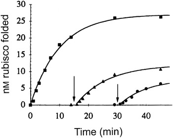
Yet a number of earlier studies carried out at 23°C and above have also observed situations where the substrate proteins Rubisco and MDH fail to reach the native state while free in solution and yet remain accessible to it upon subsequent addition of the chaperonin system. For example, following neutralization, acid-unfolded Rubisco failed to refold while free in solution over periods of 15 or 30 min, but upon addition of the chaperonin system, a fraction of the Rubisco (decreasing with time) proceeded to the native state (with identical kinetics) (Todd et al. Reference Todd, Lorimer and Thirumalai1996, reproduced in Fig. 8). This presumably involves recruitment of kinetically trapped monomers, unable to proceed to the native state, to open GroEL rings, followed by cis folding. Given the observations of Lin & Rye at 4°C, one must presume that binding to GroEL effectively stretched the Rubisco monomers, releasing them from a kinetic trap. Similar experiments have been reported as well for MDH (Peralta et al. Reference Peralta, Hartman, Hoogenraad and Høj1994; Ranson et al. Reference Ranson, Dunster, Burston and Clarke1995). This suggests, once again, that, even while there is little or no stable secondary structure in the trapped non-native states, their topologies can become sufficiently non-native that thermal motions will not provide sufficient energy to drive productive folding and that the action of multivalent hydrophobic binding by an open ring is able to relieve the trapped state.
Folding of Rubisco by GroEL added at different times after dilution from denaturant. Acid-denatured Rubisco was diluted directly into refolding buffer containing GroEL, GroES, and ATP (squares) or into buffer with GroES and ATP, with GroEL added at 15 (triangles) or 30 min (circles). In each case, the rate of recovery of Rubisco activity was the same, although the yield decreased with time before GroEL addition. In the absence of added GroEL, however, no Rubisco activity was recovered. This suggests that GroEL is binding a metastable, kinetically trapped intermediate and returning it to a folding pathway. Redrawn from Todd et al. (Reference Todd, Lorimer and Thirumalai1996), with permission. Copyright 1996, National Academy of Sciences, USA.

A more recent study has also observed that polypeptide binding by GroEL can be associated with passive stretching, using as substrate a protein called DM-MBP (double mutant V8G/Y283D of the mature maltose-binding protein; Sharma et al. Reference Sharma, Chakraborty, Müller, Astola, Tang, Lamb, Hayer-Hartl and Hartl2008). Several different pairs of fluorophore-labeled cysteine-substituted positions were used in ensemble FRET kinetic studies. There was a rapid rise of FRET efficiency within milliseconds of dilution from denaturant, whether GroEL was present or not, reflecting collapse of the polypeptide. In the presence of GroEL, however, this was followed by a rapid drop in FRET efficiency, exhibiting a t ½ of ~100 ms, which corresponds to a typical rate of substrate protein binding. This drop was attributed to an expansion of the substrate associated with GroEL binding.
2.4.2 Single-molecule FRET studies of stably associated substrate proteins reveals heterogeneity of substrate topology
Seeking to address the behavior of stably GroEL-bound substrate are two recent single-molecule FRET experiments, one examining rhodanese, the other examining DM-MBP. Both studies imply that stably bound substrates behave as a heterogeneous set of conformations. In the rhodanese study, Schuler and co-workers placed Alexa Fluor 488 and 594 FRET reporters at two different pairs of positions on rhodanese and measured energy transfer efficiency at the single-molecule level under denaturing conditions, after binding to GroEL, and after ATP–GroES-mediated refolding (Hillger et al. Reference Hillger, Hänni, Nettels, Geister, Grandin, Textor and Schuler2008). Both in denaturant and after refolding, they observed a relatively narrow distribution of transfer efficiency that corresponded in the former case to the distance along the primary structure separating the fluorophores, and in the latter to the distance determined from the X-ray structure of the native state. In the denatured state, studies indicated rapid intramolecular dynamics, as might be expected, on the timescale of ~70 ns. When the rhodanese was bound by SR1, a broad distribution of transfer efficiencies was observed, reflecting a heterogeneous ensemble of distances, with a maximum that resembled the distance that would be observed in the native state, suggesting possibly a bias toward a native topology. The dynamics for rhodanese in the binary complex, measured by several means, corresponded to that of GroEL itself, suggesting that rhodanese, or at least the fluorophore-bearing parts of the chain, is relatively static while bound (on a timescale up to 100 ms). Studies of singly labeled molecules indicated large increases of fluorescence anisotropy upon binding to SR1, with the rate of anisotropy decay corresponding to the rotational correlation time of SR1, suggesting orientational restriction under these conditions. Thus, it seems possible that bound rhodanese is exhibiting a static heterogeneity being reported by the fluorophores. Alternatively, however, the fluorophores themselves might be driving static heterogeneity of rhodanese by affixing to the cavity wall. The findings are somewhat at difference with the more rapid local dynamic mobility of at least the portion of the rhodanese chain that is visible in the NMR studies described above. Further studies, including those with NMR, should be of help to resolving the static vs. dynamic behavior of the various apical-associated and non-associated regions of bound substrate polypeptide.
A single-molecule study of GroEL-bound DM-MBP, as with the study of bound rhodanese, observed a broad distribution of FRET efficiency in the GroEL-bound state (Sharma et al. Reference Sharma, Chakraborty, Müller, Astola, Tang, Lamb, Hayer-Hartl and Hartl2008). In this case, however, there was a bimodal distribution where both highly expanded and more compact states were populated. The highly expanded states were suggested to comprise multivalently bound substrate.
In sum, the FRET studies examining polypeptide binding support the idea of topological stretching occurring on the timescale of binding to an open ring, producing an ensemble of topologies in the bound state, as reflected by the heterogeneity of FRET efficiencies. Presumably, both stretching and the heterogeneous efficiencies of the bound state reflect the nature of hydrophobic interactions between non-native substrate and the apical domains. It is still to be established, as indicated above, how static such substrate–apical interactions might be. It remains, however, that, in the absence of measurable secondary structure in physiologic substrates, such topology effects, both the selection by GroEL from the ensemble of states by thermodynamic partitioning and stretching action mediated by multivalent binding, comprise the major unfolding actions associated with polypeptide binding, and both are apparently passive actions.
3. ATP–GroES-triggered encapsulation and commencement of folding
3.1. Order of events
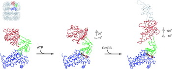
During the step of ATP–GroES-triggered encapsulation, polypeptide substrate becomes displaced from the hydrophobic binding sites of an open ring as these sites are removed from facing the cavity by apical domain movements. The released substrate then commences folding in a GroES-encapsulated and now hydrophilic cis chamber that is a stable end-state (Figs 1 and 2). The precise order of events during this step has not been entirely clear, but reports of the past few years are beginning to resolve this transition from a binding-proficient to a folding-active state, carried out in approximately a second. Normally, during a cycling reaction, this step would involve the open ring of an asymmetric ADP–GroEL–GroES complex (Figs 1 and 2). It has been shown that binding of ATP and substrate polypeptide to an open ring precedes GroES binding and substrate encapsulation (Rye et al. Reference Rye, Burston, Fenton, Beechem, Xu, Sigler and Horwich1997, Reference Rye, Roseman, Furtak, Fenton, Saibil and Horwich1999). It has also been established that GroES binding, which is dependent on ATP, involves two steps. First, ATP directs an initial small elevation and 20° counterclockwise twist of the apical domains, observable by EM (Fig. 9; Ranson et al. Reference Ranson, Farr, Roseman, Gowen, Fenton, Horwich and Saibil2001). This likely comprises the ATP-dependent state that forms initial contacts with the mobile loops of GroES, which proffer a hydrophobic IVL sequence in one edge of the loop (Xu et al. Reference Xu, Horwich and Sigler1997; Shewmaker et al. Reference Shewmaker, Maskos, Simmerling and Landry2001), to produce a putative collision complex. A large, forceful, and cooperative rigid body movement then follows, in which the apical domains elevate to 60° and rotate clockwise by 120° to produce the stable domed GroES-bound state that has been observed at high-resolution crystallographically (Chaudhry et al. Reference Chaudhry, Farr, Todd, Rye, Brunger, Adams, Horwich and Sigler2003; Xu et al. 1997). Whereas non-native polypeptide substrate does not appear to be mobilized during the initial collision with GroES (Cliff et al. Reference Cliff, Limpkin, Cameron, Burston and Clarke2006; see below), during the subsequent rigid body movements, it is released from the apical domains into the folding chamber.
Motions of a GroEL subunit on binding ATP and GroES. One subunit of GroEL, oriented as indicated in the thumbnail on the upper left, is shown in a set of Cα traces based on the crystal structure 1GRL (left; Braig et al. Reference Braig, Otwinowski, Hegde, Boisvert, Joachimiak, Horwich and Sigler1994), EM structure 1GR6 (middle; Ranson et al. Reference Ranson, Farr, Roseman, Gowen, Fenton, Horwich and Saibil2001), and crystal structure 1AON (right; Xu et al. Reference Xu, Horwich and Sigler1997). When ATP (gold) binds to the equatorial domain (blue) (middle), small changes in the intermediate domain (green) lead to a slight elevation and 20° counterclockwise twist of the apical domain (red). Subsequent GroES (gray) binding (right) produces a much larger additional elevation (50°) of the apical domain, accompanied by a 120° clockwise twist that removes the apical polypeptide binding site from facing the cavity and permits GroES to bind stably to the apical domain via its mobile loop. The forceful movement of the apical domains driven during the second step releases polypeptide into the chamber to begin folding.

The uncertainties concerning cis complex formation involve, first, the question of the order of arrival of ATP and polypeptide ligands to the open ring. A certain order would specify possible mechanisms that could influence the conformation of the polypeptide. For example, if polypeptide arrives first at an open ring, then apical movements directed by subsequent binding of ATP could be used to exert forced stretching/unfolding of the substrate. If, by contrast, ATP binds first and the ATP-directed apical movements have already occurred by the time of substrate binding, then polypeptide will not be subject to ATP-mediated mechanical effects.
A further aspect of uncertainty has concerned the nature of binding of GroES to an ATP–polypeptide-bound ring. This must occur in such a manner that polypeptide does not become prematurely displaced from GroEL into solution during the encapsulation step. This is a significant concern because the mobile loops of GroES become associated with the same apical hydrophobic surface in the GroES-bound end-state that is occupied by substrate polypeptide in an open ring, leaving open the possibility that GroES effectively ‘competes off’ the substrate protein (Fenton et al. Reference Fenton, Kashi, Furtak and Horwich1994). A means of obviating this potential leakage of polypeptide must therefore be operative and appears to lie in a phase during which the partly mobilized apical domains share their hydrophobic surface with both bound substrate protein and the GroES mobile loops. This state is implied by recent kinetic data, discussed below, and by the study of mutant GroEL molecules, but so far, it has not been directly observed.
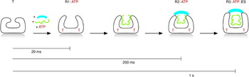
Finally, associated with the subsequent large movements of the GroEL apical domains, polypeptide is forcefully ejected in a concerted fashion, from all of the apical domains at once, into the cis cavity. There was a suggestion from an early tritium exchange study of Rubisco that a forced unfolding action might occur during this step (Shtilerman et al. Reference Shtilerman, Lorimer and Englander1999), but a subsequent exchange study failed to confirm the presence of stable protected secondary structure either of a Rubisco metastable intermediate free in solution before binding or in the GroEL-bound state (Park et al. Reference Park, Fenton and Horwich2005), obviating any ability in such a study to inspect for loss of structure during subsequent encapsulation. Moreover, two recent FRET studies have observed compaction of polypeptide during this phase (Lin et al. Reference Lin and Rye2004; Sharma et al. Reference Sharma, Chakraborty, Müller, Astola, Tang, Lamb, Hayer-Hartl and Hartl2008). We have adapted a scheme from Cliff et al. (Reference Cliff, Limpkin, Cameron, Burston and Clarke2006), showing the putative phases of folding activation in relation to their kinetic fluorescence studies (Fig. 10) and we describe the bases for and implications of this model.
Timeline of formation of a cis folding-active complex. The formation of a cis GroEL–GroES–ATP–polypeptide complex is diagrammed based on the phases and their rates of formation as reported by Cliff et al. (Reference Cliff, Limpkin, Cameron, Burston and Clarke2006). Only one GroEL ring is shown for simplicity. First, an unliganded ring (T state) rapidly binds ATP to form the R1:ATP state with somewhat twisted apical domains (cf. Fig. 9, middle). If non-native polypeptide is present, it likely binds to this state after ATP binds. GroES then interacts (with or without polypeptide) to form the R2:ATP state, essentially a collision complex where GroES and polypeptide share the apical binding sites, which are still exposed in the cavity. In a somewhat slower step, the R2:ATP state converts to the R3:ATP:GroES state, during which large rigid body apical domain movements occur (see second step of Fig. 9), releasing polypeptide into the central chamber to fold.

3.2 Fluorescence changes of GroEL W485 and W231 as monitors of steps of cis complex formation
The kinetic study by Cliff et al. (Reference Cliff, Limpkin, Cameron, Burston and Clarke2006) monitored fluorescence of a tryptophan substituted at position 485 in the equatorial domain of GroEL, taking advantage of the fact that GroEL and GroES are naturally devoid of Trp. An additional kinetic study from Taniguchi et al. (Reference Taniguchi, Yoshimi, Hongo, Mizobata and Kawata2004) monitored a tryptophan substituted at position 231 at one edge of the face of the apical domain, both in GroEL and SR1, observing the same results. These two studies thus offer parallel views of the steps taking place during the binding-to-folding transition of a ring. First, there is a rapid enhancement of fluorescence produced by ATP over ~20 ms, termed T→R1, that is unaffected by the presence of GroES and, as discussed below, is far faster than substrate polypeptide binding (see Fig. 10). This rapid initial phase is observed for tryptophan at both positions, indicating that the effect of ATP binding in the equatorial domains is rapidly transmitted to the apical domains. The rate of a second phase, termed R1→R2, observed as fluorescence quenching at both positions, is also not affected by GroES, but occurs on a timescale, ~200 ms, that would accommodate substrate polypeptide binding as determined from measured bimolecular rate constants (e.g. Ranson et al. Reference Ranson, Burston and Clarke1997; Rye et al. Reference Rye, Roseman, Furtak, Fenton, Saibil and Horwich1999; Tyagi, unpublished observations). A structural transition occurs during these first two phases as indicated by time-dependent small-angle X-ray scattering studies of Kuwajima and co-workers (Inobe et al. Reference Inobe, Arai, Nakao, Ito, Kamagata, Makio, Amemiya, Kihara and Kuwajima2003). This probably corresponds to the small apical domain elevation and counterclockwise twist observed in EM when GroEL is exposed to ATP alone for several seconds (Ranson et al. Reference Ranson, Farr, Roseman, Gowen, Fenton, Horwich and Saibil2001), albeit that the structural bases of the two distinguishable phases remains to be resolved. Effects of the presence of GroES on the amplitude of the R1→R2 transition assign the initial interaction of GroES with GroEL to the R2 state (Fig. 10). The rate of a third phase of fluorescence quenching of W485 (enhancement at W231), R2→R3, is substantially affected by the presence of GroES, and the presence of GroES produces a further quench of W485, R3→R3*, followed by an enhancement of fluorescence intensity to reach an end-state, R4. These latter steps are associated with measurable tightening of affinity of GroEL for GroES. In the study of Cliff et al., when changes in a pre-bound fluorescently labeled substrate (dansylated RCMLA, reduced and carboxymethylated α-lactalbumin) were monitored after ATP–GroES addition, they did not occur until the R2→R3 transition, a point by which GroES was already associated with the GroEL ring, indicating that GroES collision occurs before substrate protein begins to be released (Fig. 10, fourth panel). Such an order of events assures that polypeptide becomes encapsulated during the transition.
3.3 GroES–GroEL collision state
The foregoing fluorescence studies imply that there is a brief phase when the apical hydrophobic surface of GroEL might be occupied with both substrate polypeptide and the mobile loops of GroES. This could potentially occur on the same subunits, for example, with polypeptide occupying helix I, as it has been observed to do by X-ray and EM studies of bound peptides and polypeptides (e.g. Fig. 4), while the GroES mobile loop forms an initial contact with helix H at the rim of the cavity. Alternatively, because substrate protein may typically occupy only three to four of the seven apical domains of a ring, the GroES mobile loops may favor contacting the three to four unoccupied apical domains of a ring. This latter arrangement seems somewhat supported by a recent study of engineered covalent GroES molecules bearing different numbers of mutant binding-defective mobile loops, which indicates that five to six functional mobile loops are required in order to form a stable folding-active GroEL–substrate–GroES complex (Nojima et al. Reference Nojima, Murayama, Yoshida and Motojima2008). Yet, in the absence of structural information, the topology of polypeptide and GroES in sharing the apical domains remains speculative.
Two different mutants of GroEL appear, however, to behave as if stalled at the R2→R3 transition, unable to effectively dissociate substrate protein from a GroES-encapsulated state into the folding chamber. A mutant isolated by Kawata and co-workers, C138W, in the ascending limb of the intermediate domain, behaved in a temperature-sensitive manner, unable to productively refold bound substrate upon addition of GroES and ATP at 25°C, trapping it in a GroES-encapsulated complex where it was protected from exogenous protease (Kawata et al. Reference Kawata, Kawagoe, Hongo, Miyazaki, Higurashi, Mizobata and Nagai1999; Miyazaki et al. Reference Miyazaki, Yoshimi, Furutsu, Hongo, Mizobata, Kanemori and Kawata2002). Remarkably, upon elevation of temperature to 37°C, the trapped substrate was released into the folding chamber and proceeded to the native state. Similarly, an equatorially altered GroEL molecule studied by Rye and co-workers, with pyrene attached to S43C, appeared likewise to be stalled at the R2→R3 transition, with bound Rubisco substrate trapped underneath GroES but unable to be released to undergo conformational changes (Madan et al. Reference Madan, Lin and Rye2008). Thus, in both cases, while encapsulation occurred normally, the subsequent ATP–GroES-directed powerstroke needed to release substrate and trigger folding appeared to be blocked.
3.4 Order of binding ATP and polypeptide to an open trans ring
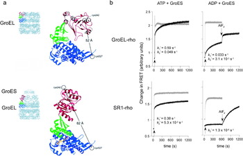
It is actually the steps immediately preceding encapsulation that seem the least resolved, namely, the order of arrival of ATP and polypeptide ligands to an open trans ring before GroES encapsulation. Two fluorescence studies of the past year assume that polypeptide arrives first and that subsequent ATP binding performs a stretching action on the bound substrate protein (Lin et al. Reference Lin, Madan and Rye2008; Sharma et al. Reference Sharma, Chakraborty, Müller, Astola, Tang, Lamb, Hayer-Hartl and Hartl2008), but such a conclusion seems premature in the absence of any resolution of the order of addition. Nevertheless, Rye and co-workers have argued for arrival of polypeptide first, before ATP, with an item of experimental evidence. They observed that the rate of ADP release from a discharged cis complex was significantly slower than the rate of release of GroES (the latter <1 s) (Madan et al. Reference Madan, Lin and Rye2008). Release of radioactive ADP as measured by a spin column separation technique seemed to require as long as 15–20 s, raising the question of whether ADP release could be a limiting step relative to entry of ATP into an ‘open’ trans ring. That is, ADP from a past cis ternary ring (which has become an open trans ring) would have to leave the nucleotide pocket before ATP could arrive, thus potentially allowing substrate to arrive first if ADP departure were slow enough. Whether such a delay is indeed long enough to span the entirety of a reaction cycle carried out on the other ring is unclear. If such a delay were in fact long enough, it would conceivably become the rate-limiting step in the reaction cycle, to date understood to be cis ATP hydrolysis. Indeed, such a step of ADP release is proposed by Rye and co-workers to comprise a ‘throttle’ on cis hydrolysis.
Yet recent data would argue against the foregoing order of events. The rate of ATP binding to a trans ring has been measured using a nonperturbing fluorescent reporter placed adjacent to the nucleotide pocket (Tyagi, unpublished observations). Fluorescence intensity changes report a rate of ATP binding to either an open ring of unoccupied GroEL or the open trans ring of an ADP asymmetric complex of ~2×10⁵ M−1 s−1 (equivalent to 100 s−1 at 0·5 mm ATP and 1 μm GroEL) in agreement with an earlier measurement taken with the GroEL mutant F44W (Yifrach & Horovitz, Reference Yifrach and Horovitz1998). In addition, an apical domain adjustment, measured with an apical-attached fluorescent reporter, occurs at the same rapid rate, in agreement with the earlier observations of Taniguchi et al. (Reference Taniguchi, Yoshimi, Hongo, Mizobata and Kawata2004) using R231W GroEL. By contrast, FRET measurements of substrate binding to the open ring of an asymmetric ADP complex, examining three different polypeptides, show that the rate, ~1 s−1 at 1 μm polypeptide and 1 μm asymmetric complex (agreeing with earlier measurements from both Ranson et al. Reference Ranson, Burston and Clarke1997 and Rye et al. Reference Rye, Roseman, Furtak, Fenton, Saibil and Horwich1999), is far slower than the rate of ATP binding and is not affected by co-mixing of ATP (Tyagi, unpublished observation). Thus, in these tests, polypeptide must arrive later than ATP, and its rate of binding is not affected by the earlier-occurring ATP-induced apical adjustments. Yet, of course, these complexes differ from those of a cycling reaction in that the trans ring is devoid of ADP produced from a previous cycle. On the other hand, the results were unaffected when the same reactions were carried out in the presence of ADP in the starting mixture, to which ATP was then added. Thus, it does not appear that access of ADP to the open trans ring can compete successfully against the rapid entry of ATP. Furthermore, in a cycling reaction, where ATP arrives before substrate protein and apical movements appear to have occurred already before polypeptide arrives, no substrate stretching event is likely to occur. These considerations suggest that the stretching observed to date upon ATP addition is largely a consequence of the investigators' choice of binding polypeptide to GroEL first, before adding ATP. Further, in a functional test of order of addition, measuring the production of native Rubisco by an SR1 reaction, the rate of refolding by either order was similar and, interestingly, the yield of the folding reaction was greater when ATP was added first (Tyagi, unpublished observation).
It should also be commented that the folding of stringent substrate proteins appears to be able to occur productively without the need for an ATP-mediated stretching event. In one experiment, refolding of DM-MBP occurred with the same kinetics as in a cycling reaction when DM-MBP complexed with SR1 was mixed with ADP-AlFx and GroES, yet here no rapid stretching was observed, only a relatively slow loss of FRET efficiency (Sharma et al. Reference Sharma, Chakraborty, Müller, Astola, Tang, Lamb, Hayer-Hartl and Hartl2008). In a second experiment, SR1 was observed to produce substantially less ATP-mediated stretching of bound Rubisco than GroEL (Lin et al. Reference Lin, Madan and Rye2008), yet the ultimate rates and extent of recovery of the native protein by SR1 in the presence of GroES were not substantially different from those of GroEL–GroES. (Note, however, that the initial rate of recovery of native Rubisco seemed to be greater if it was allowed to become further passively stretched on SR1 by longer incubation [5 min or more] before addition of ATP–GroES; even so, apparently Rubisco on its own can escape whatever kinetic impediment is relieved by such a step once it is released into the long-lived SR1–GroES cis cavity.)
In sum, on the one hand, it seems unlikely that polypeptide binds before ATP to an open ring in a cycling reaction, and on the other, a stretching event before the cis encapsulation step does not seem to be required for productive folding. But at a further level, considering all of the foregoing experiments, it seems that productive folding can occur with either order of addition of ATP and polypeptide to an open GroEL ring. This perhaps reflects that the action of ATP is critical to recruiting GroES and producing cis encapsulation but not to directing action on polypeptide substrate beyond driving the forceful apical movements that release it into the cis chamber, as next discussed.
3.5 Release of polypeptide during encapsulation step
3.5.1 A power stroke dependent on the γ-phosphate of ATP drives release
The evidence for forceful release of substrate protein into the cis chamber derives from the work of Motojima et al. (Reference Motojima, Chaudhry, Fenton, Farr and Horwich2004), who followed the rate and extent of apical movement during GroES encapsulation by monitoring FRET between a fluorophore placed on the stable equatorial ‘base’ of GroEL and a fluorophore placed on the mobilizable apical face (Fig. 11a). A displacement between these points from 52 Å in an open ring to 82 Å in the GroES bound end-state was measured from the respective X-ray structures. When either ATP–GroES or ADP–GroES was incubated with labeled GroEL or with labeled SR1, both the rate and extent of excursion of the apical domains were the same, occurring in <1 s (Fig. 11b, gray traces). If polypeptide substrate was present, however, as would be the case at this phase of rigid body movement during cis complex formation (R3 and beyond, see Fig. 10), the rate of movement produced by ATP–GroES was only slightly slowed relative to the absence of polypeptide, still completing within a second, but ADP–GroES required many seconds to reach completion (Fig. 11b, black traces). In the case of SR1+ADP–GroES, this movement was unable to be completed. The result with ADP–GroES correlates with the inability of ADP–GroES to release substrate protein from the cavity wall, as measured by fluorescence studies (Weissman et al. Reference Weissman, Rye, Fenton, Beechem and Horwich1996), and with failure of any productive folding to occur despite its ability to produce an encapsulated, protease-protected substrate protein. Remarkably, polypeptide release and productive folding by such ADP–GroES complexes could be subsequently triggered by addition of aluminum fluoride, simulating the γ-phosphate of ATP, triggering both an immediate further excursion of the apical domains as detected by FRET and immediate commencement of production of the native state (Chaudhry et al. Reference Chaudhry, Farr, Todd, Rye, Brunger, Adams, Horwich and Sigler2003; Motojima et al. Reference Motojima, Chaudhry, Fenton, Farr and Horwich2004; see Fig. 11). Thus, the γ-phosphate of ATP is apparently not required in order to form a potentially productive complex in which GroES and polypeptide share apical hydrophobic surface, and this state, produced in ADP, appears to be stable over many minutes. Whether it is identical to the collision state formed on the ATP-driven pathway remains to be established. The structure of this state is unknown, and in particular, whether the ADP–GroEL–GroES–polypeptide complex has elevated apical domains producing a fully domed state, is unknown. It is unlikely that a full excursion of the apical domains could proceed, as this would completely remove the hydrophobic surface from the cavity, perforce releasing substrate protein. Thus, it would seem that at least some residual hydrophobic surface must remain exposed to retain polypeptide substrate. Whatever the extent of excursion may be, it is clear that aluminum fluoride, binding at the position of the γ-phosphate as observed crystallographically by Chaudhry et al. (Reference Chaudhry, Farr, Todd, Rye, Brunger, Adams, Horwich and Sigler2003), drives a productive release of substrate protein. Perhaps, if the complex is already substantially domed in the ADP state, this is tantamount to an energetic shaking of the cis ring, releasing the substrate protein. In energy measurements performed by Chaudhry et al. (Reference Chaudhry, Farr, Todd, Rye, Brunger, Adams, Horwich and Sigler2003), it was observed that the binding of aluminum fluoride to an ADP complex (absent substrate protein) released ~40 kcal mol−1 of free energy per ring, a total energy equal to that already produced by ADP–GroES binding. So it appears possible that a power stroke in the same direction as that mediated by ATP releases the substrate in this context.
Polypeptide acts as a load on the apical domains, restricting their rate of movement when ADP and GroES are added. (a) Ribbons representations of a subunit of unliganded GroEL (top) and a subunit of the cis ring of a GroEL–GroES–ADP–AlFx complex (bottom), colored as in Fig. 10 (note that GroES is not shown in the bottom panel). The thumbnails at the left show the position of the subunits in the intact complexes. Indicated are the two Cys residues that were labeled with a FRET reporter pair, fluorescein and tetramethylrhodamine, to monitor movements of the apical domain relative to the equatorial domain; the distances shown were measured between the β-carbons of these residues in the crystal structure (Chaudhry et al. Reference Chaudhry, Farr, Todd, Rye, Brunger, Adams, Horwich and Sigler2003). (b) Stopped-flow measurements of FRET changes on addition of GroES and ATP to GroEL–rhodanese or SR1–rhodanese complexes (left) or on addition of GroES and ADP (right). Gray traces show FRET changes of the respective chaperonins in the absence of substrate protein. The rates of change in FRET (with presence of polypeptide) were fit to the sum of two exponentials, as indicated in the individual panels. ATP supports a rapid movement of the apical domains even with polypeptide bound, with half-times (1–2 s) that are physiologically reasonable, given the ~10-s lifetime of the folding-active complex. With ADP, however, the rates are at least 20-fold slower (when substrate protein is present) and the extent of movement is reduced. When AlFx is added to the ADP complexes to form a folding-active ATP analog in the nucleotide binding site (right), the expected extent of the FRET change is achieved at a rate consistent with the refolding rate under these conditions. From Motojima et al. (Reference Motojima, Chaudhry, Fenton, Farr and Horwich2004). Copyright 2004, National Academy of Sciences, USA.

3.5.2 Concerted substrate release
The power stroke of rigid body movement that releases substrate from the apical hydrophobic surface is virtually certainly concerted in character, involving all of the apical domains at once. This is particularly enforced by GroES binding, which, beyond the collision state, involves tightening of the interaction of all of its mobile loops in 1:1 contacts with the apical domains of the bound ring, simultaneously moving all of the apical domains. As pointed out by Ma & Karplus (Reference Ma and Karplus1999), who carried out normal mode analyses, the excursion is so substantial that steric collisions would eventuate unless there was coordinated movement of all of the domains at once. The quantitative requirement for ATP binding at the level of a ring in order to trigger (with GroES) such movements has recently been determined using a small molecule inhibitor that selectively binds in the equatorial ATP pocket of a variant GroEL subunit, blocking ATP binding. Mixed rings were formed, composed of varying numbers of variant and wild-type GroEL subunits, with only wild-type subunits able to bind ATP in the presence of compound. In order to drive substrate release and productive folding, ATP was required to occupy at least five or six and, for full efficiency, all seven equatorial domains of a ring (Chapman et al. Reference Chapman, Farr, Fenton and Horwich2008).
The symmetry of the GroEL system and its concerted behavior contrasts with the type II eukaryotic cytosolic chaperonin, CCT, which has a heterooligomeric subunit composition and a ‘built-in’ lid attached to the apical domain of each subunit. In the CCT system, a sequential as opposed to concerted release of substrate domains may occur, enabling sequential domainwise folding of such proteins as actin and tubulin (Rivenzon-Segal et al. Reference Rivenzon-Segal, Wolf, Shimon, Willison and Horovitz2005). Interestingly, GroEL binds non-native forms of these proteins and GroES encapsulates them as well, but they never reach the native state, cycling on and off the bacterial chaperonin in a futile fashion (Tian et al. Reference Tian, Vainberg, Tap, Lewis and Cowan1995).
Recent fluorescence experiments have suggested that ATP–GroES-driven polypeptide release from the apical domains occurs in an ordered fashion, with locally less hydrophobic regions of polypeptide mobilizing first and more hydrophobic segments leaving last (Sharma et al. Reference Sharma, Chakraborty, Müller, Astola, Tang, Lamb, Hayer-Hartl and Hartl2008). This, of course, would parallel the relative affinities of the hydrophobic binding surface of the GroEL apical domains for such regions of the chain. The notion that hydrophobic collapse may occur last does not really seem a likely mechanistic requirement, given the myriad of in vitro studies diluting unfolded proteins from denaturant where ‘indiscriminate global collapse’ occurs yet productive folding nevertheless ensues. Considering the experiments discussed in the next section, collapsed ensembles of GroEL substrates, however arrived at (i.e., discharged from the hydrophobic cavity wall or diluted from denaturant into free solution) seem to take the same subsequent kinetic course to the native state. In sum, the mechanism of collapse seems to have little to do with kinetic outcome, at least in the case of the substrate proteins studied to date.
3.5.3 No evidence for unfolding during the step of ATP–GroES-driven release of substrate
Finally, polypeptides undergoing release from the forcefully moving apical domains might have been subject to stretching or ‘forced unfolding’ before final release, but, as mentioned earlier, hydrogen exchange studies carried out to date do not detect significant secondary structures in bound substrates, making it difficult to evaluate whether unfolding action occurs at the level of secondary structure. But FRET studies examining topology of Rubisco (Lin et al. Reference Lin and Rye2004) and DM-MBP (Sharma et al. Reference Sharma, Chakraborty, Müller, Astola, Tang, Lamb, Hayer-Hartl and Hartl2008) actually have observed compaction and not expansion of these polypeptides during this phase of cis complex formation.
4. Folding in the GroES-encapsulated cis chamber, an ‘Anfinsen cage’
Following release from the ATP–GroES-mobilized apical domains, substrate protein attempts to reach the native state in the cis cavity during what is the longest phase of the reaction cycle, ~10 s. Release from the apical domains during cis complex formation is surely attended by a rapid collapse of substrate, as the FRET studies showing compaction would indicate. But what follows during folding in the cis chamber? Is this a reaction ‘supervised’ by the nearby cavity walls or is it more a reaction pursued as if there were no interference, e.g. as if folding at infinite dilution? In 1993, John Ellis coined the term ‘Anfinsen cage’ to describe the latter model for folding in the cis chamber (Saibil et al. Reference Saibil, Zheng, Roseman, Hunter, Watson, Chen, Auf der Mauer, O'Hara, Wood, Mann, Barnett and Ellis1993). He proposed that polypeptide in the cis chamber would follow the energy landscape guided by its primary structure but without the complication of multimolecular aggregation that could occur in the bulk solution. Such a model was then challenged a decade later by studies that proposed a model that the chamber could actively accelerate folding to the native state under so-called ‘permissive’ conditions where productive folding can occur either by the chaperonin system or in free solution (Brinker et al. Reference Brinker, Pfeifer, Kerner, Naylor, Hartl and Hayer-Hartl2001; Tang et al. Reference Tang, Chang, Roeben, Wischnewski, Wischnewski, Kerner, Hartl and Hayer-Hartl2006). By ‘closely confining’ the substrate protein and providing specific clusters of negative charge in the cavity walls (Fig. 12), it was proposed that the cis cavity would alter the energy landscape of a non-native monomeric protein, as compared with that in free solution, in particular by closing off access to kinetic traps.
The electrostatic and hydrophilic nature of the cis folding chamber. A cutaway view of a space-filling model of the cis chamber of a GroEL–GroES complex (1AON), with residues colored according to their nature: acidic (red), basic (blue), neutral polar (green), hydrophobic (yellow), and main chain (white). Parts of three subunits are shown. Y71 near the top is a GroES residue. Adapted from Farr et al. (2007), with permission. Copyright 2007, National Academy of Sciences, USA.

The acceleration of folding to native form by the chaperonin system was first reported in 1990 by Lorimer and co-workers in studies with Rubisco (Viitanen et al. Reference Viitanen, Lubben, Reed, Goloubinoff, O'Keefe and Lorimer1990). This followed their initial reconstitution of chaperonin-mediated folding of the same protein, which employed nonpermissive conditions, 25°C and ~100 nm substrate concentration, where folding in solution resulted in wholesale aggregation without recovery of native form, contrasted with full recovery by the chaperonin system (Goloubinoff et al. Reference Goloubinoff, Christeller, Gatenby and Lorimer1989). In this subsequent study, when lower temperature (15°C or below) was employed, productive folding could now occur both with the chaperonin system and free in solution. Under these conditions, the rate of recovery of the native state was up to 10-fold faster when mediated by the chaperonin system. This was interpreted as indicating that the chaperonin system somehow prevented aggregation, whereas in free solution, reversible aggregation contributed to slowing of the rate of recovery of the native state. By this model, in contemporary terms, confinement to a cis chamber simply eliminates a host of off-pathway steps of reversible multimolecular aggregation that can occur in solution and slow the overall reaction (Fig. 13). The curtailing of such steps could thus directly explain the acceleration of folding by the use of the cis cavity. We have recently reviewed evidence that supports this latter mechanism (Horwich et al. submitted), so we will only summarize here a number of observations that bear on the mechanism of acceleration.
Folding in the GroEL–GroES chamber vs. folding in solution. (a) An unfolded polypeptide substrate (U), released from the hydrophobic apical binding sites upon ATP–GroES addition, folds in isolation in the cis chamber to produce native protein, with no possibility that aggregation can intervene, and at a rate that is essentially the intrinsic folding rate of the polypeptide. (b) A non-native polypeptide diluted from denaturant into solution can fold to the native state or it can undergo aggregation, often in a stepwise manner with low-order aggregates forming initially. If this initial aggregation step is reversible, the overall rate of folding to the native state will be reduced relative to the intrinsic rate, although the yield may not be affected. Irreversible aggregation, by contrast, will not affect the rate of renaturation but will reduce the yield. Folding in the chaperonin chamber avoids these complications on the folding landscape by preventing aggregation altogether.

Two substrate proteins whose folding is accelerated 5- to 10-fold by the chaperonin system under permissive conditions have been recently examined (Apetri & Horwich, Reference Apetri and Horwich2008), Rubisco and a DM-MBP (V8G/Y283D). In the case of Rubisco, it was noticed that all of the previous studies of folding under permissive conditions had included bovine serum albumin (BSA). Here, omission of BSA completely inhibited recovery of Rubisco folding in solution but had no effect on recovery from the chaperonin reaction. This strongly suggested that aggregation was occurring during the solution reaction. This was directly observed by dynamic light scattering measurements. Even with BSA present in solution, aggregation was detected within seconds of dilution of Rubisco into buffer. Yet, aggregation in solution was reversible, i.e. low-order aggregates could reversibly produce native Rubisco as directly observed by gel filtration chromatography carried out at various times during the Rubisco solution reaction. At early time, a collective of species of several hundred kilodaltons was observed, but these then gave rise at later times to native Rubisco (100 kDa dimer of 51 kDa subunits) and to apparently irreversible aggregates of several megadaltons in size. Thus, the directly observable reversible aggregation of Rubisco must be contributing to the relative slowing of folding in free solution. By contrast, the chaperonin reaction, which isolates Rubisco as a monomer in the cis chamber, is free of such kinetic complications (see Fig. 13).
The results with DM-MBP were also revealing. Here also, the solution reaction produced light scattering. In addition, the rate of recovery of native DM-MBP was inversely concentration dependent, a tell-tale sign of protein aggregation. In contrast, wild-type MBP, which exhibits neither a rate difference in folding at chaperonin vs. solution nor aggregation while folding in solution, showed no such concentration dependence of its rate of refolding. Most striking in the case of DM-MBP folding free in solution, when chloride ions in the refolding buffer were replaced with acetate, the aggregation in the solution reaction was entirely abrogated, and now the rate of folding to native form became identical to that by the chaperonin system. This once again demonstrates that it is the kinetic complication of reversible aggregation while folding free in solution that explains the relative acceleration of folding by the chaperonin system. Further, the observation that the folding rate becomes equal between solution and the cis cavity once aggregation is abolished argues that, at least for this substrate, the folding landscapes for the monomer are likely to be virtually identical inside the cavity and outside in solution.
Additional studies support the similarity of the landscapes. A hydrogen–deuterium exchange/NMR study of human DHFR folding inside the cis cavity of SR1–GroES vs. outside in solution observed virtually identical positions and kinetics of acquired protection during folding (Horst et al. Reference Horst, Fenton, Englander, Wüthrich and Horwich2007). Yet the recovery of native DHFR in free solution was only ~50% of the input (vs. near-quantitative recovery in the chaperonin reaction), with about half of the molecules lost to rapid and, in this case, irreversible aggregation. This further reiterates the role of cis encapsulation to prevent multimolecular aggregation.
More generally, in light of these recent observations, permissive folding conditions can now be considered again, as they were in 1990, to represent a simple extension of nonpermissive ones, insofar as these conditions, typically lowered concentration of substrate protein and lowered temperature (reducing hydrophobic interactions that direct multimeric aggregation), are such that aggregation is less favored (see also Buchner et al. Reference Buchner, Schmidt, Fuchs, Jaenicke, Rudolph, Schmid and Kiefhaber1991). In sum then, folding of monomers inside the cis chamber appears to proceed on the same landscape as that which monomers pursue in free solution except that, in the cavity, they proceed without the complication of off-pathway multimeric aggregation that comprises, in effect, a portion of energy landscape that is no longer accessible inside the ‘Anfinsen cage’.
5. Acknowledgements
We thank George Farr for help with preparation of the figures, and Howard Hughes Medical Institute for generous support of our work on chaperonins over many years.