Introduction
In Japan, two subspecies of wild boars exist: Japanese wild boars (Sus scrofa leucomystax) in most parts of the mainland of Japan (Honshu, Shikoku and Kushu), and Ryukyu wild boars (Sus scrofa riukiuanus) in the south-western islands of Japan, such as Ryukyu Islands and Amami Island (Watanabe et al., Reference Watanabe, Okumura, Ishiguro, Nakano, Matsui, Sahara and Komatsu1999). Although these two subspecies of wild boars are supposed to have partly different helminth faunas, depending on their different geographical distributions, they serve as definitive or paratenic hosts for some zoonotic helminths, such as Gnathostoma doloresi, Onchocerca dewittei, Paragonimus westermani, and Macracanthorhynchus hirudinaceus (Miyazaki & Habe, Reference Miyazaki and Habe1976; Kamiya et al., Reference Kamiya, Suzuki and Hayashi1977; Miyazaki et al., Reference Miyazaki, Terasaki and Iwata1978; Nawa & Imai, Reference Nawa and Imai1989; Uni et al., Reference Uni, Bain, Takaoka, Miyashita and Suzuki2001; Takaoka et al., Reference Takaoka, Yanagi, Daa, Anzai, Aoki, Fukuda, Uni and Bain2005). Trichinella spp. have never been found in wild boars in Japan, whereas wild boars in Europe are known to play an important role in the sylvatic cycle of Trichinella spp., with recent outbreaks of trichinellosis in hunters after consumption of wild boar meat (Pérez-Martín et al., Reference Pérez-Martín, Serrano, Reina, Mora and Navarrete2000; Gari-Toussaint et al., Reference Gari-Toussaint, Tieulié, Baldin, Dupouy-Camet, Delaunay, Fuzibet, Le Fichoux, Pozio and Marty2005). Records of focused or limited parasite species in wild boars in Japan are prevalent (Yamaguti, Reference Yamaguti1935, Reference Yamaguti1943, Reference Yamaguti1961; Noda, Reference Noda1973; Hatsushika et al., Reference Hatsushika, Shimizu, Kawakami and Sawada1978; Chinone et al., Reference Chinone, Itagaki, Hoshino and Imaoka1984; Kagei et al., Reference Kagei, Uga and Gondo1984; Nawa & Imai, Reference Nawa and Imai1989; Uni et al., Reference Uni, Bain, Takaoka, Miyashita and Suzuki2001; Morita et al., Reference Morita, Haruta, Shibata-Haruta, Kanda, Imai and Ike2007); however, only a few studies providing general views of visceral helminths of wild boars exist (Shoho & Machida, Reference Shoho and Machida1979; Uchida et al., Reference Uchida, Uchida and Itagaki1984).
Recently, increasingly frequent appearances of endemic wildlife, such as wild boars, deer, monkeys and bears, in addition to feral alien raccoons, coypus, squirrels, etc., in areas of human residence or agricultural fields have become a major social problem in Japan. For their management, assessment of their potential roles in transmission of zoonoses or veterinary diseases might be of major value. From this viewpoint, the present survey of helminths in Japanese wild boars was initiated along with helminthological surveys on other wild mammals (Sato & Suzuki, Reference Sato and Suzuki2006; Sato et al., Reference Sato, Suzuki, Osanai, Kamiya and Furuoka2006, Reference Sato, Torii, Une and Ooi2007).
Materials and methods
Examined samples
Twenty-nine Japanese wild boars collected in the hunting seasons of 2005 and 2006 (15 November–15 March for wild boars) were examined for their visceral helminths. Twenty wild boars (11 females and nine males; nine boars with body weight of more than 40 kg, seven boars with 20–40 kg body weight, and four boars with less than 20 kg body weight) were trapped in Tanabe City, Wakayama Prefecture (33°44–50′N, 135°20–30′E), and the rest (three females, five males and one unrecorded; no information on their body weights) came from Sasayama City (35°00–10′N, 135°04–23′E) and Sayou Town (35°02′N, 134°06′E), Hyogo Prefecture, Japan. In addition to the 29 wild boars mentioned above, the stomach and large intestine of 20 and 27 wild boars, respectively, caught in Tanabe City were also examined for their parasitic helminths. Muscles of the diaphragm of 24 wild boars from Tanabe City and Sasayama City were also examined. Fresh rectal faeces were collected from 14 wild boars hunted in Tanabe City, and sent under cool conditions to the parasitology laboratory to be examined for Strongyloides infection by faecal culture.
Parasitological examination
Viscera in the thoracic and peritoneal cavities were individually frozen, labelled and sent to the parasitology laboratory. After thawing, each organ was checked by the naked eye, and placed in separate large glass dishes. Digestive tracts were cut open longitudinally, and immersed in tap water. After leaving the materials for 1 h, the mucosa was carefully scraped. Then, these materials were washed repeatedly by simple sedimentation in tap water. Sediments were checked thoroughly under a dissection microscope, and collected parasites were fixed in 10% neutral-buffered formalin or 70% alcohol. Muscles of the diaphragm, weight range 52–230 g (mean ± SD: 133 ± 57 g; n = 24), were individually digested in an artificial gastric juice containing 0.8% pepsin (1 : 10,000; NACALAI TESQUE, Kyoto, Japan) and 0.8% HCl at 37°C for 4–6 h. After repeated simple sedimentation in physiological saline, the sediment was checked under a dissection microscope. Morphological examinations were performed under a light microscope, with reference to respective publications for each species. Parts of nematodes were cleared in glycerin–alcohol when microscopic examinations were made. Figures were drawn with the aid of camera lucida. Some measurements were made using these drawn figures.
Faecal samples were examined using the Petri dish faecal culture method with an unglazed tile. Faecal cultures were kept at 28°C for 7–10 days, and the appearance of filariform larvae in culture water was checked under a dissection microscope.
Collected helminth specimens were deposited in the National Science Museum, Tokyo, Japan under specimen numbers NSMT-As 3114–As 3444, and NSMT-Pl 5568–Pl 5587.
Results
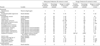
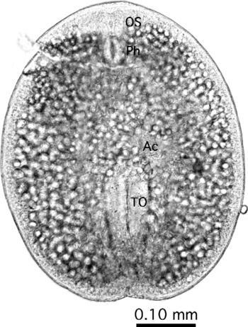
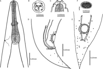
Worm recovery is summarized in table 1. Two or more related species were found in the lungs (four Metastrongylus spp.), small intestines (two Globocephalus spp.), and large intestines (two Oesophagostomum spp.). Most of the helminth species listed in the table were highly prevalent in wild boars caught in both localities, except for a few species, including Oesophagostomum dentatum, which was not found in wild boars caught in Hyogo Prefecture. Plagiorhynchus ogatai (Fukui and Morisita, 1936) from a single boar caught in Wakayama Prefecture should be referred to as pseudoparasitism. Ten adults of Morgascaridia kugii sp. n. (see figs 2 and 3) were detected in the large intestine of a female wild boar with a body weight of 59 kg, caught on 28 December 2006 in Wakayama Prefecture. From 20 additional stomachs and 27 large intestines, no more specimens of Morgascaridia kugii sp. n. were collected. Muscle digestion using an artificial gastric juice of the diaphragm detected only one diplostomulum, probably of Pharyngostomum cordatum, 0.52 mm long by 0.42 mm wide (fig. 1).
Helminth parasites recovered from wild boars in the western part of Japan (Wakayama Prefecture and Hyogo Prefecture).

a Not examined (NE) apart for one animal.
A diplostomulum encapsulated in the muscle of the diaphragm of a Japanese wild boar. Unstained, formalin-fixed specimen. OS, oral sucker; Ph, pharynx; Ac, acetabulum; and TO, tribocytic organ. Note numerous calcareous corpuscles distributed throughout the whole body.

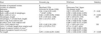
Parasitic females of Strongyloides ransomi (Schwartz & Alicata, Reference Schwartz and Alicata1930) were found in 6 of 20 wild boars (30%) in Wakayama Prefecture and 1 of 9 wild boars (11%) in Hyogo Prefecture. Faecal culture detected filariform larvae in 8 of 14 samples (57%) from Wakayama Prefecture, indicating that parasitological examination of the small intestine only partially detected infecting parasitic females. As shown in table 2, the filariform parasitic adults were 2.55–3.69 mm in length and 0.048–0.056 mm in maximum width. The stoma in the apical view was dumb-bell shaped and both the anterior and posterior branches of the ovary ran spirally with the intestine. The tail was conical, tapered gradually, with a more or less blunt tip. Although major morphological features were shared by S. ransomi of different origins, our specimens from wild boars were smaller compared to specimens from domestic pigs (table 2).
Comparison of actual and relative lengths of different features of parasitic females of Strongyloides ransomi from pigs and wild boars (in mm)a.

a Values are expressed as mean ± SD with range in parentheses.
b No information.
c Statistically not significant.
d Distance from the posterior end of the oesophagus.
e Distance from the anus.
Morgascaridia kugii sp. n
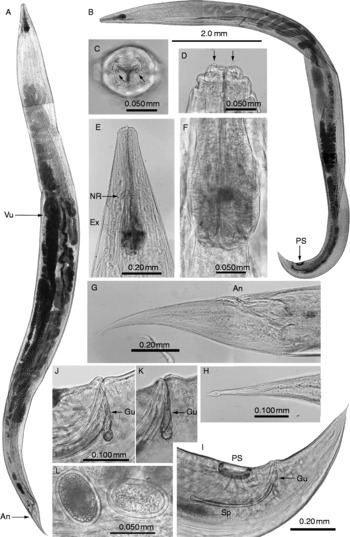
General description. Small, spindle-shaped nematodes approximately 9–12 mm in body length, having a maximum body width of 0.4–0.9 mm at the middle of the whole body length. Cuticle thin and finely striated. Three lips without denticles on the inner surface. Head with two distinctly large amphids protruding anteriorly and four pairs of sessile papillae situated at the four right angles of a square around the stoma. Oesophagus club-shaped, expanding near the posterior end. Luminal surface of the oesophagus at the maximum diameter serrate. Males having a circular precloacal sucker with a definite cuticular rim, an evenly pointed tail, and various numbers of small sessile papillae in the area from the anterior rim of the precloacal sucker to the tail. Left and right spicules equal in size and shape with bluntly spatulate distal ends. Gubernaculum having a sharply pointed distal end. Tail of females having a lancet-shaped end. Vulva pre-equatorial. Developed, but not embryonated, eggs in the uterus were thick-shelled, measuring 0.067 ± 0.003 (0.063–0.071) mm by 0.045 ± 0.001 (0.044–0.046) mm (mean ± SD (ranges); n = 10) (figs 2 and 3).
Morgascaridia kugii sp. n. in the large intestine of a Japanese wild boar. Unstained, formalin-fixed specimens. (A) Entire view of a female adult with developed eggs in the uterus. Photographs of A and B are at the same magnification. (B) Entire view of a male adult with a precloacal sucker. (C) En face view of the anterior end. Arrows indicate amphids protruding anteriorly. (D) Lateral view of the anterior end. Arrows indicate amphids protruding anteriorly. (E) Lateral view of the anterior end in the oesophageal region. (F) High magnification of the posterior region of the oesophagus. Luminal surface at the maximum diameter appears serrate. (G) Lateral view of the posterior end of a female adult. (H) Ventral view of the posterior tip of the tail of a female adult. (I) Lateral view of the posterior end of a male adult. (J) and (K) Lateral view of the gubernaculum. (L) Eggs in the uterus. An, anus; Ex, excretory pore; Gu, gubernaculum; NR, nerve ring; PS, precloacal sucker; Sp, spicule; and Vu, vulva.

Morgascaridia kugii sp. n. in the large intestine of a Japanese wild boar. (A) Lateral view of the anterior end. (B) En face view of the anterior end. (C) Lateral view of the anterior end. (D) Lateral view of the posterior end of a male adult. (E) Ventral view of the posterior end of a male adult. (F) Eggs in the uterus.

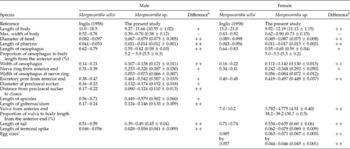
In the following sections, measurements of the holotype and allotype are shown in millimetres, and those of all specimens, including paratypes, are shown in table 3.
Comparisons of actual and relative lengths of different features of Morgascaridia spp. (in mm)a.

a Values are expressed as range with mean ± SD in parentheses when possible. Blank means no value available.
b Because Inglis (Reference Inglis1958) gave only range values, statistical analysis was not conducted. Instead, differences between the two species are assessed as follows: ++, no overlap of ranges; +, limited overlap of ranges; –, larger overlap of ranges.
c Given as length by width.
Male (holotype). Body length 11.38 and maximum width at the middle area of the body 0.69. Width of head 0.079 and length of pharynx 0.014. Length of club-shaped, muscular oesophagus 0.59, corresponding to 5.2% of total body length and its maximum width near the posterior end 0.12. Nerve ring from the anterior end 0.287 and the width of oesophagus at nerve ring 0.067. Excretory pore from the anterior end 0.54. Diameter of precloacal sucker 0.152 and distance between the sucker and the cloaca 0.112. Length of spicules 0.461 and gubernaculum 0.129. Length of tail 0.438, with the terminal spike 0.037.
Female (allotype). Body length 12.19 and maximum width at the middle area of the body 0.90. Width of head 0.087 and length of pharynx 0.014. Length of club-shaped, muscular oesophagus 0.65, corresponding to 5.3% of total body length and its maximum width near the posterior end 0.14. Nerve ring from the anterior end 0.348 and the width of oesophagus at nerve ring 0.084. Excretory pore from the anterior end 0.50. Vulva at 4.78 from the anterior end, corresponding to 39.2% of the body length from the anterior end. Length of tail 0.635.
Taxonomic summary
Type host. Japanese wild boars (Sus scrofa leucomystax Temminck and Schlegel).
Site of infection. Large intestine.
Prevalence. This species was found in one out of 56 samples of the large intestine.
Intensity. Ten adults (four females and six males) were found in a female wild boar, caught on 28 December 2006, weighing 59 kg.
Type locality. Tanabe City, Wakayama, Japan (33°46′58″N, 135°26′35″E).
Specimens deposited. The National Science Museum, Tokyo, Japan; NSMT-As 3445 (holotype), As 3446 (allotype) and As 3447 (paratypes).
Etymology. The species was named after Dr Giiti Kugi, who has carried out extensive helminthological surveys on a variety of wild birds and mammals, including Japanese wild boars.
Remarks
Taxonomy followed Inglis & Chabaud (Reference Inglis and Chabaud1960) and Chabaud (Reference Chabaud1978). To date, the only recorded species of the genus Morgascaridia Inglis, Reference Inglis1958 was M. sellsi (Morgan, Reference Morgan1927) Inglis, Reference Inglis1958, collected from a wild pig of unknown species in Uganda (Morgan, Reference Morgan1927) and a red river hog (Potamochoerus porcus Linnaeus) in Congo (Vuylsteke, Reference Vuylsteke1956), and thenceforth, no recovery of the species has been recorded. Morgascaridia kugii sp. n. differed from M. sellsi by smaller body dimensions, posteriorly positioned excretory pore near the border between the oesophagus and the intestine, closer position of the precloacal sucker with the cloaca, smaller sizes of the copulatory spicules and the gubernaculum, and smaller sizes of eggs (see table 3).
Discussion
Numerous helminth species with high prevalences of respective parasites were found in Japanese wild boars, and several species were shared with wild boars in other countries (Henne et al., Reference Henne, Nickel and Hiepe1978; Barutzki et al., Reference Barutzki, Schoierer and Gothe1990; Eslami & Farsad-Hamdi, Reference Eslami and Farsad-Hamdi1992; Mennerich-Bunge et al., Reference Mennerich-Bunge, Pohlmeyer and Stoye1993; De-la-Muela et al., Reference De-la-Muela, Hernández-de-Luján and Ferre2001; Fernandez-de-Mera et al., Reference Fernandez-de-Mera, Gontazar, Vicente, Höfle and Fierro2003; Solaymani-Mohammadi et al., Reference Solaymani-Mohammadi, Mobedi, Rezaian, Massoud, Mohebali, Hooshyar, Ashrafi and Rokni2003). Physocephalus sexulata, Strongyloides ransomi, Trichuris suis, Morgascaridia kugii sp. n., and a diplostomulum are new host records for Japanese wild boars. Ooi et al. (Reference Ooi, Kamiya, Kudo, Hashimoto and Kitazawa1984) detected a diplostomulum of Pharyngostomum cordatum in the muscle of a raccoon dog (Nyctereutes procyonoides) in Japan. Diplostomula of P. cordatum have been reported from frogs and snakes in the western part of Japan (Kurimoto, Reference Kurimoto1976; Uchida et al., Reference Uchida, Inoue and Itagaki1977), and Shin et al. (Reference Shin, Chai and Lee2001) reported migration of the diplostomula to the lungs, intercostal muscles, heart and brain of experimental rodents. In addition to 21 helminth species listed in table 1, at least Paragonimus westermani and Paragonimus ohirai have been reported from Japanese wild boars as visceral helminth (Miyazaki & Hirose, Reference Miyazaki and Hirose1976; Ashizawa et al., Reference Ashizawa, Habe, Nosaka and Tateyama1977; Miyazaki et al., Reference Miyazaki, Terasaki and Iwata1978).
Detection of Morgascaridia kugii sp. n. from a Japanese wild boar was intriguing. The genus Morgascaridia was created by Inglis (Reference Inglis1958) for Paraspidodera sellsi Morgan, Reference Morgan1927 and P. sellsi var. zadi Vuylsteke, Reference Vuylsteke1956, containing only one species, M. sellsi, to date. Now the genus Morgascaridia is placed with genera Schneidernema Travassos, 1927 and Ascaroterakis Vicente, 1965 in the subfamily Schneidernematinae, family Schneidernematidae, order Ascaridida (Chabaud, Reference Chabaud1978). Morgan (Reference Morgan1927) described M. sellsi as Paraspidodera sellsi using materials from the Helminthological Collection of the London School of Hygiene and Tropical Medicine, with only the following information: the parasite had been collected by Dr Sells from a wild pig in Uganda, with no indication of the organ the parasites were isolated from. Vuylsteke (Reference Vuylsteke1956) described M. sellsi as Paraspidodera sellsi var. zadi using materials collected by E. Dartevelle in 1937 from the stomach of a female red river hog (Potamochoerus porcus (Linnaeus, 1758)) in the Congo. When redescription of the species was made, Inglis (Reference Inglis1958) stated that specimens examined by Morgan (Reference Morgan1927) and Vuylsteke (Reference Vuylsteke1956) were in rather poor condition. Our specimens were not in poor condition, and collected from a host belonging to the family Suidae, similarly to M. sellsi. However, the collection of only ten worms after examination of 56 samples of the large intestine and 49 samples of the stomach is strange. Furthermore, the latest collection of M. sellsi was made in 1937 (Vuylsteke, Reference Vuylsteke1956), and since then nobody has found M. sellsi again, or a relative species, until the present study. Accordingly, it is highly possible that nematodes of the genus Morgascaridia take an unknown animal host other than suids as the natural host, and the recovery of the parasites from suids indicates pseudoparasitism or accidental infection.
As mentioned above, the subfamily Schneidernematinae consists of three genera, Schneidernema, Ascaroterakis and Morgascaridia (Chabaud, Reference Chabaud1978). Reported hosts of genera Schneidernema and Ascaroterakis are anteaters, sloths or armadillos belonging to the superorder Xenarthra Cope, distributed only in the American continents. Therefore, an estimation of the natural host, when the Japanese wild boar is considered only as an accidental host for M. kugii sp. n. or a host of its pseudoparasitism, is difficult. At present, at least we know geographical locations where M. sellsi or M. kugii sp. n. were found. More surveys of the wildlife would determine whether the wild boar serves as the natural host for Morgascaridia spp., adding a proper phylogenetic interpretation for this unique and rare nematode.
Acknowledgements
We are indebted to anonymous hunters and trappers who collected material for our parasitological examinations. The present work was supported in part by a research grant from Fujiwara Natural History Foundation and a grant-in-aid (No. 19580355) from the Japanese Society for the Promotion of Science.