

Introduction
Neurosurgery, in some form, has been practiced for millennia. Indeed, archeological evidence suggests it began prior to the dawn of human civilization (Reference André1). While we can only speculate as to the characteristics of these early pioneers, the neurosurgeon of recent history is a natural innovator. Our training mandates we develop as physician-scientists; as soldiers battling to unlock the boundless secrets of the nervous system: cognition, emotions, consciousness, neurochemistry and physiology, neurovascular form and function, neuro-oncology, even the basis of the soul itself. Beyond this, we are also its stewards. Neuroscience is one of the most poorly understood fields of biology, and as such, the neurosurgeon’s charge to restore the brain, spine, and nerves to a healthy state requires operating within this darkness. It is our duty to constantly evolve our field, moving the dial of knowledge to provide better care for our patients. With only 50,000 practicing neurosurgeons in the world currently, and fewer than 3,500 in the United States, we cannot do this alone. Whether consultation with an ancient shaman or healer in prehistory to target a trepanation, or collaboration with an engineer and investor of today to bring a novel and complex device to the operating room, a team-based approach was and always will be essential. It is this integration of minds that brought us from crude chisels and hammers to minimally invasive, robotic surgical machines, from humors and spirits to three-dimensional augmented-reality intraoperative imaging. Together, we stand at the precipice of a new age of neurosurgery, one in which the explosion of technological and scientific advances will drive neurosurgical innovation to create the neurosurgeon of the future. Hereafter, we start in the Paleolithic era on a journey from neurosurgery’s ancient origins and into the modern period, passing the torch to those who will shepherd us into the coming age.
Ancient Origins
Trepanation, or the intentional creation of a permanent opening in a person’s skull, is the earliest known neurosurgical procedure. The oldest confirmed trephined skull was discovered in Neolithic France, dated to 7000 BCE (Figure 1.1). However, evidence exists that the practice was performed since at least 10,000 BCE or prior in Europe, Africa, Asia, and Meso and South America (Reference André1). Indications are thought to have been broad, depending on the culture and time period, ranging from the spiritual, to clinical, to a combination of these. In many series, up to 10% of certain populations were shown to be trephined, with an estimated survival rate of 40% (Reference Faria2). Despite primitive understanding of human anatomy and physiology, the practice remained widespread for millennia.
A trepanned neolithic skull; the growth of new bony tissue around the lesion suggests the patient survived the procedure.

Equally, if not more, important than procedural technique and tools is an understanding of neuroanatomy. The earliest written evidence of neuroanatomical study is the Edwin Smith Papyrus of ancient Egypt, dating from 1600 BCE (3), which documented anatomical relationships with neuro-trauma. In ancient Greece, Galen is most famous for linking neuroanatomy and physiology, publishing most of his work in the second century AD (Reference Rocca4). Limited evidence exists of simple neurosurgical procedures being performed to repair skull fractures during the Roman Empire, but the Dark Ages saw a decline in scientific inquiry and knowledge; one notable exception is al-Zahrawi of al-Andalus on the Iberian Peninsula, who performed a variety of neurosurgical procedures in the late tenth century AD, and is known as the greatest surgeon of the Middle Ages (Reference Krebs5). Over 1,000 years passed until Andreas Vesalius, Leonardo da Vinci, and others re-investigated neuroanatomical study in the Renaissance. Vesalius’s monumental work “De humani corporis fabrica” dissected human neuroanatomy with unparalleled detail at the time, setting new standards for accuracy and meticulous observation. Indeed, the Renaissance marks a turning point where scientific curiosity began to bridge the gap between ancient practices and modern understanding. Enkindled by this knowledge, the blueprint of the modern neurosurgeon begins to take shape.
The Birth of Modern Neurosurgery
The nineteenth century heralded a period of transformative change, characterized by significant advancements in anesthesia, aseptic techniques, and the comprehension of neurological disorders. It was during this time that pioneering figures such as Sir Victor Horsley, Sir William Macewen, and Sir Rickman Godlee emerged, leaving an indelible mark on the trajectory of neurosurgery. In 1879, Macewen achieved a milestone by successfully removing a brain tumor, localized by neurologic signs alone, a feat previously deemed inconceivable. These procedures were soon repeated by Godlee, Horsley, and others.
War and Neurosurgery
The bellicosity around the turn of the century saw a global incidence in neuro-trauma, and as such, the turbulence of the American Civil War and World War I brought neurosurgery to the forefront of medical practice, accelerating its evolution as a specialized field. At the outset of the American Civil War, considerable knowledge regarding closed and penetrating head injury was known, and the development of anesthesia in the 1840s allowed major surgery to be conducted in an organized and tolerable fashion for the first time. However, antisepsis was not widely understood or practiced, and as such, most physicians were reluctant to perform major neurosurgical procedures due to extreme morbidity from post-operative infection (Reference Kaufman6). However, the large numbers of cases during wartime saw a refinement in the knowledge of traumatic neurologic injury during this time period; advancements in categorization of injury, natural progression, and triage are still relevant today. While principles of neurologic localization were yet to be fully elucidated, and due to a lack of medical imaging, successful treatment of many neurologic injuries was precluded. Despite this, observations of hematoma and abscess formation and progression, and limited experiences of their surgical intervention, were described in detail in several manuals and textbooks circulated at the time. Importantly, the germ theory of disease was expanded greatly by Louis Pasteur in the 1850s. Because antisepsis was not yet utilized in surgery, the recognition of pathogens as the cause of infections caused surgeons at this time to be less aggressive with surgical indications compared with the prior century, due to fear of newly understood pathogenesis of infection (Reference Kaufman6). Given the relatively fledgling medical knowledge at the time, controversy was widespread about surgical indications, and even use of bloodletting and other pseudo-scientific approaches.
The unprecedented number of soldiers afflicted with head injuries in World War I prompted the establishment of dedicated neurosurgical units for the first time (Reference Rainone, Zelmanovich, Laurent and Lucke-Wold7). This pivotal moment marked the transition of neurosurgery from an emerging discipline to an indispensable medical specialty. This period saw the rise to prominence of Dr. Harvey Cushing, the most famous neurosurgeon and the “Father of Modern Neurosurgery.” Cushing’s meticulous documentation of cases, emphasis on patient-centered care, and innovative surgical techniques elevated neurosurgery to new heights. At the outset of the Great War, mortality for neurosurgical treatment of wounds involving the brain exceeded 60% in some studies – through his experience over two years near the French front, he reduced this by more than half to 28%, mainly through refinement of operative technique. Intraoperative antisepsis, use of tools including catheters and suction, techniques for formal craniectomy as an advancement from trepanation techniques, and wound closure were all cited as contributory to his success (Reference Carey8). His groundbreaking work “The Surgery of the Central Nervous System” laid the foundation for neurosurgical practices, emphasizing the importance of precision and methodical exploration. The crucible of war catalyzed the growth of neurosurgery and fostered a culture of collaboration and innovation that continues to shape the field.
Technical Advancements
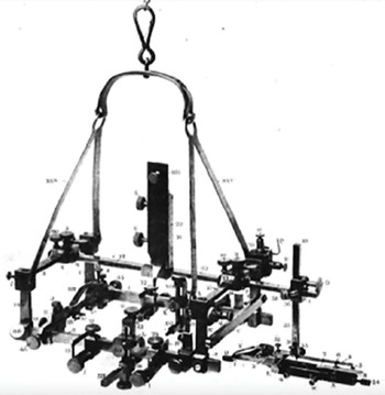
The foremost mantra of the modern physician and surgeon is “first, do no harm,” immortalized by Hippocrates in the fourth century BCE. Historically, medical physicians and surgeons were treated as two separate professions, with the Hippocratic Oath being applied to the former. Indeed, prohibition of “cutting for stone” suggests the barbaric and morbid nature of surgery at the time. And yet, neurosurgeons have adopted the tenets of this Oath into our own field; performing only those surgeries which are necessary, avoiding complications, and placing a patient’s welfare above our own are all foremost in the neurosurgeon’s practice. To this end, the rapid growth of neurosurgery in the late nineteenth and early twentieth century yielded advanced neurosurgical tools, allowing surgeons to minimize morbidity while achieving surgical goals. Most were adapted from other medical specialties, and many from fields outside of medicine entirely. Illustrative of this was Horsley, most famous for his development of stereotaxy, a term derived from the Greek “stereos” (solid) and “taxis” (arrangement), which involves the precise localization of specific anatomical targets within the brain for therapeutic interventions. In keeping with the indispensability of collaboration, the origins of stereotaxy can be traced back to the Cartesian coordinate system, developed by René Descartes in the seventeenth century. This mathematical framework provided a means of representing points in three-dimensional space using three orthogonal axes (Reference Gildenberg, Krauss, Lozano, Gildenberg and Tasker9). While not initially intended for medical applications, this system laid the foundation for later developments in stereotactic techniques, first achieved in 1908 with the Horsley–Clarke Apparatus (Figure 1.2) and improved by Lars Leksell in the 1940s. This period witnessed further breakthroughs; the surgical scalpel was developed by Morgan Parker in 1914 and used in neurosurgery immediately thereafter (Reference Brill, Harrison, Sise and Ignacio10). Dr. Fedor Krause published on the use of X-ray in neurosurgery in 1908 (Reference Elhadi, Kalb, Martirosyan, Agrawal and Preul11), which was the first application of imaging in the field. This technique involved injection of air into the ventricles, or fluid-filled central cavities, of the brain, and using the X-ray machine to visualize the orientation and shape of the air, which could be displaced by different pathologies.
The first tool for utilizing a Cartesian coordinate system for precisely localizing brain areas, the Horsley–Clarke Apparatus.

The adoption of stereotaxy in neurosurgery also paved the way for advancements in a controversial topic in the field: psychosurgery. While psychosurgery has been practiced in some form for millennia, beginning with early trepanations in the Neolithic period, the 1930s saw the rise in popularity of frontal lobotomy spurred by Dr. Walter Freeman, the father of the now-infamous “icepick lobotomy.” These treatments severed connections between various association areas in the frontal lobes which controlled emotions, the exact physiology of which is still poorly understood, which rendered patients abulic and with permanently “flat” affect. By the 1950s these techniques had fallen out of favor, but stereotactic principles allowed surgeons to precisely target interventions to alter brain function with previously unparalleled accuracy; this and many other techniques would be advanced further in the decades to come.
A Quantum Leap – Neurosurgery in the Last Century
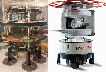
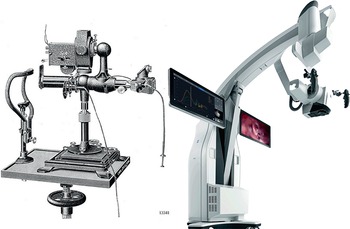
The last hundred years saw the development and refinement of the technologies perhaps most critical to the current practice of neurosurgery, including the computed tomography (CT) scan, magnetic resonance imaging (MRI) scan (Figure 1.3), the operating microscope (Figure 1.4), advanced light sources, ultrasonic aspirators, microinstruments, endoscopes, radiosurgery devices, and many others. These tools enabled clinicians to visualize and manipulate the intricate architecture of the brain non-invasively and with unprecedented detail and precision. Collaboration between the gamut of specialists, including physicists, engineers, investors, biomedical engineers, chemists, neuroscientists, anesthesiologists, neurologists, pathologists, and experts in infectious disease and critical care made these advancements possible and were instrumental to their success.
The first machine, the MRI Scanner Mark One, to be successfully used to create clinically useful images of the human body using magnetic resonance technology, by John Mallard in 1980 (reused from Wikimedia Commons under a CC BY-SA 4.0 licence from Andy Gaskell). Juxtaposition of the first portable MRI scanner, the HyperFine, pictured in 2024.

One of the first surgical operating microscopes, the Zeiss Operating Slit Lamp, from 1911. Juxtaposition with the Zeiss Kinevo, a modern surgical microscope developed in 2019.

Advancements in Imaging and Energy
The latter portion of the twentieth century saw fusion of some of these technologies to create surgical navigation systems, enhancing the precision and safety of neurosurgical procedures. Through this development, surgeons gained the ability to navigate within the brain with extraordinary accuracy in real time, reducing the risk of damage to critical structures. To do this, new technologies superimposed the patient’s brain imaging, usually an MRI or CT scan, onto a 3-dimensional map in real space in the operating room, such that the surgeon can see “inside” the brain based on this projection. In tandem with the refinement of imaging modalities came new methods of their use, including functional MRI (fMRI) – this system uses measurements of glucose metabolism and novel physical principles to measure brain activity and identify important areas unique to each individual, and diffusion tensor imaging (DTI), which maps fiber tracts, the “wires” connecting different brain areas by which they communicate. These advancements were coupled with burgeoning knowledge of human neuro-anatomy in order for the surgeon to select safe trajectories, avoiding critical structures while accessing pathologies deep within the brain (Reference Kokkinos, Chatzisotiriou and Seimenis12).
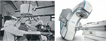
This technology has been further expanded to target more treatments, including radiosurgery, in which X-ray beams are precisely targeted to destroy lesions, or create interruptions in aberrant cerebral communication pathways (Figure 1.5). Other treatments that resulted from these advancements were focused ultrasound, in which many ultrasound beams are focused on specific brain locations to interrupt pathways or the blood–brain barrier, to treat tremor or deliver drugs or other molecules. Further, implantable lasers can be targeted with this same technology, which allows neurosurgeons to deliver intense heat to an intracranial target, destroying tumors and other lesions. Another technology that was leveraged in the twentieth century toward neurosurgical ends was transcranial magnetic stimulation (TMS). The laws of electromagnetic induction discovered by Faraday at the close of the nineteenth century made this possible, and the first experiments involving human cortical stimulation using TMS followed soon after (Reference Chail, Saini, Bhat, Srivastava and Chauhan13). This allowed practitioners to target specific brain areas to selectively interrupt activity for a variety of desired effects.
One of the first devices used to give clinically useful doses of radiation at the Royal London Hospital in 1900. Juxtaposition with the Varian Edge, a modern linear accelerator system for delivering therapeutic radiation, developed in 2016.

Decreasing Invasiveness, Improving Outcomes
This period marked a paradigm shift in the approach to neurosurgery, equipping surgeons with tools that expanded their horizons and transformed patient outcomes for the better. “Minimally invasive surgery” is a theme conceived at this juncture, and the undisputed father of this movement is Mahmut Gazi Yaşargil, arguably the greatest living neurosurgeon as of the writing of this chapter, who perfected the use of the operating microscope and developed the field of micro-neurosurgery. The late twentieth – early twenty-first century saw the refinement of this minimally invasive paradigm, with endoscopes being developed to access tiny surgical corridors, minimizing surgical trauma while achieving surgical goals. This required contributions from many fields, including mechanical and electrical engineering, optical physics, biomedical engineering, and others. Development of deep brain stimulators progressed at this time as well, which uses stereotactic principles to target electrodes in the brain with sub-millimeter accuracy, delivering electrical stimulation to neural networks to alter their function. This in turn was made possible through the work of neuroscientists, chemists, electrical, mechanical, and software engineers, physiologists, anatomists, and computer scientists. Advancements in catheter angiography of the heart were translated to neurosurgery around this time as well, allowing practitioners to access the brain via tiny wires and catheters threaded through the vascular system. The latter breakthrough was arguably one of the biggest leaps in neurosurgery in the last century. A variety of pathologies that were previously treated with extensive open surgeries are now treated entirely without incisions.
In this vein, neurosurgical problems are increasingly being treated non-invasively in some scenarios. Psychiatric drugs and cognitive–behavioral therapy are replacing lobotomies for control of mental and psychological disorders, chemotherapeutic and antimicrobial agents are shrinking tumors or infections respectively, precluding the need for surgery, or allowing more limited or less morbid procedures to be performed as adjuncts, all decreasing surgical morbidity for the patient. Examples of this include the union of neuroimaging modalities such as fMRI, DTI, and neuro-navigation with minimally invasive surgical tools such as the NICO BrainPath device, which provides a less traumatic method for surgical access to deep brain lesions (Reference Rennert, Khani, Thomas, Morris, Rodriguez and Day14). This non-invasive paradigm has led to the development of incredible technologies, including stem cell therapy, gene therapy via viral vectors and other mechanisms, and targeted molecular therapies. These fall generally into the category of “molecular neurosurgery.” The sequencing of the human genome paved the way for unprecedented understanding of human physiology and disease, and this has been harnessed to target vector-based and molecular avenues to treat neurosurgical pathology. Vector-based treatments involve utilizing a variety of delivery mechanisms that alter the DNA of specific brain regions or lesions, thereby correcting pathological processes at the molecular level. The primary paradigm for DNA delivery into human cells has been genetically engineered viruses, but nanotechnology and other avenues are actively being explored for the same ends. Also stemming from the mapping of the human genome, and the resultant discoveries regarding the proteome, or the library of protein “molecular machines” that control all life processes, are advances in molecular targeted therapies (Reference Kaplitt, Feigin, Tang, Fitzsimons, Mattis and Lawlor15). These paradigms involve designing individual molecules that target specific proteins, which alters their function to achieve a therapeutic goal while minimizing undesired side effects in other areas of the body. The role of stem cells in initiating neurosurgical disease, including tumors and certain degenerative processes, has also been elucidated in recent years. Emerging treatments often involve engineering patient-specific stem cells to regenerate damaged or degenerated brain tissues, or targeting stem cells in cancer to prevent growth or recurrence (Reference Tabar and Studer16, Reference Piao, Zabierowski, Dubose, Hill, Navare and Claros17).
Computers and the Brain
Another important paradigm born in the late twentieth century was that of the brain–computer interface, or BCI. These systems record electrical activity in the brain, often with invasive monitoring directly from brain tissue, and use the decoded output to produce external actions with the aid of a computer. This research originally began with the invention of external/non-invasive electroencephalography, or EEG, a century ago; these systems use grids on the scalp to measure gross electrical activity in brain areas non-invasively. The first invasive BCI system was implanted into a human brain in 1998, by Philip Kennedy in Georgia – interestingly, Dr. Kennedy had a BCI implanted into his own brain in 2014 to further his investigations (18). Many challenges exist in furthering this technology, including decoding and applying the boundlessly complex electrical signals from the brain, addressing signal-to-noise ratios, and dealing with the body’s rejection of delicate invasive monitors. As such, the BCI model relies heavily on technology outside of medicine – software and hardware engineering, physics, neurophysiology, robotics, chemistry, and many others. It is a technological advancement that piques the interest of scientists and lay-people alike, but its progress depends on a comprehensive collaborative approach among many different specialists (Reference Chappell19, Reference Willett, Avansino, Hochberg, Henderson and Shenoy20).
The twenty-first century has heralded further advancements, including the incorporation of virtual and augmented reality into surgical navigation devices. These took surgical navigation to its next evolution, such that the neurosurgeon can visualize internal anatomy and pathology in three dimensions in real time, seeing inside the skull with unprecedented detail. Surgical robots followed soon after, partially automating certain surgical maneuvers and processes. However, full automation of entire neurosurgical procedures has been fraught with challenges (Reference Faragasso and Bonsignorio21). Our ever-growing understanding of human neuroscience is allowing us to re-purpose existing technologies, such as focused ultrasound, transcranial magnetic stimulation, and deep brain stimulation, to treat an expanded range of pathologies including addiction, obsessive-compulsive disorder, obesity, and Alzheimer’s disease, among others. Importantly, surgical navigation, precise intervention targeting, and robotic technology all rely heavily on advances in computer science.
Advancements Outside the Operating Room
Other ideas in neuroscience that have recently gained evidentiary support include heretofore unthinkable relationships between the brain and other systems completely. The brain–gut axis is one such paradigm, which suggests that direct communication, both neural and otherwise, between one’s digestive and nervous systems is critically important for both of their functioning (Reference Appleton22). Further decoupling the nervous system from our prior understandings is the concept of the “mind–body connection,” popularized in 1997 by Dr. Candace Pert in her book Molecules of Emotions. Therein, she challenges scientific dichotomies of the objective and subjective, and provides frameworks for furthering our understanding of the complex machinations of the human central nervous system and its effects on the rest of the body (Reference Foss23). Studies of neuroplasticity and neuro-regeneration have also recently challenged ideas that the human nervous system is fixed and relatively static. In the ensuing decades, discoveries of cytokines, endorphins, and other molecules present putative mechanisms in part describing the above theories (Reference Bourgognon and Cavanagh24).
Another important contribution of computer science to neurosurgery relates to the latter part of the Information Age. In 1999, the Neurosurgery Journal went completely online, including the process of submission, review, and subscriber consumption. This milestone took subscriptions from 8,500 to a reach of 16 million readers overnight, and academic neurosurgery was never the same (Reference Apuzzo and Hodge25). The unprecedented upgrade in inter-provider communication in the field meant that the “marketplace of ideas” in neurosurgery had entered its final evolution, with reverberating consequences of improved patient care on a global scale.
This embarkation into the information age had consequences for medical education and competency-building as well. Neurosurgeons suddenly had a wealth of resources at their fingertips with which to further their career, including text, images, videos, virtual reality systems, artificial intelligence, and more. This has undoubtedly yielded a positive effect on training the physicians of the future, and will continue to do so as this technology advances.
As individual discoveries in all neuroscience-related fields amalgamate to form an ever-expanding, holistic body of knowledge, it is becoming increasingly clear that a focus on overall brain health is of paramount importance. Prevention of neurologic pathologies through diet, aerobic and cognitive exercise, and sleep health is now understood to be as important as their treatment. This shifting focus toward the overall function and health of the brain will continue to benefit our patients in the coming age.
In parallel with the development of neurosurgery is the changing landscape of medical economics. Technological advancements come at great cost, and training a single neurosurgeon today costs well over $1,000,000 (Reference Garba, Fadalla, Sarpong, Suliman, Rolle and Ammar26). With this in mind, cost-saving in innovation is increasingly a driving force in the trajectory of progress, adding a new challenge for neurosurgical pioneers to factor into their mission. What is an acceptable cost to deliver humankind from injury and disease of the brain? The answer is complex, with stakeholders in every facet of society.
The message of this entire work is to posit that neurosurgeons do not exist in a vacuum, far from it. As such, advancement of our allied clinical fields including neurology, neurocritical care, physical, occupational, and speech therapy, radiology, psychiatry, neuropsychology, and many, many more are crucial to bringing our field into its next iteration. While these fields rely on some of the same breakthroughs and discoveries as neurosurgery, they have their own unique needs and dependencies as well, which will by proxy elevate all of the clinical neurosciences, and medicine as a whole, when they are met.
New Horizons – The Way Forward
The technological advancements used in neurosurgery today were born of the sweat and tears of countless scientists, engineers, inventors, developers, visionaries, and investors. Every step forward requires yet more collaboration and cooperation. Thus, non-neurosurgeons are increasingly important to bringing the field of neurosurgery to its next evolution. Pushing the boundaries of what currently is into the realm of science fiction is the modus operandi that breeds innovation. Increasing minimally invasiveness toward non-invasiveness, advancing direct communication between the mind and machines through so-called “brain–computer interfaces,” perfecting treatments for previously untreatable conditions such as spinal cord injury through developments of exoskeleton and related technology, curing metastatic cancer through combinations of surgical, medical, and radio-therapies; these are just a few exciting breakthroughs that are on the horizon of neurosurgery. However, they will not be possible without herculean efforts in a variety of different allied fields. Particularly important will be advancements in molecular biology and genomics, proteomics and organic chemistry, neurochemistry, brain imaging, immunochemistry, robotics, mechanical and electrical engineering, miniaturization and design, computer science and software development, virtual and augmented reality, and basic neuroscience, among others. We hope that should the vision and ambition in these chapters excite you, you would consider having a hand in crafting the next generation of neurosurgical innovation.