Introduction
Bovine viral diarrhea virus (BVDV) is a single-stranded RNA virus, which belongs to the family of pestiviruses (McGovan et al., Reference McGowan, Kirkland, Richards and Littlejohns1993). BVDV infection has been associated with enteric pathology, mucosal disease and reproductive failure. Field strains of BVDV exist as two biotypes, the cytopathic (CP) and noncytopathic (NCP), distinguished by their effects in cell culture (Brownlie et al., Reference Brownlie, Clarke and Howard1984). The CP biotype induces rapid cytoplasmic vacuolization and apoptotic cell death (Fair et al., Reference Fair, Hyttel and Greve1995). In contrast, the NCP biotype can replicate in cultured cells without any adverse effects. BVDV can be passed from infected cow to the fetus and it is unknown which stages of embryo development are susceptible to BVDV infection and whether transmission through the female germline is possible. According to Gilula et al. (Reference Gilula, Epstein and Beers1978), BVDV may be able to infect oocytes as they mature within the developing follicle. Potter et al. (Reference Potter, Corstvet, Looney, Fulton, Archibald and Godke1984) and Sing et al. (Reference Singh, Eaglesome, Thomas, Papp-Vid and Hare1982) showed that BVDV could not penetrate the zona pellucida of in vivo-derived embryos, but CP BVDV strains cause the embryonic death of hatched embryos (Brock & Stringfellow, Reference Brock and Stringfellow1993).
Whereas the majority of studies on the susceptibility of in vivo-derived embryos to BVDV were conducted in in vitro experiments, there was one report of an intrauterine inoculation of day 7 embryos with BVDV. In this study, Archibald et al. (Reference Archibald, Fulton, Seger and Godke1979) observed an inhibition of embryonal development in eight out of 12 embryos collected 3 days after virus inoculation. Besides a possible pathogenic effect of BVDV on embryos, the authors observed a change in uterine environment caused by the BVDV infection, which may have caused suppression of embryonic development and embryo degeneration. In contrast, other researchers (Bielanski & Hare, Reference Bielanski and Hare1988; Bielanski et al., Reference Bielanski, Dubuc and Hare1992) reported that in vitro inoculation of in vivo embryos with CP BVDV had no adverse effect on survival and embryonic development. It has been demonstrated that BVDV may have direct access to embryonic cells when the zona pellucida is damaged following flushing or as a results of freezing–thawing (Subramanian et al. Reference Subramanian, Devanjan and Mohaman1990).
In vitro-produced embryos have a higher risk becoming infected compared with in vivo-produced embryos, due to manipulations in culture conditions (Van Soom et al., Reference Van Soom, Vanobdeobosch, Mahmoudzadeh and de Kruif1996). Vanroose et al. (Reference Vanroose, Nauwynek, Van Soom, Vanopdenbosch and de Kruif1998) showed that both CP and NCP BVDV are able replicate into embryonic cells of in vitro-produced ZP-free bovine embryos and that the production of infectious virus is related to both the stage of embryo development and the virus biotype. In contrast, no virus replication or signs of embryonic degeneration were detected in ZP-intact in vitro-derived embryos (Vanroose et al. Reference Vanroose, Nauwynek, Van Soom, Vanopdenbosch and de Kruif1998).
Vanroose et al. (Reference Vanroose, Nauyuck, Van Soom, Vanopdenbosch and de Kruif1999) demonstrated that, when CP or NCP BVD viruses were presented in each steps of an IVP system, significantly lower rates of cleavage and blastocysts formation of virus-exposed embryos were observed, in comparison with control embryos (Vanroose et al. Reference Vanroose, Nauyuck, Van Soom, Vanopdenbosch and de Kruif1999). In all experiments, virus replication was not detected in embryonic cells, but was clearly demonstrated in an oviductal cell co-culture system, resulting in degeneration of these cells.
Viral morphogenesis of BVDV using electron microscopy was described in detail in several tissues of infected animals and bovine cell cultures (Ohmann & Block, Reference Ohmann and Bloch1982; Ohmann, Reference Ohmann1990). However, ultrastructural morphology of bovine embryos infected with BVDV virus has not yet been described. Available data indicate that BVDV virus does not enter the embryo, which is surrounded with intact undamaged zona pellucida.
Therefore, the aim of our study was to evaluate development and ultrastructural morphology of bovine preimplantation embryos exposed in vitro to BVDV. In order to avoid the barrier of zona pellucida for virus penetration, we inoculated BVDV suspension under the zona pellucida using a microinjection technique.
Materials and Methods
Determination of viral titre by using a cytotoxicity test on bovine ovarian granulosa cells
Virus stock of CP BVDV was taken at a concentration 105.16 TCID/ml. Granulosa cells were aspirated from 2–5 mm diameter ovarian follicles and cultured on coverslips in the DME/F12 medium added with 10% FCS and 1% antibiotic–antimycotic solution (Sigma) during 5 days to form confluent monolayer. After preculture, the BVDV stock, diluted in the incubation medium at 1:4, 1:8, 1:16 and undiluted virus stock were added to the cells and these were cultured for 24 h. Afterwards the cells were stained with the Trypan blue vital dye for 10 min. The rest of dye was flushed out with the saline and coverslips with cells were attached to microslides with the Glycergel mounting solution (DAKO). For viral titre determination, the ratio of dead cells (trypan blue stained) to total cells was calculated.
Collection of bovine embryos
Holstein-Friesian (HF) donor cows were superovulated between the 10th and 12th days of the previous oestrus cycle by intramuscular administration of 24 mg FSH (Folicotropin inj. ad us. vet., Spofa, Prague, Czech Republic) given in a series of decreasing doses over a 4 day period. Oestrus was induced by intramuscular administration of 0.75 mg prostaglandin F2α, i.e. 750 μg cloprostenolum (Oestrophan inj. ad us. vet., Léčiva, Prague, Czech Republic) in the morning and evening of the third day of FSH treatment. Oestrus detection was performed twice per day beginning 24 h after the first prostaglandin F2 alpha injection. Donor cows were artificially inseminated 12 and 24 h after first standing of oestrus with the semen from a sire of proven fertility. Embryos were recovered by uterine flushing with Dulbecco's phosphate-buffered saline (PBS) supplemented with 1% bovine serum after slaughter of donor cow on day 6 of the cycle (day 0 = first standing oestrus). The embryos were classified according to general criteria of developmental competence and quality evaluation at 100× magnification using a stereomicroscope (Wright, Reference Wright, Stringfellow and Saidel1998) and only compact morulae were selected for further experiments.
Microinjection of bovine viral diarrhoea virus (BVDV) under the zona pellucida of bovine embryos
BVDV was directly inoculated by microinjection of approx. 20 pl of viral suspension (105.16 TCID50/ml virus suspension was diluted 1:4) under the zona pellucida using manual micromanipulation units (Alcatel, France) connected to an automatic microinjector (Eppendorf) under the Olympus (IMT-2) microscope. Briefly, embryos were put into TCM-199 medium + GlutaMAX I (Gibco BRL, Lofer, Austria) in the micromanipulation chamber and secured by glass holding pipette. The zona pellucida was penetrated, the BVDV was injected subzonally and the injection pipette was quickly withdrawn. The embryos were washed three times in SOF medium and cultured for 24–48 h at 39 °C in an atmosphere of 5% CO2, 5% O2, and 90% N2.
Detection of viral particles using immunocytochemistry technique
The embryos were processed at 24 or 48 h following microinjection and analysed for the presence of BVDV. Briefly, the embryos were washed four times in PBS–PVP, fixed in 4% (w/v) paraformaldehyde in PBS (pH 7.4) for 30 min at room temperature and permeabilized in 0.5% (v/v) Triton X-100 for 15 min. After washing in PBS–PVP (2 min) and blocking in PBS with 1% bovine serum albumin (20 min), the embryos were incubated with anti-BVDV–FITC antibody (dilution 1:100) for 60 min. Following three washes in PBS–PVP (2 min/wash), the embryos were mounted on slides with 5.5 μl of Vectashield mounting medium (Vector Laboratories, Burlingame, CA, USA) and evaluated under the Leica fluorescence microscope equipped with a DFC-480 digital camera.
Electron microscopy
Bovine embryos (control and microinjected with BVDV) were fixed in aldehyde mixture (2.5% glutaraldehyde and 2% paraformaldehyde in 0.15 M cacodylate buffer, pH 7.1–7.3) for 1 h and washed in cacodylate buffer. One hour following fixation in 1% osmium tetroxide in cacodylate buffer, the embryos were rinsed in distilled water, dehydrated in acetone series (30, 50, 70, 95 and 100%) and embedded in Durcupan ACM (Fluka). The blocks with embryos were cut on a LKB–Nova ultramicrotome into semithin sections (1–2 μm) and stained with toluidine blue. Thin sections were contrasted with uranyl acetate and lead citrate and viewed in a JEM 100 CX II (Jeol, Japan) electron microscope operating at 80 kV.
Negative-staining for BVDV virus
A BVDV suspension was mixed with an equal volume of the negative dye, 2% phosphotungstic acid (PTA, pH 6.0–7.0) and put onto a nickel-covered electron microscope grid covered with formvar (Agar Scientific Ltd). After 30 s excessive fluid was sucked off and the preparation was viewed under a JEM 100 CX II (Jeol) microscope at accelerating voltage 60–80 kV.
Results
Determination of viral cytotoxicity using bovine ovarian granulosa cells
Treatment of cells with BVDV at the dilution of 1:4 (from virus stock at 105.16 TCID50/ml) caused the appearance of plaques (empty sites) in the centre of the monolayer, and these occupied 50% of the monolayer area. Staining with trypan blue revealed an increased number of stained (dead) cells in wells with 1:4 viral dilution. At a dilution of 1:8 or 1:16 the number of dead cells and plaques was substantially lower and comparable with control cells that had been cultured without virus.
Preimplantation development of BVDV-exposed embryos
The evaluation of the developmental potential of intact control embryos cultured up to day 8, showed that 39.34% of embryos reached the expanded blastocyst stage and 30.33% of embryos reached early blastocyst stages. About 30.33% of embryos were arrested at the morula stage and did not develop further.
Among BVDV-microinjected embryos, only 12.50% of embryos developed to the blastocyst and 4.17 % to the expanded blastocyst stages, whilst the remaining embryos (83.33%) were arrested and degenerated (at p < 0.001, versus 30.33% in the control, by chi-squared test).
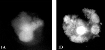
Immunofluorescence analysis localized the BVDV–FITC signal to the blastomeres of the microinjected embryos (Fig. 1a). Control embryos (non-microinjected with BVDV) showed only a slight non-specific background (Fig. 1b).
Immunofluorescent analysis of bovine ZP-surrounded blastocysts after the microinjection of the cytopathic BVDV strain suspension subzonally. (a) Control embryos (non-microinjected with BVDV) showed only a slight non-specific background. (b) BVDV–FITC signal localized inside the microinjected embryos.

Ultrastructure of intact or BVDV-exposed embryos using TEM
Negative staining revealed that viral particles of the cytopathic BVDV strain are enveloped into spherical particles approximately 50 nm in diameter (40–60 nm) containing single-stranded positive-sense RNA. The virion envelope has 10–20 nm ring-like subunits on its surface. To date, the structure or symmetry of the core has not been characterized by negative staining.
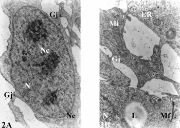
The embryos from the control group (with intact ZP) showed undamaged blastomeres and a transparent perivitelline space. The ZP was about 10–15 μm in width and consisted of two layers – external (8–10 μm) and internal (3–5 μm). Embryoblastic nuclei were round in shape, whilst trophoblastic nuclei were oval and euchromatic with several reticular nucleoli. The nucleus exhibited normal morphology; the nucleolus had a reticular structure with matured fibrillar centres (Fig. 2a). Microvilli on trophoblast cell surface were intact. The cytoplasm of trophoblastic and embryoblastic cells contained numerous mitochondria, a granular endoplasmic reticulum and a Golgi complex without changes (Fig. 2b).
Ultrastructural morphology of bovine intact blastocyst (control). (a) Normal reticular architecture of typical nucleolus (Nc) with fibrillar centres. Nuclear envelope (Ne) is normal (×15 000). (b) Morphologically normal mitochondria (Mi) and endoplasmic reticulum (ER). Gj, gap junctions; L, lipids; Mf, microfilaments; N, nucleus.

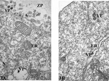
Bovine preimplantation embryos microinjected with BVDV showed the presence of virus-like particles that were related to BVDV. Following 24 h of exposure to the virus, the embryos showed degenerative signs – trophoblast and embryoblast cells often exhibited a dilated endoplasmic reticulum and the presence of small, round electron-dense virus-like particles in different areas of the mural trophoblast cell (Fig. 3a, b).
Ultrastructural morphology of bovine ZP-surrounded blastocysts 24 h after the microinjection of the CP BVDV strain suspension subzonally. (a) Dilated endoplasmic reticulum (ER) and small, round electrondense virus-like particles (VP), as seen here in different areas of the mural trophoblast cell and in the perivitelline space (×40 000). (b) Virus-like particles (VP) are seen in cytoplasma of trophoblast cell (×75 000). GA, Golgi apparatus; .M, mitochondria; N, nucleus; ZP, zona pellucida.

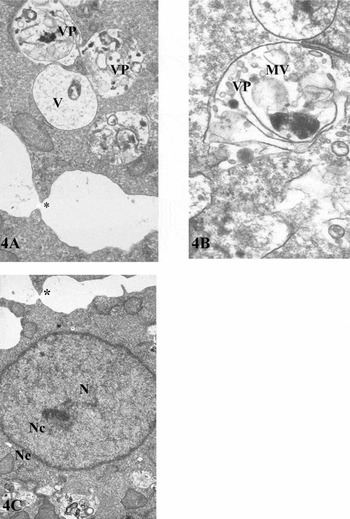
Following 48 h post-incubation, virus-like particles were observed in the dilated endoplasmic reticulum and in cytoplasmic vacuoles of the trophoblast and embryoblast cells (Fig. 4a, b). We have observed a loss of microarchitecture and the presence of abnormal disintegrated nuclei with no the heterochromatin area. The nucleolus characteristically had a loss of reticular structure. In all degenerated embryos, nuclear structure was altered and the microarchitecture of the nucleolus had disappeared when compared with the nuclei of control embryos. After exposure to BVDV the dilatation of the intercellular space and the loss of the intercellular gap junctions were often observed in bovine embryos. The abnormal disintegrated nucleus lacked a heterochromatin area, the nucleolus characteristically had no reticular structure and the nuclear envelope was disintegrated and dilated (Fig. 4c).
Ultrastructural morphology of bovine early blastocyst 48 h after the microinjection of the CP BVDV strain suspension subzonally. (a) Loss of intercellular gap junctions (asterisk); virus-like particles (VP) are appeared as granules in cytoplasmic vacuoles (V) of the embryoblast (×29 400). (b) Cytoplasmic multivesicular vacuoles (MV) with virus–like particles (VP) (×75 600). (c) Abnormal disintegrated nucleus (N) without heterochromatin area. Nucleolus (Nc) is characterized by the loss of reticular structure. Nuclear envelope (Ne) is disintegrated and dilated (×15 000).

Discussion
In our study we examined whether BVDV virus transferred under the zona pellucida can induce alterations in ultrastructural morphology in the cellular organelles of bovine embryos.
Reports concerning the potential transmission of BVDV via in vivo- and in vitro-produced embryos are contradictory (Deregt & Loewen, Reference Deregt and Loewen1995; Bolin & Ridpath, Reference Bolin and Ridpath1996; Leconte et al., Reference Leconte, Navetai, Harners, Lamôot, Scheider, Cabanie and Pestroet1996). An early study (Archibald et al. Reference Archibald, Fulton, Seger and Godke1979) validated the use of embryo washing procedures for removal of viruses after the exposure of embryos to the CP strain of BVDV. Later a study showed that the washing procedure differed in its effectiveness for removal of various isolates of BVDV (Dereggt & Loewen, Reference Deregt and Loewen1995).
Available reports concerning the ultrastructure of bovine BVDV-infected embryos and on the properties of the zona pellucida that prevent BVDV penetration (Vanroose et al., Reference Vanroose, Nauwynek, Van Soom, Vanopdenbosch and de Kruif1998; Reference Vanroose, Nauvynck, Van Soom, Ysebaert, Charlier, Van Oostveldt and de Kruif2000) using electron microscopy are limited No previous studies that have evaluated ultrastructural alterations in cell organelles after BVDV infection are known.
The mechanism of how the pathogen binds to the ZP is unknown. However, it has been also demonstrated that pathogenic agents adhere more easily to the surface of the ZP of in vitro-produced embryos comparing to the in vivo-fertilized embryos (Bak et al., Reference Bak, Callesen, Myeling and Greve1992). Furthermore, different conditions for the production of these two types of embryos may lead to changes in their morphology and to differences in the interaction of ZP with pathogens.
Concerning a risk of infection of embryonic cells with BHV-1 or BVDV, Vanroose et al. (Reference Vanroose, Nauvynck, Van Soom, Ysebaert, Charlier, Van Oostveldt and de Kruif2000) studied the outer pores of the ZP, which could be ports of entry for viruses. They showed that pores on the surface of the ZP are large enough to allow entry of BHV-1 (180–200 nm) and BVDV (45–55 nm). In all embryonal stages investigated, more than 96% of the outer pores were large enough for entry of BVDV. They also observed in some fertilized zygotes, excavation or fissures with the exact dimensions of a sperm head (which was probably caused by enzymatic digestion). These holes could provide a point of entry for the virus at the fertilization, besides serving as a potential binding place for the virus. Virus resident in such a cavity might be protected against eventual sanitary measures, e.g. washing and trypsin treatment.
Vanroose et al. (Reference Vanroose, Nauwynek, Van Soom, Vanopdenbosch and de Kruif1998) showed that CP BVDV strain had an inhibitory effect on the development of in vitro produced morulae to blastocyst stage in combination with cytopathic effect, resulting in the degeneration of embryos. They used morphology of the hatching blastocysts (HB) as an indicator of embryo quality and showed that the production of infectious virus is related to the stage of embryo development and the virus biotype. None of the HB in their study was classified as excellent quality and 75% of them were collapsed or degenerated. Similar results were reported by Brock & Stringfellow (Reference Brock and Stringfellow1993) for in vivo-derived HB inoculated with CP BVDV. In our study, most of the embryos (83%) was arrested at morula stage and subsequently degenerated. None of the embryos developed to the hatching blastocyst stage. However other authors (Potter et al., Reference Potter, Corstvet, Looney, Fulton, Archibald and Godke1984; Bielanski & Hare, Reference Bielanski and Hare1988) reported that CP BVDV had no adverse effect on embryo survival or development within 48 h of culture. Several BVDV strains may have either a lower affinity or no adherence ability to the receptors on embryonal cells. Another explanation may be the use of different inoculum. Moreover, Bielanski & Jordan (Reference Bielanski and Jordan1996) and Trachte et al. (Reference Trachte, Stringfellow, Riddell, Gulik, Riddell and Wright1997) reported that washing and trypsin treatment was ineffective for removal of NCP BVDV from ZP of in vitro produced embryos.
Although Schlafer et al. (Reference Schlafer, Gillespie, Foote, Quick, Pennow, Dougherby, Schiff, Allen, Powers and Hall1990) and Gillespie et al. (Reference Gillespie, Schlafer, Foote, Quick, Dougherty, Schiff and Allen1990) demonstrated that detection of the viral particles of BHV-1 or BVDV using morphological evaluation of the early bovine embryos by electron microscope is not always successful and unambiguous, it is assumed, that embryos with a damaged or cracked zona pellucida, perhaps due to sperm penetration, can be infected after the exposure to CP BVDV strain.
In our experiments we detected ultrastructural changes in embryos microinjected with BVDV subzonally. TEM of BVDV-microinjected embryos revealed injuries of almost all organelles (nucleus, nucleolus, endoplasmic reticulum, mitochondria) and disorders of intercellular junctions. We detected severe vacuolization in most of trophoblast and embryoblast cells with accumulation of virus-like particles in vesicles and dilated endoplasmic reticulum. Virus-like particles were seen in the cytoplasm of trophoblast cells and in cytoplasmic vacuoles of the embryoblast and trophoblast cells. Sometimes viral particles formed granular-like nucleocapsids in cytoplasm. We observed a loss of microarchitecture of nucleus and nucleolus.
Consequence of viral infection on the embryo viability may depend also on the inoculum titres of virus. Bielanski et al. (Reference Bielanski, Sapp and Lutze-Wallace1998) showed, that when the viral titre was increased about 100 times in comparison with the titre used in previous study (Bielanski & Hare, Reference Bielanski and Hare1988) CP and NCP BVDV were able to replicate in embryonic cells of in vitro-produced embryos without ZP with an increasing susceptibility of the embryos in accordance with their developmental stage.
Taking together these observations provide evidence for adverse effect of BVDV virus on the development of bovine embryos, which associated with irreversible alterations in ultrastructural morphology.
Acknowledgments
This research was supported by the Science and Technology Assistance Agency, contract No. APVT-51-002-602 and NAZV Agency of Czech Republic, contract No. 1B-44034. We thank to Mrs. Safarova for preparation and processing of samples for electron microscopy.