Introduction
The increasing use of endoscopic approaches for tumour resection is associated with potential donor morbidity for autologous tissue used in skull base reconstruction. Ensuring post-operative separation between the intracranial and sinonasal cavities is important, not only to avoid cerebrospinal fluid (CSF) leak, but also in preventing future meningitis, ventriculitis and intracranial abscess.Reference Harvey, Smith, Wise, Patel, Frankel and Schlosser 1 A number of materials have been utilised in a variety of combinations and layers for skull base reconstruction, often based on surgeon preference or local experience.
Materials most commonly used in the reconstruction layers are: inlay grafts, including autologous fat,Reference Bernal-Sprekelsen, Rioja, Ensenat, Enriquez, Viscovich and Agredo-Lemos 2 – Reference Sanders-Taylor, Anaizi, Kosty, Zimmer and Theodosopoulos 6 fascia,Reference Bernal-Sprekelsen, Rioja, Ensenat, Enriquez, Viscovich and Agredo-Lemos 2 , Reference Amit, Margalit, Abergel and Gil 7 – Reference Villaret, Yakirevitch, Bizzoni, Bosio, Bignami and Pistochini 10 boneReference Emanuelli, Milanese, Rossetto, Cazzador, d'Avella and Volo 11 , Reference Germani, Vivero, Herzallah and Casiano 12 and cartilage;Reference Germani, Vivero, Herzallah and Casiano 12 engineered allografts (i.e. acellular dermal matrixReference Germani, Vivero, Herzallah and Casiano 12 – Reference Ismail, Costantino and Sen 15 ); and synthetic materials (porous polyethylene plate,Reference Liebelt, Huang and Baskin 16 hydroxyapatite cementReference Chung, Nam, Park, Kim and Kong 17 ). These layers are frequently followed by the application of mucosal or vascularised grafts, fibrin sealants, and absorbable and/or non-absorbable packing.Reference Harvey, Smith, Wise, Patel, Frankel and Schlosser 1 , Reference Harvey, Parmar, Sacks and Zanation 18 Overall, successful repair of skull base defects has been reported in the literature to be over 90 per cent, regardless of the materials or technique used.Reference Oakley, Orlandi, Woodworth, Batra and Alt 19
The use of vascularised overlay grafts can lead to improved outcomes compared to free mucosal grafts in the case of larger defects or high-flow CSF leaks.Reference Harvey, Parmar, Sacks and Zanation 18 , Reference Soudry, Turner, Nayak and Hwang 20 With respect to inlay grafts, however, it is unclear if any material is superior to another in achieving successful repair. Fat and fascia autografts are frequently used, but are associated with donor site morbidity. Allografts and xenografts that have established utility in skull base surgery include acellular dermal matrix (AlloDerm; LifeCell, Branchburg, New Jersey, USA),Reference Germani, Vivero, Herzallah and Casiano 12 – Reference Ismail, Costantino and Sen 15 , Reference Citardi, Cox and Bucholz 21 , Reference Lorenz, Dean, Hurley, Chuang and Citardi 22 porcine small intestine (BioDesign®)Reference Illing, Chaaban, Riley and Woodworth 23 and collagen matrix products,Reference Braca, Marzo and Prabhu 24 – Reference Shorter, Connor, Thakur, Gardner, Nanda and Guthikonda 26 but these carry additional costs.
Although commonly utilised, the literature on collagen matrix in endoscopic endonasal skull base surgery is limited. We describe the short- and long-term outcomes of a collagen matrix used as a dural inlay, with a single mucosal graft layer, in endoscopic skull base reconstruction.
Materials and methods
A retrospective case series was performed of consecutive patients from a tertiary rhinology practice who underwent endoscopic skull base reconstruction using DuraGen collagen matrix (Integra Neurosciences, Plainsboro, New Jersey, USA) as an inlay graft.
Data collected from the patient population included age, gender, smoking status, history of chronic rhinosinusitis, asthma, post-operative radiation therapy, benign or malignant pathology, and origin of pathology. Smokers were defined as any patient currently smoking or who had ceased within the last 12 months. Chronic rhinosinusitis was defined as per the European Position Paper on Rhinosinusitis and Nasal Polyps (‘EPOS’) guidelines, with both symptoms and evidence of non-tumour related sinus inflammation on endoscopy (with or without computed tomography evidence).Reference Fokkens, Lund, Mullol, Bachert, Alobid and Baroody 27 Asthma status was determined by a positive spirometry result on challenge testing or β-agonist use, or if currently using regular inhaled bronchodilator or corticosteroid therapy. Although all surgery was transdural, the origin of pathology was defined as either intracranial or paranasal sinus in origin.
All patients had collagen matrix as the first layer of their skull base reconstruction, placed intracranially as an inlay graft in the subdural or arachnoid space (Figure 1). This was followed by a free mucosal graft (usually for defects less than 1 cm), a vascularised mucoperiosteal flap or a combination. Porcine gelatine sponge was used to cover the reconstruction, and a bolster of either absorbable (NasoPore; Polyganics, Groningen, Netherlands) or removable packing (Bismuth iodine paraffin embedded gauze or Foley balloon) was used to keep the repair in place.
(a) Pre-operative, coronal, T1-weighted, post-contrast fat-saturation magnetic resonance imaging scan of a left olfactory groove meningioma in a 43-year-old male. (b) Intra-operative view of the collagen matrix inlay as the first layer of the skull base reconstruction following resection of this tumour. This was followed by a nasoseptal flap overlay.

A standard post-operative management protocol was followed, which consisted of at least 1 night in the intensive care unit, with bed rest for 48 hours, then mobilisation. Intravenous antibiotics were administered while the patient was in hospital, followed by 10 days of oral antibiotics and twice daily saline irrigations upon discharge.
Peri-operative morbidity
Immediate peri-operative outcomes (occurring three months or less post-operatively) were assessed, including CSF leak, meningitis, other intracranial infections (ventriculitis or cerebritis), intracranial bleeding, epistaxis and seizures. The diagnosis of CSF leak was based on surgeon assessment according to clinical evidence of CSF leaking from the surgical site. Any CSF leaks that occurred within days of the surgery did not require confirmation with a β−2 transferrin test and were treated with re-operation. Meningitis was diagnosed for any patient with positive CSF cultures or clinical symptoms consistent with the diagnosis, such as fever, headache, neck stiffness, photophobia and leukocytosis. Diagnosis of ventriculitis was based on clinical symptoms, magnetic resonance imaging findings and positive CSF cultures. Intracranial bleeding consisted of any collection of blood under the skull base repair, or otherwise in the epidural, subdural or intraventricular space, which necessitated medical or surgical intervention to manage, such as a return to the operating theatre for evacuation or medical treatment of intracranial pressures. Epistaxis was defined as persistent, brisk bleeding from the nose that required nasal packing, electrocautery or other surgeon intervention to manage. Seizure diagnosis was based on witnessed seizure activity meeting clinical criteria.
Delayed complications
In addition, delayed complications (occurring more than three months post-operatively) were examined, which included delayed healing, meningoencephalocele formation, prolapse of reconstruction, delayed CSF leak and ascending meningitis. Delayed healing consisted of any ongoing sinus dysfunction, in the form of persistent epithelial disruption, ulceration, granulation tissue, serous weeping or bleeding, abnormal crusting, or dehiscence of the vascularised flap–sinus mucosal margin. Meningoencephalocele was defined as protrusion of meningeal and brain tissue into the paranasal sinus cavity. Prolapse of reconstruction involved the extension of both the reconstructive layers and the adjacent underlying intracranial contents beyond the bony margin of the skull base, with or without dislodgment of the reconstruction. Delayed CSF leak and ascending meningitis were diagnosed as described above, but occurred outside the three-month post-operative timeframe.
Statistical analysis
Statistical assessment was performed using SPSS version 22 software (IBM, Armonk, New York, USA). Descriptive data are presented as percentages and means ± standard deviations (SDs) for parametric data and medians (interquartile ranges) for non-parametric data. The chi-square test and Fisher's exact test were used for the analysis of nominal values. The student's t-test (two-tailed) was employed for the comparison of parametric data. Results with a p value of less than 0.05 were considered significant.
Results
A total of 120 patients (mean age of 51.0 ± 17.5 years, 41.7 per cent female), presenting from October 2008 to September 2015, were assessed. Of these, 4.7 per cent were smokers and 11.4 per cent had a pre-operative diagnosis of asthma. A total of 28.3 per cent of skull base reconstructions were the result of malignant pathology and 53.3 per cent of all pathology was of intracranial origin. Post-operative radiation therapy was necessary in 28.7 per cent of patients. Average follow-up duration was 39.0 months (range, 23.0–58.8 months).
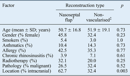
Following placement of the collagen matrix inlay, a nasoseptal flap was used as an overlay graft in 69.2 per cent of cases. Patients with pathology of intracranial origin were statistically more likely to receive a nasoseptal flap (62.7 vs 32.4 per cent, p < 0.05). There were otherwise no differences between the patients receiving nasoseptal flaps versus those with non-vascularised repairs (Table I).
Baseline demographic and treatment factors*

*Comparison between patients with nasoseptal flap based reconstruction versus those without. † n = 83; ‡ n = 37. SD = standard deviation
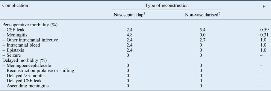
There was an all-inclusive peri-operative complication rate of 12.7 per cent, in which one complication occurred in 10.8 per cent of patients (n = 13) and two complications (CSF leak and other intracranial infection) occurred in 0.8 per cent of patients (n = 1). Of these complications, 3.3 per cent were CSF leaks, 3.3 per cent were meningitis, 2.5 per cent were other intracranial infections, 1.7 per cent were intracranial bleeding and 1.7 per cent were epistaxis. There were no peri-operative seizures. Delayed complications (delayed healing, meningoencephalocele, prolapse of reconstruction, delayed CSF leak and ascending meningitis) did not occur in any patients.
Factors associated with complications
There was no significant difference between patients who had a CSF leak and those who did not with respect to age (58.5 ± 11.9 vs 50.8 ± 17.6 years, p = 0.39), gender (50.0 vs 41.4 per cent female, p = 1.0), smoking (0.0 vs 4.9 per cent, p = 1.0), asthma (50.0 vs 9.9 per cent, p = 0.06), chronic rhinosinusitis (0.0 vs 5.0 per cent, p = 1.0), radiation therapy (33.3 vs 28.6 per cent, p = 1.0), tumour character (25.0 vs 28.4 per cent malignant, p = 1.0), tumour origin (75.0 vs 52.6 per cent intracranial, p = 0.62) or use of a nasoseptal flap (50.0 vs 69.8 per cent, p = 0.59) after collagen matrix inlay. Whether or not a nasoseptal flap was used following collagen matrix inlay had no effect on the rate of any of the peri-operative complications (Table II).
Comparison of complication rates for nasoseptal flap versus non-vascularised repair groups*

*Comparison based on whether or not a nasoseptal flap was used as part of skull base reconstruction following placement of collagen matrix layer. † n = 83; ‡ n = 37. CSF = cerebrospinal fluid
Discussion
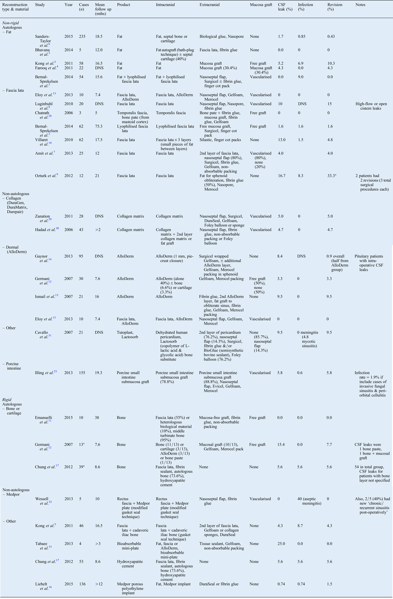
Collagen matrix is effective as an inlay graft in endoscopic skull base reconstruction. Collagen has been assessed in skull base reconstruction in the open repair of middle cranial fossaReference Braca, Marzo and Prabhu 24 , Reference Shorter, Connor, Thakur, Gardner, Nanda and Guthikonda 26 and posterior cranial fossa defects,Reference Narotam, Qiao and Nathoo 25 with similar peri-operative outcomes as those in this study. When it comes to graft materials selected and number of layers used, no reconstructive strategy appears to be superior to others. Comparative studies are uncommon and most publications are retrospective case series, with heterogeneity of reconstructive methods. There is heterogeneity in terms of patient characteristics, graft material, number of layers used, overlay graft selection and peri-operative management employed. A structured summary of the various underlay materials described in the literature for endoscopic skull base reconstructions in the last 10 years is presented in Table III.Reference Bernal-Sprekelsen, Rioja, Ensenat, Enriquez, Viscovich and Agredo-Lemos 2 – Reference Chung, Nam, Park, Kim and Kong 17 , Reference Illing, Chaaban, Riley and Woodworth 23 , Reference Chatrath and Saleh 28 – Reference Tabaee, Kamat and Shrivastava 33
Summary of underlay materials described for endoscopic skull base reconstructions in last 10 years

*Represents the relevant subset of the study population (rather than the overall study numbers). Mths = months; CSF = cerebrospinal fluid; DNS = data not shown
Autologous tissue is an established underlay material. Common autografts include abdominal or thigh fat, fascia, cartilage, and bone. Fat has proven useful in obliterating dead space, whether intracranially or in the sphenoid sinus. Fascia lata also can be used successfully either as an individual underlay or in multiple layer fashion.Reference Luginbuhl, Campbell, Evans and Rosen 8 Both of these autologous materials have the benefit of being readily available on a patient, with no risk of foreign body reaction or disease transmission. However, they both require a separate donor site, which carries with it potential morbidity, such as infection, haematoma, seroma, cosmetic concerns and possibly increased operative time for harvest. Abdominal haematoma has been reported to occur in 0.9–3.2 per cent of cases.Reference Sanders-Taylor, Anaizi, Kosty, Zimmer and Theodosopoulos 6 , Reference Sanna, Taibah, Russo, Falcioni and Agarwal 34 For autologous fat, a 1 per cent complication rate, in a series of 974 patients who underwent skull base reconstruction, has been reported.Reference Taha, Almefty, Pravdenkova and Al-Mefty 35 These complications included leakage of liquefied fat from a fistula, delayed CSF leak after radiation, and lipoid meningitis. Two cases of lipoid meningitis were also reported in a series in which fat graft was used for translabyrinthine craniotomies, requiring CSF diversion.Reference Hwang and Jackler 36
Autologous cartilage or bone are often utilised to provide a rigid buttress to the repair. One method that has been described is the ‘gasket seal’ method, in which fascia lata is placed in the defect followed by a bony buttress, which is then countersunk, wedging its edges beyond the bony defect edges, holding it firmly in place.Reference Leng, Brown, Anand and Schwartz 37 Although it has proven effective, whether or not this type of rigid support is needed is not known. This study's authors have removed bone and titanium sequestra from skull base reconstructions performed elsewhere at three months and beyond. Such a repair introduces the potential for sequestra formation; furthermore, it still needs to be combined with another soft tissue material, and is of questionable additional benefit when vascularised mucosal graft already improves the outcome for large dural defects and high-flow CSF leaks.Reference Harvey, Parmar, Sacks and Zanation 18 , Reference Soudry, Turner, Nayak and Hwang 20 These rigid autologous grafts can be taken from resected sinonasal tissue that would otherwise be discarded, such as the septum or turbinates, making them convenient options. However, their availability is dependent on involvement in the primary pathology. Therefore, they are not a consistent or reliable option in skull base reconstructions. Rigid and non-rigid allografts and synthetic materials, on the other hand, are readily available materials that can be customised to a given defect intra-operatively, but these risk rejection under a thin mucosal surface.
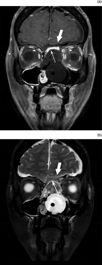
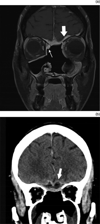
DuraGen is a collagen-based dural replacement product that is derived from freeze-dried ground bovine Achilles tendon. This collagen foam is processed to remove any cellular or immunogenic components, and chemically crosslinked, giving it its porosity and matrix structure. In a canine model, Zerris et al. compared the physico-mechanical properties, structure and performance of DuraGen and two other available collagen-based dural replacement products, Dura-Guard (Synovis, Saint Paul, Minnesota, USA), derived from bovine pericardium, and Durepair (TEI Biosciences, Boston, Massachusetts, USA), derived from fetal bovine dermis. DuraGen was demonstrated to be a much more delicate material than Durepair or Dura-Guard, have a larger pore size (100 µm in DuraGen, vs 10–20 µm in Durepair and 5 µm in Dura-Guard), and be much more quickly incorporated into and replaced by native host tissue (four weeks vs six months).Reference Zerris, James, Roberts, Bell and Heilman 38 In endoscopic CSF leak repair, remucosalisation time was shown to occur significantly sooner with collagen matrix products (DuraGen and DuraForm) compared to acellular dermal matrix (AlloDerm) (6.6 vs 11.7 weeks), but second to mucosal grafts (4.9 weeks).Reference Prickett, Wise and Delgaudio 39 No significant foreign body reaction was noted for any of the bovine collagen grafts.Reference Zerris, James, Roberts, Bell and Heilman 38 Figure 2 demonstrates early post-operative imaging characteristics of collagen matrix underlay. Radiologists need to be aware of the expected appearance of this reconstructive layer to avoid confusion with residual or recurrent disease or signs of an infective process (Figure 3), an error that has occurred in the past.Reference Walsh, Illing, Riley, Cure, Srubiski and Harvey 40
(a) Coronal, T1-weighted, post-contrast fat-saturation magnetic resonance imaging (MRI) scan of a patient at post-operative day 1 following resection of sinonasal squamous cell carcinoma with collagen matrix underlay as part of the skull base reconstruction. The hypointensity of this underlay (thick arrow) can be observed in the anterior cranial fossa abutting the brain, under which the enhancing nasoseptal flap overlay is seen (thin arrow), followed by absorbable packing and a Foley catheter balloon in the nasal cavity. (b) This same reconstruction observed on a T2-weighted, fat-saturation MRI scan.

(a) Coronal, T1-weighted, post-contrast fat-saturation magnetic resonance imaging scan of the same patient as in Figure 2, five weeks following squamous cell carcinoma resection. Note the enhancement of the collagen matrix underlay (thick arrow), which could be easily confused with meningeal enhancement from extension of the malignancy. The pedicle of the nasoseptal flap overlay can also be observed (thin arrow). (b) Coronal computed tomography brain scan, with contrast, of a patient six weeks following endoscopic resection of an anterior cranial fossa meningioma, showing hypodensity of the collagen matrix underlay (arrow), which could be mistaken for a fluid collection on imaging.

Acellular dermal matrix (AlloDerm) is a commonly used allograft made from cadaveric human skin that has been specially treated to remove all cellular and immunogenic components. Gaynor et al. compared transsphenoidal pituitary surgery reconstructions using acellular dermal matrix and fat graft in the event of intra-operative CSF leak, and found equivalent success between them.Reference Gaynor, Benveniste, Lieberman, Casiano and Morcos 14 There were 160 cases of intra-operative CSF leak, with 8.4 per cent in the acellular dermal matrix group versus 15.2 per cent in the fat graft group with reported post-operative leak (p = 0.34). Several other studies have reported over 90 per cent success rate of skull base reconstruction with the use of acellular dermal matrix.Reference Germani, Vivero, Herzallah and Casiano 12 , Reference Ismail, Costantino and Sen 15 , Reference Citardi, Cox and Bucholz 21 , Reference Lorenz, Dean, Hurley, Chuang and Citardi 22 , Reference Leong, Citardi and Batra 41
-
• Ensuring post-operative separation between the intracranial and sinonasal cavities is important to prevent significant morbidity
-
• There are various inlay graft materials for endoscopic skull base reconstruction available to the surgeon
-
• These include autologous fat, fascia, bone, cartilage, engineered allografts and synthetic materials
-
• Collagen matrix is an effective inlay graft; it provides a robust endoscopic skull base repair, is easily obtainable and avoids donor site morbidity
Collagen matrix presents similar advantages to acellular dermal matrix, in that there is no donor site morbidity, it is readily available and it can be sized to the defect. With either allografts or xenografts, there is a theoretical risk of transmissible spongiform encephalopathies, such as Creutzfeldt–Jakob disease. However, the World Health Organization have designated these substances as not having any detectable infectivity; they are processed to eliminate any potential infectivity, and there have been no reports of disease transmission in more than two million uses.Reference Zerris, James, Roberts, Bell and Heilman 38 , 42 , Reference Warren, Medary, Dureza, Bellotte, Flannagan and Oh 43
This study has the advantages of a large study population, a long follow-up time, and a variety of sinonasal and skull base pathology and subsequent skull base defects. Nevertheless, it remains limited by its retrospective nature and an inability to compare to other reconstructive methods.
Conclusion
Various reconstruction strategies are available. Collagen matrix, in a simple two-layer reconstruction, is an effective option for endoscopic skull base repair. It is readily obtainable and without donor site morbidity. It provides a robust reconstruction that prevents ascending infection and delayed breakdown, and it is rigid enough to withstand intracranial pressure forces. While it carries additional cost, this is weighed against the surgical consumables used to harvest, repair and dress the donor site.