Introduction
Uvulopalatopharyngoplasty (UPPP) is a common basic surgical treatment for obstructive sleep apnoea hypopnea syndrome (OSAHS). This surgery can eliminate retropalatal obstruction effectively; however, severe apnoea and hypopnea remain widespread in a notable proportion of OSAHS patients who receive UPPP alone.Reference Friedman, Ibrahim and Joseph1, Reference Li, Wu and Shi2 Uvulopalatopharyngoplasty cannot completely relieve symptoms in these OSAHS patients. These patients suffer from multi-level obstruction, especially that of the retroglossal area.
The retroglossal area is a commonly obstructed site, in addition to the retropalatal area.Reference Singh, Al-Reefy, Hewitt and Kotecha3, Reference Tanyeri, Serin, Ayanoglu, Polat and Cuhadaroglu4 Compared with nasal, nasopharyngeal and retropalatal obstruction severity, retroglossal obstruction severity is more difficult to diagnose and predict. Many measuring approaches, including the Friedman tongue position, airway computed tomography, drug-induced sleep endoscopy and upper airway–oesophagus pressure monitoring, have been utilised to evaluate retroglossal obstruction, but are individually limited.
Hyoid suspension is a classical surgery for retroglossal obstruction.Reference Vilaseca, Morelló, Montserrat, Santamaría and Iranzo5, Reference Richard, Timmer, van Tinteren and de Vries6 This method moves the hyoid and tongue base toward the mandible, to expand the cross-sectional area and shorten the length of the glossopharyngeal airway. This surgery also secures the tongue to prevent glossoptosis.
A previous study found that the hyoid bones of patients who underwent UPPP alone with insufficient curative effect are located more inferiorly than in patients with complete curative effects.Reference Cao, Ye, Zhang, Tan and Dong7 The hyoid bones of paediatric mouth breathers with OSAHS are significantly more anterior and inferior than those of paediatric nasal breathers.Reference Vieira, Itikawa, de Almeida, Sander, Fernandes and Anselmo-Lima8 These findings indicate a correlation between abnormal hyoid bone position and retroglossal obstruction. This study aimed to confirm the possibility of this correlation and evaluate the predictive value of the correlation to retroglossal obstruction severity.
Materials and methods
Patients
Subjects were recruited from the Otolaryngology Department of the General Hospital of Shenyang Military Area Command from October 2015 to February 2017. All the patients were diagnosed with OSAHS by polysomnography, with an apnoea-hypopnoea index equal to or greater than 5 events per hour. Alcohol, tea, coffee and any other drugs that can disturb the sleep architecture were forbidden during examination. None of the patients underwent any surgical procedures for OSAHS prior to recruitment. All patients were keen to undergo surgical treatment. Informed consent was obtained.
The exclusion criteria included: (1) definite pulmonary or metabolic diseases; (2) significant rhinal or nasopharyngeal obstructive diseases; (3) definite deformations such as retrognathia; and (4) a retroglossal space occupied by a hypopharyngeal tumour, hypertrophied palatine tonsils or lingual tonsils.
Image acquisition
Lateral films of the cervical vertebra were obtained in all enrolled patients to visualise the hyoid body, cervical vertebra and mandible. The patients were required to keep their mouth closed and breathe quietly during the radiography, without swallowing or chewing. The radiographs were then transferred to a computer workstation for cephalometric measurement.
Cephalometric measurements
These measurements included the distances of the anterior edge of the hyoid to: the mental tubercle, the prevertebral plane, the mental tubercle coronal plane and the mental tubercle horizontal plane. The hyoid to the horizontal plane distance represented a vertical position of the hyoid in relation to the mandible. The hyoid to the coronal plane distance represented a horizontal position of the hyoid in relation to the mandible. The hyoid to the prevertebral plane distance represented a horizontal position of the hyoid in relation to the post-pharyngeal space.
The hyoid to coronal plane distance plus hyoid to horizontal plane distance value represented a simple two-dimensional direction of the hyoid. Given the diversity of post-pharyngeal space size in these patients, compound parameters, such as the hyoid to mental tubercle distance/hyoid to prevertebral plane distance value, and the (hyoid to coronal plane distance plus hyoid to horizontal plane distance)/hyoid to prevertebral plane distance value, could provide a two-dimensional direction of the hyoid in the upper respiratory tract more precisely. The detailed measurement method is shown in Figure 1.
Diagram of cephalometric tracing. ‘HM’ is the distance between the anterior edge of the hyoid and the mental tubercle. ‘HP’ is the distance between the anterior edge of the hyoid and the prevertebral plane, which is a plane passing through the anterior superior edges of the third and fourth vertebrae body. ‘HC’ is the distance between the anterior edge of the hyoid and the mental tubercle coronal plane. A vertical line is drawn up from the mental tubercle to the prevertebral plane. This vertical line is considered a nearly horizontal plane passing through the mental tubercle. ‘HH’ is the distance between the anterior edge of the hyoid and this mental tubercle horizontal plane.

Polysomnography before and after tube insertion
The Alice 5 polysomnography system (Philips Respironics, Murrysville, Pennsylvania, USA) was used for monitoring sleep. The American Academy of Sleep Medicine standards were employed to monitor sleep and classify OSAHS grade.Reference Iber, Ancoli-Israel, Chesson and Quan9 Patients were categorised by apnoea-hypopnoea index into mild (5.00–14.99 events per hour), moderate (15.00–29.99 events per hour) and severe (30.00 or greater events per hour) grades. The patients diagnosed with OSAHS underwent nasopharyngeal tube polysomnography examinations.
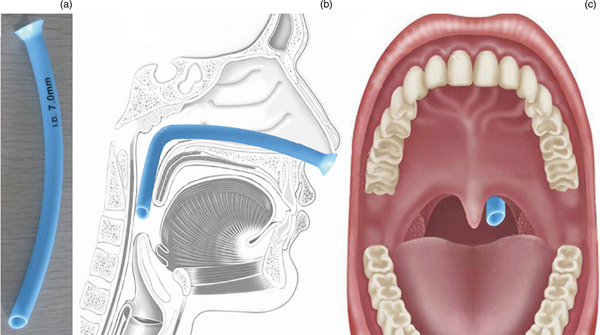
Nasal spray with 1 per cent ephedrine solution and 1 per cent tetracaine solution was applied thrice, to decongest and anaesthetise the nasal mucous membrane. A soft and elastic silicon nasopharyngeal tube (inner diameter of 7 mm; Figure 2a), with surface lubrication of liquid paraffin, was gently inserted into the wider anterior nostril until it reached the oropharyngeal cavity, via the nasal and nasopharyngeal cavities (Figure 2b). The position of the distal tube end was slightly beyond the free edge of the soft palate (Figure 2c). The nasopharyngeal tube was fixed with a length of new bandage at the anterior naris to prevent dislocation during sleep.
Diagram of nasopharyngeal tube insertion. (a) Soft, elastic silicon nasopharyngeal tube (inner diameter of 7 mm). (b) The tube was inserted into wider anterior nostril until it reached the oropharyngeal cavity, via the nasal and nasopharyngeal cavities. (c) The position of the distal tube end was slightly beyond the free edge of the soft palate.

Polysomnography was repeated after nasopharyngeal tube insertion and immobilisation. The examination procedure and data recording were conducted similarly to the first polysomnography. Nasopharyngeal tube insertion could eliminate the obstruction of the nasal cavity, nasopharyngeal cavity and retropalatal region during sleep. Thus, the apnoea-hypopnoea index for nasopharyngeal tube polysomnography was used as the diagnostic criteria for glossopharyngeal obstruction in all the patients.Reference Li, Wu, Bao and Shi10
Statistical analysis
Statistical analysis was performed using SPSS® software, version 16.0. Correlations between the hyoid cephalometrics and apnoea-hypopnoea index results of routine polysomnography and nasopharyngeal tube polysomnography were explored using simple linear correlation. Pearson correlation was performed when the linearity, independence, normality and homogeneity of the variances of the variables were met; otherwise, Spearman's coefficient was employed. Multiple linear regression analysis was used to further determine the relationship between hyoid cephalometrics and the apnoea-hypopnoea index.
Results
Patients’ demographics
A total of 96 adult patients (15 females and 81 males) with typical OSAHS symptoms, who were diagnosed with OSAHS, participated in the study. There were 5 patients with mild OSAHS, 23 with moderate OSAHS and 68 with severe OSAHS. Based on the polysomnography results, none of the patients had ‘positional OSAHS’. The data on age, body mass index and the apnoea-hypopnoea index for routine polysomnography are listed in Table 1.
Patients’ characteristics*

*n = 96. N/A = not applicable; SD = standard deviation; BMI = body mass index; AHI = apnoea-hypopnoea index.
Hyoid parameters
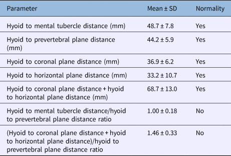
The single parameters of the hyoid, including the distances of the hyoid to the mental tubercle, prevertebral plane, mental tubercle coronal plane and mental tubercle horizontal plane, are presented in Table 2. The average value of the hyoid to mental tubercle distance was greater than that of the other single hyoid parameters. The data on these single parameters all fitted the normal distribution. The compound parameters, namely the (hyoid to coronal plane distance plus hyoid to horizontal plane distance)/hyoid to prevertebral plane distance value, and the hyoid to mental tubercle distance/hyoid to prevertebral plane distance value, did not fit the normal distribution.
Hyoid bone parameters*

*n = 96. SD = standard deviation.
Apnoea-hypopnoea index before and after tube insertion
The apnoea-hypopnoea index of all the patients decreased to varying degrees after nasopharyngeal tube insertion (Table 1). The OSAHS symptoms, such as feelings of sleepiness and fatigue, improved in most patients (71 out of 96). After nasopharyngeal tube insertion, there were 15 patients with mild OSAHS, 30 with moderate OSAHS and 36 with severe OSAHS. The remaining 15 patients no longer met the diagnostic criteria for OSAHS after nasopharyngeal tube insertion.
Correlation between hyoid parameters and apnoea-hypopnoea index
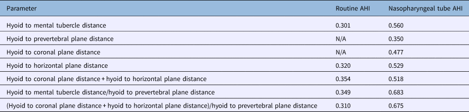
Simple linear correlation showed that the hyoid to mental tubercle distance/hyoid to prevertebral plane distance value had the most significant positive correlation with the apnoea-hypopnoea index for nasopharyngeal tube polysomnography (r = 0.683). The (hyoid to coronal plane distance plus hyoid to horizontal plane distance)/hyoid to prevertebral plane distance value was also significantly correlated with the apnoea-hypopnoea index for nasopharyngeal tube polysomnography (r = 0.675).
Among the single parameters, hyoid to mental tubercle distance had the most significant positive correlation with the apnoea-hypopnoea index for nasopharyngeal tube polysomnography (r = 0.560). The coefficients for the correlations of the other hyoid parameters with the apnoea-hypopnoea index are shown in Table 3.
Correlations between apnoea-hypopnoea index and hyoid bone parameters

Data represent r values. AHI = apnoea-hypopnoea index; N/A = not applicable.
The apnoea-hypopnoea index for nasopharyngeal tube polysomnography could be converted into a normal distribution with logarithmic transformation. Multiple stepwise linear regression revealed that the logarithmically transformed apnoea-hypopnoea index for nasopharyngeal tube polysomnography was associated with the hyoid to mental tubercle distance (b = 0.047; p < 0.001) and hyoid to prevertebral plane distance (b = −0.016; p < 0.05). The hyoid to coronal plane distance and hyoid to horizontal plane distance were excluded. Simple regression showed that the hyoid to mental tubercle distance/hyoid to prevertebral plane distance value (b = 1.275; p = 0.001) was associated with the logarithmically transformed apnoea-hypopnoea index for nasopharyngeal tube polysomnography. The hyoid to mental tubercle distance revealed a strong correlation with the hyoid to coronal plane distance plus hyoid to horizontal plane distance value (r = 0.931; p < 0.001).
Discussion
Retroglossal obstruction is mainly caused by glossoptosis or upper airway stenosis of the tongue base region, or both. The genioglossus is the major upper airway dilator that maintains sufficient ventilation in the retroglossal area. The genioglossus is a fan-shaped extrinsic tongue muscle that originates from the mental spine of the mandible, and inserts into the hyoid bone and tongue bottom. A more inferior and posterior hyoid bone is assumed to represent a more inferior and posterior genioglossus and tongue base. With the loss of genioglossus tone, patients with abnormal hyoid bone locations seem more prone to retroglossal stenosis during sleep than those with normal hyoid bone locations.
In order to prove this hypothesis, one must first eliminate the retropalatal obstruction. Thus, a nasopharyngeal tube made of soft silicone was used in this study. After nasopharyngeal tube insertion, an artificial upper airway passage was established. The new passage ensured the patency of the path from the nasal cavity to the nasopharyngeal and retropalatal airway. If the patient still obtained a high apnoea-hypopnoea index for nasopharyngeal tube polysomnography and experienced obvious obstructive sleep apnoea, the site of airway obstruction could be deduced as the retroglossal region, just below the tip of the nasopharyngeal tube. With sufficient decongestion and anaesthesia of the nasal mucous membrane, patients could tolerate nasopharyngeal tube insertion well.
A previous study showed that nasopharyngeal tube insertion could improve sleep efficiency and architecture as a result of the improvement of apnoea and hypoxemia during sleep.Reference Li, Wu, Bao and Shi10 Nasopharyngeal tube polysomnography can be used as a simple and effective method for localising glossopharyngeal airway obstruction.Reference Li, Wu, Bao and Shi10, Reference Li, Wu, Bao and Shi11 Moreover, the method can predict the therapeutic effect of UPPP.Reference Li, Wu, Bao and Shi10 In this study, the decrease of apnoea-hypopnoea index after nasopharyngeal tube insertion was similar to that reported in the literature.Reference Li, Wu, Bao and Shi10, Reference Li, Wu, Bao and Shi11
Significant correlations were found between the apnoea-hypopnoea index for nasopharyngeal tube polysomnography and all the cephalometric measurements of the hyoid bone. However, the (hyoid to coronal plane distance plus hyoid to horizontal plane distance)/hyoid to prevertebral plane distance value had a higher correlation coefficient than the distances of the hyoid to the mental tubercle coronal plane, the mental tubercle horizontal plane and the prevertebral plane. Compared with the single hyoid cephalometric parameters, the ratio depicted a more accurate location of the hyoid bone in the upper respiratory tract. This more accurate method of description might be the main reason for the stronger correlation.
In theory, an inferior and posterior hyoid bone also represented a longer distance from the hyoid to the mental tubercle. The strong correlation (r = 0.931) between the hyoid to mental tubercle distance and the hyoid to coronal plane distance plus hyoid to horizontal plane distance value confirmed this hypothesis. This observation may explain why the hyoid to the mental tubercle distance/hyoid to prevertebral plane distance also had a strong significant correlation with the apnoea-hypopnoea index for nasopharyngeal tube polysomnography, as had the (hyoid to coronal plane distance plus hyoid to horizontal plane distance)/hyoid to prevertebral plane distance value.
A higher (hyoid to coronal plane distance plus hyoid to horizontal plane distance)/hyoid to prevertebral plane distance value, and a higher hyoid to mental tubercle distance/hyoid to prevertebral plane distance value, both represented more inferior and posterior hyoid bones. They also implied a narrow retroglossal area and a long glossopharyngeal airway. The strong correlations showed that a hyoid bone with a more inferior and posterior position usually indicated a higher apnoea-hypopnoea index for nasopharyngeal tube polysomnography. In other words, OSAHS patients with a more inferior and posterior hyoid bone position seemed highly prone to retroglossal stenosis and obstruction. However, this result was not consistent with the findings of a previous study,Reference Vieira, Itikawa, de Almeida, Sander, Fernandes and Anselmo-Lima8 which showed that a more anterior and inferior position of the hyoid bone was more likely to lead to a respiratory disorder in children. This inconsistency may be because of the different age groups of the subjects.
Given the movement of the hyoid and tongue base toward the mandible, hyoid suspension could lengthen the hyoid to prevertebral plane distance, and shorten the hyoid to coronal plane distance, hyoid to horizontal plane distance and hyoid to mental tubercle distance in OSAHS patients. The values of (hyoid to coronal plane distance plus hyoid to horizontal plane distance)/hyoid to prevertebral plane distance, and hyoid to mental tubercle distance/hyoid to prevertebral plane distance, would then decrease. Based on the findings, these cephalometric changes provide us with a good explanation of how hyoid suspension alleviated the retroglossal obstruction. In this study, all the patients with a high nasopharyngeal tube apnoea-hypopnoea index underwent both UPPP and hyoid suspension. The pulling force of the suture that made the hyoid bone and mandible closer was different. The patients with a high hyoid to mental tubercle distance/hyoid to prevertebral plane distance value, and (hyoid to coronal plane distance plus hyoid to horizontal plane distance)/hyoid to prevertebral plane distance value, always received a stronger pulling force in hyoid suspension surgery.
We found a strong but not complete correlation between the compound parameters and the apnoea-hypopnoea index for nasopharyngeal tube polysomnography. The incomplete correlation indicates that the hyoid bone and tongue base locations do not solely affect retroglossal obstruction. The size of the lingual tonsils and lower portion of the palatine tonsils, the degree of genioglossus tone loss, the small transversal diameter of the retroglossal region, and some other unknown abnormities, are all influencing factors. In this study, we could only exclude the known and definite influencing factors, such as hypertrophic lingual tonsils, lower palatine tonsils and hypopharyngeal tumours. These severely abnormal anatomical factors could considerably affect the study results. Electronic laryngoscopy or indirect laryngoscopy was required to exclude the patients with the retroglossal abnormalities mentioned above prior to the evaluation.
In most OSAHS patients, the retropalatal region is the narrowest upper airway section. The apnoea-hypopnoea index is mainly determined by retropalatal obstruction and secondarily affected by retroglossal obstruction. Our data showed a weak correlation (r = 0.310) between the cephalometric measurements of the hyoid bone and the apnoea-hypopnoea index for routine polysomnography. Given the weak correlation, hyoid bone cephalometrics could not be used to predict the apnoea-hypopnoea index associated with retropalatal obstruction. Cephalometric hyoid bone measurements are only suitable for evaluating retroglossal obstructions.
Lateral films can show the structural and morphological changes of the soft and bone tissues surrounding the glossopharyngeal airway, and provide a reference for treatment planning. However, in light of the weak but extra radiation, lateral films and cephalometrics are not recommended for patients with a low routine or nasopharyngeal tube apnoea-hypopnoea index. These patients rarely suffer from significant retroglossal obstruction.
In summary, the compound hyoid cephalometric parameters evaluated – namely, the hyoid to mental tubercle distance/hyoid to prevertebral plane distance, and the (hyoid to coronal plane distance plus hyoid to horizontal plane distance)/hyoid to prevertebral plane distance – are valuable for predicting retroglossal obstruction severity in OSAHS patients.
• Hyoid bone position is significantly correlated with retroglossal obstruction in obstructive sleep apnoea hypopnea syndrome (OSAHS) patients
• The OSAHS patient with a more inferior and posterior hyoid bone position is more prone to retroglossal stenosis and obstruction
• The OSAHS patient with a longer hyoid to mental tubercle distance is more likely to have retroglossal obstruction as well
• The OSAHS patient who has a longer hyoid to mental tubercle distance also has a hyoid bone with a more inferior and posterior position
Acknowledgement
Research for this paper was supported by The Natural Science Foundation of Liaoning Province (grant number 2015020441).
Competing interests
None declared