

Introduction
Heliura Butler, Reference Butler1876 belongs to the subtribe Ctenuchina (Lepidoptera: Erebidae: Arctiinae: Arctiini) (Jacobson and Weller Reference Jacobson and Weller2002; Zahiri et al. Reference Zahiri, Kitching, Lafontaine, Mutanen, Kaila and Holloway2010) and comprises 40 valid species, many of them endemic to the Amazonian forest. The genus has never been revised, and it is clearly polyphyletic (personal observation). Hampson (Reference Hampson1898) characterised and divided it in two sections, but most of the species that are currently placed in Heliura were described only in the second decade of the 20th century.
During the revision of Heliura (L.R.P., in preparation) and preparation of manuscripts on the Ctenuchina from French Guiana (J.-A.C., in preparation), five new species from the Amazon were identified as belonging to this genus. They are here described to facilitate the upcoming revision and phylogenetic studies on Heliura.
Material and methods
Specimens were studied following standard methodologies (Winter Reference Winter2000; see also Pinheiro and Duarte Reference Pinheiro and Duarte2010). Genitalia of both sexes were dissected and immersed in a solution of 10% potassium hydroxide (KOH) at room temperature for ~16 hours. The abdominal pelts and genitalia were stored in small plastic vials with the same number added to the specimen, except in one case for which we prepared a slide, following the methodology described in the literature (Robinson Reference Robinson1976). Photographs of genitalia were taken in a stereomicroscope Zeiss Discovery V20 with a camera attached, using the Axiovision Vs40 (Zeiss International, Jena, Germany) and CombineZP (Alan Hardley, United Kingdom) software.
The acronyms used in this study are the following: (AMNH) American Museum of Natural History, New York, New York, United States of America; (AS) private collection of Anthony Seguna, Naxxar, Malta; (BH) private collection of Bernard Hermier, Rémire, French Guiana; (BMNH) Natural History Museum, London, United Kingdom; (JAC) private collection of J.-A. Cerda, Régina, French Guiana; (LACM) Los Angeles County Museum of Natural History, Los Angeles, California, United States of America; (ML) private collection of Michel Laguerre, Léognan, France; (MPEG) Museu Paraense Emílio Goeldi, Belém, Brazil; (MNHN) Muséum National d’Histoire Naturelle, Paris, France; (PhC) private collection of Philippe Collet, Caen, France; (PS) private collection of Paul Sammut, Rabat, Malta; (RMNH) Naturalis Biodiversity Center, Leiden, The Netherlands. The types are deposited at the institutions of origin.
Some morphological terms were abbreviated as follows: abdominal segment (A); abdominal tergite (T); abdominal sternite (S). The nomenclature of genitalia follows Klots (Reference Klots1956). The verbatim label data of each specimen is separated by semicolons, while commas separate different lines in the same label, and a forward slash separates different labels from the same specimen. Whenever additional information was added to label data, this was done between square brackets.
In Ctenuchina, males are much more common in collections than females. For this reason, and because of the known sexual dimorphism that occurs in many genera, they were preferably used for the description of the new species.
Barcoding methodology. One species, Heliura beneluzi new species was barcoded. The procedure followed to the protocols described in Vaglia et al. (Reference Vaglia, Haxaire, Kitching, Meusnier and Rougerie2008) and Decaëns and Rougerie (Reference Decaëns and Rougerie2008). We also provided the barcode index number (BIN) for this species, as proposed by Ratnasingham and Hebert (Reference Ratnasingham and Hebert2013).
Results and discussion
A preliminary investigation has revealed that Heliura is involved in a complex polyphyletism linking many genera of Ctenuchina, with species incorrectly placed in the genus, and others placed in other genera that seem to belong in it. Comparative analysis of morphological characters revealed that a few species of Delphyre Walker, 1854, Eucereon Hübner, [1818], Telioneura Felder, 1874, Ptychotricos Schaus, 1892, and Mesocerea Hampson, Reference Hampson1898 could be more closely related to Heliura rhodophila (Walker, 1856) than to the type species of the genera in which they are currently placed. However, with the possible exceptions of Mesocerea and Ptychotricos, which contain two and three species, respectively, the type species of these genera clearly do not belong to Heliura. As discussed in Pinheiro and Duarte (Reference Pinheiro and Duarte2013), the type species of Delphyre is probably not an arctiine, but a Lithosiinae. This may also be the case of the type species of Telioneura, T. glaucopis Felder, 1874, which is apparently an Arctiina. The case of Eucereon is more complicated, because it comprises hundreds of species, the habitus of most of which – including of E. archias (Stoll, 1790), its type species – is somewhat similar to that of Heliura. However, a comparative analysis failed to find any particular affinities of both genera beyond wing and abdomen colour patterns. The male genitalia of species of both genera is particularly dissimilar: most species of Eucereon, including E. archias, have a characteristic pair of dorsal projections between tegumen and uncus, which are absent in Heliura.
Few characters may be considered exclusive to Heliura, all of them from the genitalia: long lateral setae on the proximal portion of the uncus; finger-like paired projections arising from the sacculus region of the valvae, with a few short setae on their tips; T9 of females fully sclerotised; and the intersegmental membrane seven to eight of males unmodified. These are present in most species of Heliura already assigned to species groups within the genus. There are, however, species ambiguously placed in Heliura that seem to belong to other genera. Their placement has not yet been determined.
So far, three species groups of Heliura include new species: the H. marica (Cramer, 1775), H. thysbe (Möschler, 1877), and H. thysbodes Dognin, 1914 species groups. They are characterised below, together with the description of their new species. Three of the five new species were described solely based on males. In one case (H. laguerrei new species) the female is unknown. In all other cases, however, this happened because the association of males and females was impossible to determine on morphological grounds. In fact, we found more species represented only by females than only by males, but the uncertainty of the associations prevented us from describing the females of certain species. These species with problematic sex association belong to the Heliura stolli Rothschild, Reference Rothschild1912 species group, as discussed below.
Heliura marica species group
This species group includes six species – H. marica, H. amazonica (Rothschild, Reference Rothschild1912), H. postcoerulea (Rothschild, Reference Rothschild1912), H. quadriflavata Kaye, 1919, H. flavopunctata (Dognin, 1911), and H. gigantea (Druce, 1900). These species are clearly linked by their habitus, and, except for H. flavopunctata, which could not be dissected, recharacterised by having long valvae, with the proximal end cylindrical and a long aedeagus with few terminal cornuti. Additionally, except for H. quadriflavata and H. gigantea, they also have peculiar green scales found in no other species of Heliura. A new species for this group is described below.
Heliura laguerrei Pinheiro and Cerda, new species
Holotype. Male: FRENCH GUYANA, Regine, Rte. a Belizon, km 24, el. 250 m. 8 February 1991, coll. C. Snyder (at lights) (AMNH). Four paratypes, males: FRENCH GUYANA, / Mtgne de Kaw, Patawa PL, 02.i.1998, J.-A. Cerda / , / 4765 / , / Gen. no. 512 / (one male) (MNHN); / Mtgne de Kaw, 30.vi.1992 Pk 47, PL J.-L.Giuglaris, coll. J.-A. Cerda / , / 4766 / (one male); 12.viii.[19]94, piste de Kaw, PK38, M. Laguerre leg. / , / gen. ML982 / (one male) (ML); BOLIVIA: / 5-XI-2000, 800 m, Rte Caranavi-Quiquibey, Environ de la Cascada, M. Laguerre leg. / , / gen. ML2128 / (one male) (ML).
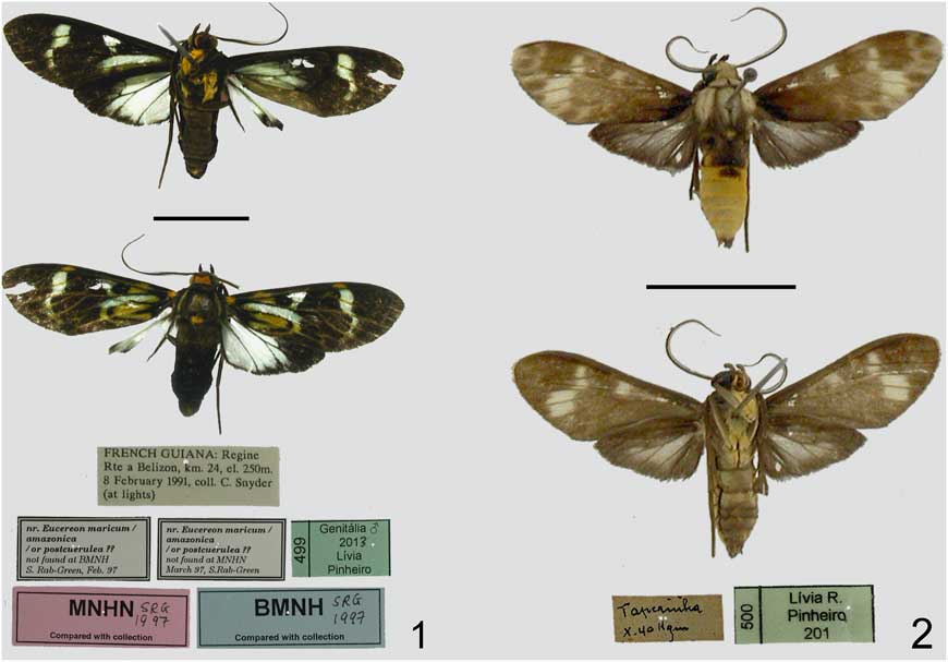
Diagnosis ♂. Heliura laguerrei is closely related to Heliura marica (Cramer, 1775), H. postcoerulea, and H. amazonica (the two last species were illustrated in Draudt 1915, plate 24, row i). Heliura laguerrei differs from the other species by having darker forewings and genitalia, frontoclypeus and scape entirely brown, and without sparse green and/or orange scales, and dorsal axillary scales on the forewings orange instead of green.
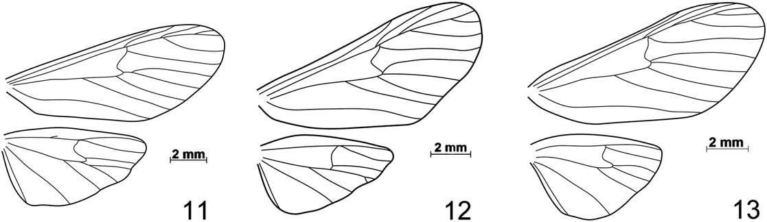
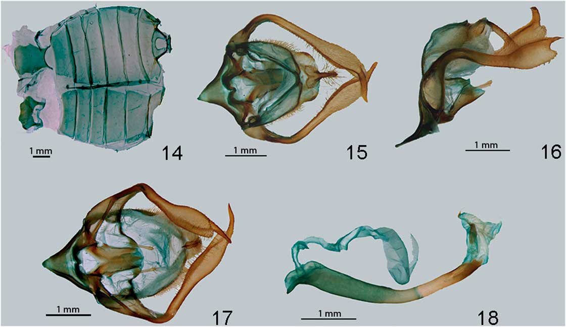
Description ♂. Head. Proboscis light brown. Labial palpi entirely brown, exceeding vertex. Frontoclypeus approximately as high as wide, entirely brown, smooth-scaled ventrally and rough-scaled dorsally. Vertex entirely brown. Postocciput and dorsal cervical area with orange scales. Scape, pedicel, and flagellomeres uniformly brown. Thorax. Almost entirely brown dorsally, with dark yellow scales at the posterior end of mesoscutellum. Metascutellum brown. Patagiae predominantly brown, anterior margin green. Tegulae predominantly brown with a green longitudinal stripe. Epimera and episterna predominantly white, brown at the dorsal end. Procoxae roseate anteriorly and light orange laterally. Mesocoxae and metacoxae brown anteriorly and light yellow laterally. Profemora and mesofemora almost entirely brown, with sparse white scales. Metafemora white internally and brown on the remaining surfaces. Tibiae and tarsi of all legs entirely brown, except for the metatibiae, with a light spot on each proximal end. Forewings. Entirely scaled. Dorsal surface: axillary scales orange. Veins predominantly covered with brown scales, some parts with green or orange scales. Proximal part of the wings with green, hyaline and dark brown areas; distal part mainly dark brown, with a hyaline area between cells R5–M1 and M3–CuA1. Ventral surface dark brown with the same hyaline areas as dorsal surface. Venation shown in Figure 11: R1 arising near the distal end of the discal cell. R2 arising very close to R3. M2 and M3 arising from a common stalk. Forewings. Entirely scaled. Veins covered with dark brown scales. Cells M1–M2, M2–M3, and M3–CuA1, and outer margin of cells CuA1–CuA2, CuA2–1A, and 1A–2A also with dark brown scales. The rest of the wing surface with white-blue scales. Venation as in Figure 11; Sc vestigial; M2 arising at a small distance from the stalk of M3 and CuA1. M3 and CuA1 many times longer than their stalk. Abdomen. Entirely brown. T8 and S8 sclerotised. Intersegmental membrane seven to eight well developed, forming a structure probably of glandular function. Male genitalia. Aedeagus of approximately constant width, smooth, curved dorsally, and somewhat to the right. Ejaculatory duct inserted dorsally. Coecum turned downwards, somewhat long. Vesica with a small number of spiniform cornuti. Aedeagus approximately as long as the genital capsule when the vesica is everted. Saccus wide and acute, symmetrical. Tegumen with setae, considerably wider than vinculum, with the ventral end not covering the proximal portion of valvae. Anterior margin of tegumen with an indentation. Uncus simple, laterally compressed, with the basis with a few setae, and having approximately three times the width of the lobe, apex acute. Transtilla sclerotised. Juxta sclerotised, glabrous, somewhat like the second abdominal sternite. Valvae subequal, exceeding uncus, with setae and scales. Sacculus with one pilose projection each side. Cucullus and valvula distinct, the former longer and more sclerotised than the former. Female genitalia. Unknown.
Etymology. This species is dedicated to Michel Laguerre, who kindly shared information with the first author on Heliura from French Guiana.
Heliura thysbe species group
The H. thysbe species group is here established with three other species: H. thysbodes Dognin, 1914, H. ockendeni Rothschild Reference Rothschild1912, and H. beneluzi new species. It is probably closely related to the H. stolli species group (see more details below), and its species are characterised by the presence of grey scales on the vertex, and a complete vein Sc on the hindwings. A new species for the group is described below.
Heliura beneluzi Pinheiro and Cerda, new species
Holotype male: BRAZIL, Pará, [Fazenda] Taperinha, / x.[19]40, Hagmann / , / LRP 500 / (MPEG) (one male); Nine paratypes, five males and three females. BRAZIL, Acailandia, Reserva Biologica Integral de Gurupi Lt -4.0014° Ln -46.8372° 275 m T. Decaens, 4/17/2010 / , BC-INCT0940, / Barcode of Life, DNA voucher specimen, Smple ID: BC-INCT0940, BOLD Proc. ID: INCTA940-10 / , / Heliura beneluzi M. Laguerre det 2014 (one male) (MPEG); Pará, [Santarém], [Fazenda] Taperinha, Setembro 1942, Hagmann (MPEG) (one female); Pará, [Santarém], [Fazenda] Taperinha, x.[19]40, Hagmann (MPEG) (one female); PERU, Manu, Pakitza, 11°56'S 71°18'W, 250 m, 6–8.ix.1989, Trail 1, mkr. 8, UV light #6, D. Adamski and M. Epstein (USNM) (one male); FRENCH GUYANA, / S. L. du Maroni, P. Cr. Serpent Pk.15+7.5, 18.x.2006, J.-A. Cerda / , / 6527 (one male) (JAC); / Mtgne de Kaw, Patawa PL, 06.vi.2000, J.-A.Cerda / , / 4986 (one male) (MNHN); Bélizon, Pk. 15+15 PL, 02–03.ii.2006, F. Bénéluz, coll. J.-A. Cerda / , / 6526 / , / Gen. no. 455 / (one male) (JAC); 357 / N2 pk41 4°38'N 53°22'W, 23.v.2001, B. Hermier [piège lumineux] / , / Hermier. no. 19544 / , / Photo 05285 RL www.guianensis/ (one male) (BH); PL Patawa, 28.viii.2002, J.-A. Cerda / , / 4985 (one female) (JAC).
Diagnosis ♂ and ♀. Heliura beneluzi is a very distinctive species, with the darker elements on the dorsal side of the forewing much less defined than those found in others species of the genus. Moreover, its yellow abdomen is also remarkable in the genus.
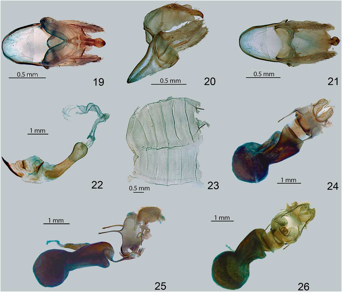
Description ♂ and ♀. Head. Proboscis light brown. Labial palpi predominantly brown, exceeding the vertex; third palpomere twice longer than wide. Frontoclypeus slightly longer than wide, ventrally smooth-scaled (brown scales), and dorsally rough scaled (grey scales). Vertex grey. Postocciput and dorsal cervical area yellow. Scales situated immediately after the antennal socket grey. Scape brown on the external and posterior margins, and grey on the anterior and internal margins. Flagellomeres predominantly brown, the proximal flagellomeres grey on the internal margin, and the most distal white dorsally. Thorax. Predominantly grey dorsally. Mesonotum brown laterally and yellow posteriorly. Metascutellum brown. Patagia grey with brown margins. Tegulae predominantly grey, with brown spots anteriorly and posteriorly. Epimera and episterna predominantly brown, dorsal edge yellow. Coxae light yellow anteriorly and laterally. Femora brown dorsally and white ventrally. Tibiae and tarsi entirely brown. Forewings. Entirely scaled. Dorsal surface: veins and cells predominantly covered with brown scales. Four hyaline areas on proximal portion of cells R5–M1 to M3–CuA1, and one inside discal cell. Cells near apex of the wing with areas more lightly coloured. Ventral surface uniformly brown, with marks of the hyaline areas described above. Venation as in Figure 12: R1 arising near the distal end of the discal cell. R2 arising slightly closer to R3 than to R1. M2 and M3 arising very near each other, without a common stalk, or with a small stalk at their base. Hindwings. Entirely scaled. Veins and cells covered with brown scales, more densely on the margins than on the centre. Venation as in Figure 12; Sc absent. M2 arising at a small distance from the stalk of M3 and CuA1. M3 and CuA1 many times shorter than their stalk. Females with two frenular bristles. Abdomen. T1–3 yellow laterally and brown dorsally; T1 predominantly brown, yellow on T3. In males, S2 white or brown ventrally and white laterally; S3–8 brown; in females, S2 white, S3–4 brown ventrally and white laterally, S5–7 brown with white spots on the anterior border; S8 brown. T8 and S8 sclerotised in males and females. Male genitalia. Aedeagus approximately same width across its length, about 1.5 the length of the genital capsule with the vesica everted. Ejaculatory duct inserted dorsally. Coecum rounded. Vesica with minute cornuti and one large apical cornutus, plus a small sclerotised plate. Saccus wide, rounded, and symmetrical. Tegumen glabrous, considerably wider than the vinculum, with its ventral edge not covering the proximal portion of valvae. Anterior margin of tegumen indented; posterior margin with two large sclerotised projections, both glabrous and smooth. Uncus simple, basis with approximately the same width as the lobe, with few setae. Transtilla and juxta sclerotised, glabrous, and without spines. Valvae subequal, reaching uncus. Sacculus with a small pilose projection on each side. Distal portion of valvae bifurcate, the dorsal projection shorter and more sclerotised than the ventral projection. Female genitalia. Papillae anales well developed. Ostium centralised, lamella antevaginalis with small setae. Ductus bursae sclerotised, without signa. Corpus bursae longer than wide, with large signa covering almost its entire internal surface. Ductus seminalis arising from the bursa.
Distribution. This species is known from the Amazonian forest, from French Guiana to the Peruvian lowland forests.
Etymology. This species is dedicated to Frédéric Beneluz, who although is mainly interested in Saturniidae, kindly collected arctiines for J. -A.C. for the past 25 years.
Heliura stolli species group
This species group is here defined comprising three valid species and three new species: H. stolli Rothschild, Reference Rothschild1912, H. parcipuncta (Hampson, Reference Hampson1914) new combination, H. pieroides (Rothschild, Reference Rothschild1912) revised combination, H. crameri new species, H. nigriventris new species, and H. pauloi new species. Based on the study of these six species, plus almost all species currently placed in Heliura and Delphyre, it seems reasonable to admit that they form a cohesive group of species characterised by the presence of membranous pouches on the internal surface of the tegumen, which is absent in all other species of Heliura.
However, as already mentioned in this paper, for many species it was not possible to ascertain the correspondence between males and females. It appears that this problem will only be solved by rearing immatures or with molecular studies involving the primary types. A total of eight species were identified as belonging to the H. stolli species group. Two of them were tentatively associated with males based on external characters. Another one is H. pieroides Rothschild, Reference Rothschild1912, known only from its female lectotype. The five left remain unnamed and undescribed, until further studies can be carried out with the aim of solving this problem. Due to the difficulty of identification in this group, a key is provided below:

Descriptions and redescriptions of the Heliura stolli species group
Heliura crameri Pinheiro and Cerda, new species
Holotype. Male: FRENCH GUYANA, / Roura, Kaw Mtn., km 45, el. 225 m, 24.i.1991, C. Snyder col. (at lights) / , / LRP557 (AMNH). Twenty five paratypes, 19 males and six females. FRENCH GUYANA, / Régine, Rte a Belizon, km 24, el. 250 m, 8.ii.1991, C. Snyder col. (at lights) / , / LRP457 (one male) (AMNH); / 228 / , / N2 pk79, 4°24'N 53°18'30''W, 23.ix.1995, B. Hermier, [piège lumineux] / , / Hermier no. 7562 / , / J.-A. Cerda coll. / , / 4836 / , / Gen. no. 448 (one male) (JAC); Mtgne de Kaw, Pk.37 PL 26.viii.2000, G. Miral / , / 4837 (one male) (JAC); Camp Changement / , / lumière, 14.xii.2003 (one male) (PhC); Camp Changement / , / lumière, 12.xii.2003 / P. Collet col. (one male) (PhC); Camp Changement / , / lumière, 25.i.2004 (one male) (PhC); Rés. des Nouragues / , / Inselberg turbine - UV, 5.ix.2010 (one male) (PhC); Cayenne, Kaw road, nr. Fourgassie 275 m., 04°39.232'N; 52°18.061'W, 18.xi.2007, P. Sammut (one male) (PS); Cayenne, Patawa 275 m., 04°32.024'N, 52° 07.684'W, 20.xi.2007, P.Sammut (two males) (PS); Cayenne, St. George de l’Oyapock, Saut Maripas, 03°48.153'N, 51° 53.138'W, 24 / 26.xi.2007, P. Sammut (one male) (PS); Cayenne, St. George – Regina rd., 04'09.834'N, 52'06.766'W, 27.xi.2007, P. Sammut (one male) (PS); Cayenne, Tonnegrande alt. 2 m, 04°49.851'N, 52°26.790'W., 2–4.xi.2007, 2×125w M.V. hg:1, A.Seguna col. (one male) (AS); Cayenne, Patawa 275 m, 04°32.024'N, 52° 07.684’W, 20.xi. 2007, A. Seguna (two males) (AS); Cayenne, Regina road, S. Cacao, 04'32.469'N, 52'22.840'W, 11.xi.2007, A. Seguna (one male) (AS); 30.vii.1991, Piste de Kaw PK 11, M. Laguerre col. / , / Genit. ML2085 (one male) (ML); 30.vii.2003, 200 m, Piste de Kaw PK 37.5+2, 4°33.691'N 52°08.391'W, M. Laguerre col. (one male) (ML); 10.ii.2013, 200 m, Piste de Kaw PK 28+2, O. Félis leg., coll. M. Laguerre (one male) (ML); Gr. Inini, Saut Batardeau PL, 29–30.ix.1997, M. Duranton, coll. J.-A.Cerda / , / 4840 (one female) (JAC); Mtgne de Kaw, Patawa PL, 02.xi.1992, J.-A.Cerda / , / 4839 (one female) (MNHN); Mtgne de Kaw, Patawa 09.vi.1994, J.-A. Cerda / , / 4835 (one female) (JAC); Cayenne, St. George, Regina road, 04º09.834'N 52º06.766'W., 27.xi.2007, 2×125w M.V. light, A. Seguna col. (one female) (AS); 20.vii.2003, 270 m, Piste de Kaw PK 37.5, 4°32.908 N 52°08.728 W, M. Laguerre col. (one female) (ML); 15.ii.1999, Piste de Bélizon PK 27, M. Laguerre col. (one female) (ML).
Diagnosis ♂ and ♀. Heliura crameri differs from most of the other species of the H. stolli complex by the yellow, instead of red, surface of the procoxae. This character is shared with H. pieroides, from which it differs mainly in the grey, as opposed to yellow, metascutellum, and the T8 and abdominal sternites entirely brown.
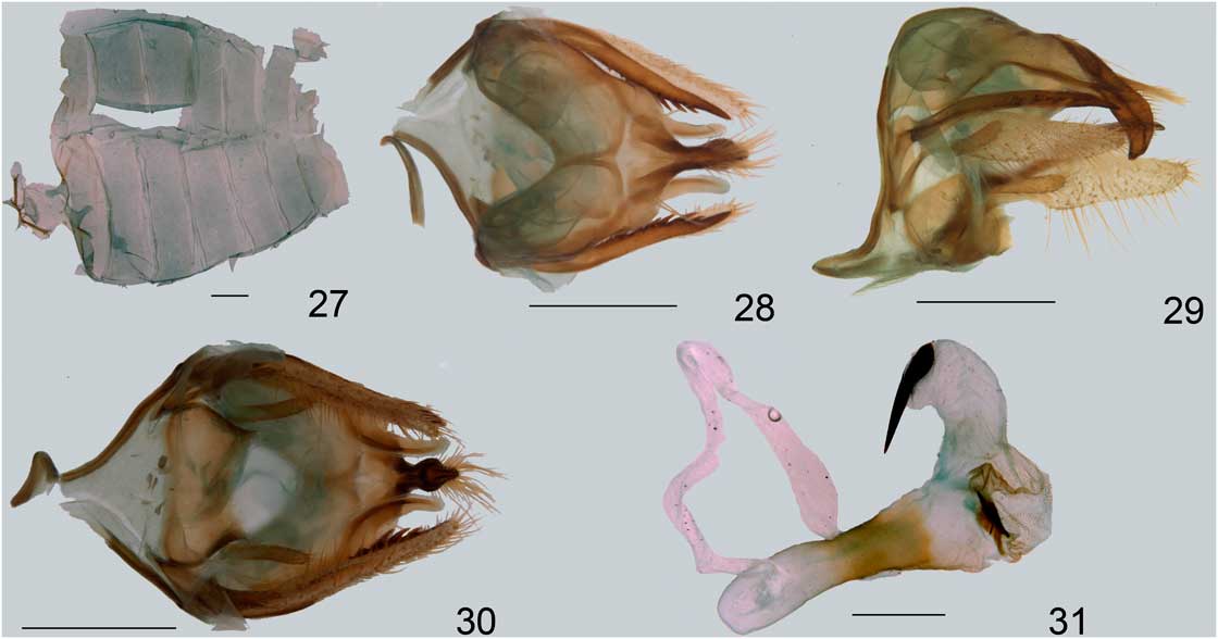
Description ♂ and ♀. Head. Proboscis light brown. Labial palpi dark brown, exceeding the vertex; third palpomere longer than wide. Frontoclypeus entirely dark brown, longer than wide, ventrally smooth-scaled, and dorsally rough-scaled. Vertex grey. Postocciput and dorsal cervical area yellow ochre. Scales posterior to the antennal socket white. Scape and pedicel predominantly brown, yellow anteriorly. Flagellomeres dark brown, except for the first two at base, grey on the inner surface. Thorax. In dorsal view, thorax brown and grey. Posterior portion of mesoscutellum light grey laterodorsally and brown dorsally. Metascutellum grey. Patagia brown, with anterior and lateral margins beige. Tegulae beige with two brown areas of approximately the same size, one on the external margin, the other on the internal margin. Epimera and episterna with long brown scales. Procoxae yellow ochre anteriorly and brown laterally. Profemora predominantly brown, yellow ochre proximally. Protibiae and tarsi brown. Mesolegs and metalegs with the same pattern, except for the outer surface of the coxae, yellow ochre instead of brown, and the proximal surface of the tibiae, with a greyish stripe. Forewings. Entirely scaled. Dorsal surface with veins predominantly covered with beige scales and dark brown spots of different shapes and sizes, forming four distinguishable groups: the most proximal with three spots; the next group with four spots; the third with 12 spots, comprising the discal area and the region immediately distal to it, and all cells of the wing; distalmost group with eight spots, separated in a subdistal group of three spots, and an apical group of five spots. External margin with dark brown scales anteriorly, and white scales posteriorly. Ventral face predominantly brown, except for four white areas between the discal cell and the apex, and a less conspicuous white area inside the discal cell. Venation: R2 arising at the same distance of the transversal vein and R3. M2 and M3 branching from the transversal vein very near each other, without a common stalk. Hindwings. Entirely scaled. Veins and most of the dorsal and ventral surfaces covered with brown scales. Discal cell and costal area immediately above it with white scales. Females with two frenular bristles. Venation; Sc present. M2 arising at a small distance from the stalk of M3 and CuA1. M3 and CuA1 slightly shorter than their stalk. Abdomen. Entirely brown. T8 and S8 sclerotised. Intersegmental membrane seven to eight of males unmodified, coremata absent. Male genitalia. Aedeagus smooth and straight, with approximately the same width across its entire length, longer than the genital capsule when with the vesica everted. Ejaculatory duct positioned dorsally. Coecum rounded. Vesica with many minute cornuti and a sclerotised plate with a few large cornuti; left surface with a sclerotised plate without cornuti. Saccus long, wide and somewhat acute, symmetrical. Tegumen considerably wider than the vinculum, with few setae, with the ventral part not covering the proximal portion of the valvae. Posterior margin of the tegumen with sclerotised projections. Internal surface of the tegumen with two membranous pouches. Uncus symmetrical, with few setae at its base and long setae at its mid portion. Apex sharp. Transtilla absent. Juxta glabrous, wider than high. Valvae subequal, reaching the apex of the uncus. Each sacculus with a small digitiform projection covered with few setae. Cucullus heavily sclerotised, bearing large spines on its inner surface. Valvula densely scaled, distinct from cucullus and longer than it. Female genitalia. Pheromone glands membranous and digitiform. Ostium centralised. Antevaginal and postvaginal lamellae sclerotised, the former covered by minute setae ventrally. Ductus bursae flat and sclerotised. Corpus bursae entirely covered by signa, those in the posterior end thinner than those positioned at the round part of the bursa. Seminal duct originating off the bursa, dorsally.
Distribution. This species is so far known only from French Guiana, though it is likely to occur in nearby areas considerably less collected, such as Suriname and the Brazilian states of Amapá and northern Pará.
Etymology. The specific epithet is homage to Pieter Cramer, noted amateur lepidopterist from the eighteenth century.
Remarks. Heliura crameri resembles H. perexcavata (Rothschild, Reference Rothschild1912), but other external and genitalic characters suggest that the latter may be the closest relative of H. excavata (Dognin, 1910), which does not belong to the H. stolli species group.
Heliura nigriventris Pinheiro and Duarte, new species
Holotype. Male: BRAZIL, / Pará, A. M. Moss / , / Rothschild bequest / (BMNH), / LRP605.
Diagnosis ♂. Heliura nigriventris has the unique combination of red anterior surface of the procoxae with completely brown abdominal sternites. It can also be distinguished from H. crameri, which also has all the abdominal sternites brown, by the dorsal lobe of the valvae shorter than the ventral lobe, and by the presence of two, instead of one, large cornutus. Additional differences in relation to other species with red procoxae are as follows: H. nigriventris presents ventral projections on the tegumen, which are absent in H. pauloi. The latter also possess the dorsal and ventral lobes of the valvae of approximately the same length. The genitalic difference between H. nigriventris and H. stolli is more subtle: the projections on the tegumen are present in both, but they are more rounded in H. stolli, and more finger-like in H. nigriventris.
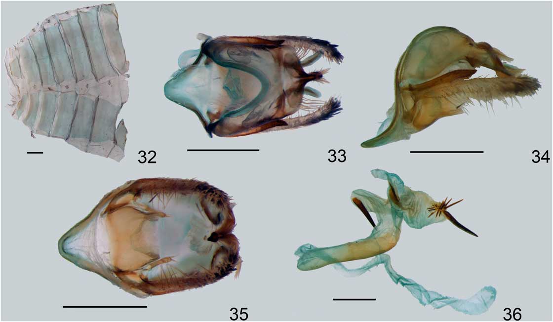
Description ♂. Head. Proboscis light brown. Labial palpi brown, exceeding the vertex; third palpomere twice longer than wide. Frontoclypeus longer than wide, ventrally smooth-scaled, and dorsally rough scaled, all of them dark brown. Vertex grey. Postocciput and dorsal cervical area light red. Scales situated posteriorly to the antennal socket grey. Scape and pedicel predominantly brown, but grey anteriorly. Flagellomeres predominantly brown, the proximal third grey on the internal margin. Thorax. Predominantly grey dorsally. Mesonotum brown and grey. Metascutellum brown. Patagia brown, with anterior and external margin beige. Tegulae predominantly beige, with the internal margin and a dorsal patch with dark brown scales. Epimera and episterna with long brown scales. Procoxae light red anteriorly. Profemora predominantly brown dorsally, and light red ventrally. Tibiae and tarsi brown. Mesolegs as prolegs, except for the tibiae, with a proximal yellow stripe. Metalegs with the following pattern: coxae light red anteriorly, femora almost entirely brown, tibiae and tarsi completely brown. Forewings. Entirely scaled. Dorsal surface predominantly covered with beige scales and dark brown spots of different shapes and sizes forming four distinguishable groups: the most proximal with three spots; the next group with four spots; the third with 12, comprising the discal area and the region immediately distal to it, and all cells of the wing; distalmost group with eight spots, separated in a subdistal group of three spots, and an apical group of five spots. External margin with dark brown scales anteriorly, and white scales posteriorly. Ventral surface predominantly brown, except for four white areas between the discal cell and the apex, and a distinct white area inside the discal cell. Venation: R2 arising at the same distance of the transversal vein and R3. M2 and M3 branching from the transversal vein very near each other, without a common stalk. Hindwings. Entirely scaled. Veins and most of the dorsal surface covered with brown scales. Discal cell and costal area immediately above it with white scales. Ventral surface brown at the margins, and white proximally and medially. Venation: Sc present. M2 arising at a small distance from the stalk of M3 and CuA1. M3 and CuA1 slightly shorter than their stalk. Abdomen. Entirely brown. T8 and S8 sclerotised. Intersegmental membrane seven to eight of males unmodified, coremata absent. Male genitalia. Aedeagus with approximately the same width across its whole length, smooth and straight, longer than the genital capsule when with the vesica everted. Ejaculatory duct positioned dorsally. Coecum rounded. Vesica with many minute cornuti, a sclerotised plate, an area with one long cornutus, and another area with a few small cornuti and one large cornutus. Saccus long, wide, and symmetrical. Tegumen glabrous, considerably wider than the vinculum, with the ventral part not covering the proximal portion of the valvae. Posterior margin of the tegumen with filiform sclerotised projections. Internal surface of the tegumen with two membranous pouches. Uncus symmetrical, with few very long setae at its base and smaller setae at its mid portion, more concentrated. Apex sharp. Transtilla absent. Juxta glabrous, wider than high. Valvae subequal, exceeding the apex of the uncus. Each sacculus with a small digitiform projection covered with few setae. Cucullus heavily sclerotised, bearing large spines on its inner surface. Valvula densely scaled, distinct from cucullus and longer than it. Female genitalia. Unknown.
Distribution. Heliura nigriventris is known only from Pará, Brazil.
Etymology. This species is named H. nigriventris because the brown abdominal sternites are the most obvious character distinguishing it from other species in the H. stolli species group with light red coxae.
Remarks. The male genitalia of H. nigriventris is remarkably similar to that of H. stolli, differing in only one character: the projections of the tegumen, which are more rounded and turned interiorly in H. stolli, while in H. nigriventris are more filiform and turned to the outside.
Heliura pauloi Pinheiro and Duarte, new species
Holotype. Male: PERU, Pasco, Pan de Azúcar, 8.vii.1961, F. S. Truxal (LACM) / dissection number LRP456. One paratype male, GUYANA, Tumatumari, Rio Potaro, iii.1912 (USNM), dissection number LRP536.
Conditions of the types. Holotype: wings slightly wore. Metathoracic legs lost. Abdomen dissected. Paratype: forewings slightly damaged. Abdomen dissected.
Diagnosis ♂. Heliura pauloi can be distinguished from H. crameri and H. pieroides by the red, instead of yellow, coxae. It also differs from H. nigriventris by the abdominal sternites, entirely brown in the latter and mostly red in H. pauloi. Moreover, the latter differs from H. stolli in the following characters: hindwings with hyaline appearance below the discal cell, dorsal lobe of the valvae as long as the ventral lobe, and ventral projections on the tegumen absent in H. pauloi, as opposed to hindwing more predominantly covered with dark brown scales, dorsal lobe of the valvae much shorter than the ventral lobe, and ventral projections on the tegumen much shorter than the ventral lobe in H. stolli
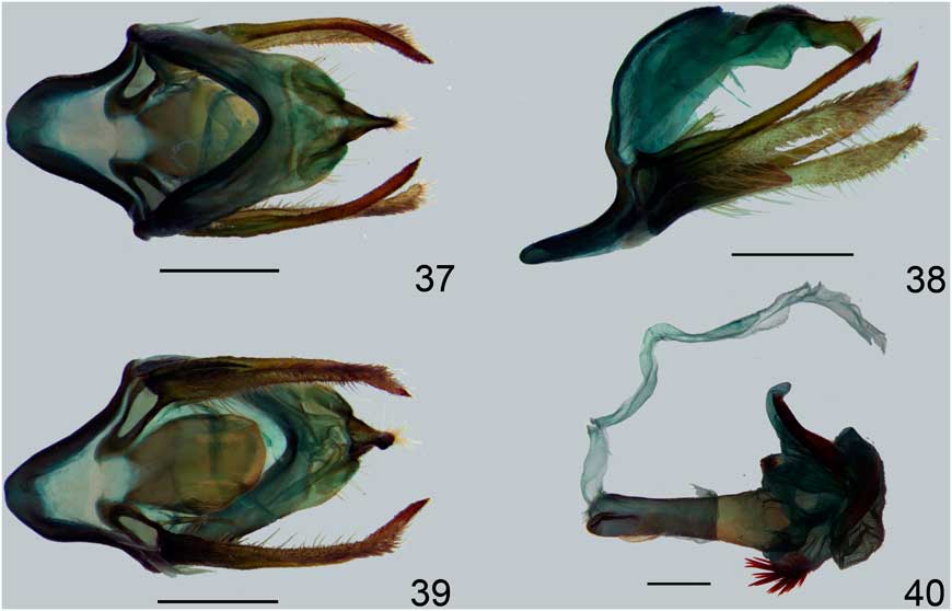
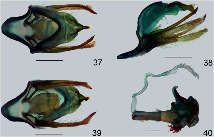
Description ♂. Head. Proboscis light brown. Labial palpi dark brown, exceeding the vertex; first palpomere sometimes with a few light red scales proximally on the ventral surface; third palpomere longer than wide. Frontoclypeus entirely dark brown, a little longer than wide, ventrally smooth-scaled, and dorsally rough-scaled. Vertex grey. Postocciput and dorsal cervical area light red. Scales posterior to the antennal socket white. Scape and pedicel predominantly brown, white anteriorly. Flagellomeres dark brown, except for the proximal quarter, grey on the inner surface. Thorax. In dorsal view, thorax brown and grey. Posterior portion of mesoscutellum light grey laterodorsally and brown dorsally. Metascutellum grey. Patagia brown, with anterior and lateral margins beige. Tegulae beige with two brown areas, one dorsally, the other on the internal margin. Epimera and episterna with long brown scales. Procoxae light red anteriorly and laterally. Profemora predominantly brown, light red proximally. Protibiae and tarsi brown. Mesolegs and metalegs with the same pattern, except for the outer surface of the coxae, light red instead of brown, and the proximal surface of the tibiae, with a yellow stripe. Forewings. Entirely scaled. Dorsal surface with veins predominantly covered with beige scales and dark brown spots of different shapes and sizes forming four distinguishable groups: the most proximal with three spots; the next group with four spots; the third with 12, comprising the discal area and the region immediately distal to it, and all cells of the wing; distalmost group with eight spots, separated in a subdistal group of three spots, and an apical group of five spots. Outer margin with dark brown scales anteriorly, and white scales posteriorly. Ventral surface predominantly brown, except for four white areas between the discal cell and the apex, and a less conspicuous white area inside the discal cell. Venation: R1 arising well before the transversal vein. R2 arising at the same distance of the transversal vein and R3. M2 and M3 branching from the transversal vein very near each other, without a common stalk. Hindwings. Entirely scaled. Veins and most of the dorsal and ventral surfaces covered with brown scales. Discal cell and costal area immediately above it with white scales. Venation: Sc present. M2 arising at a small distance from the stalk of M3 and CuA1. M3 and CuA1 slightly shorter than their stalk. Abdomen. Tergites entirely brown. S2–3 light red, S4–8 brown. T8 and S8 sclerotised. Intersegmental membrane seven to eight unmodified, coremata absent. Male genitalia. Aedeagus with approximately the same width across its length; smooth and straight, longer than the genital capsule when with the vesica everted. Ejaculatory duct positioned laterally, to the left. Coecum rounded. Vesica with four branches: a lateral rounded branch bearing a patch of cornuti, a ventral branch, also rounded, with a wider area bearing larger cornuti, a dorsal branch, long, with a sclerotised plate, and a posterior branch with small cornuti distributed on the majority of its surface, and a sclerotised area located more dorsally. Saccus long, wide and more or less rounded, symmetrical. Tegumen with few setae and scales, restricted to the posterior margin. Tegumen considerably wider than the vinculum, with the ventral part not covering the proximal portion of the valvae. Tegumen without sclerotised projections or membranous pouches. Uncus symmetrical, with few setae at its base and long setae at its lobe, which is laterally compressed. Apex sharp. Transtilla membranous, with minute setae. Juxta sclerotised, long, and narrow. Valvae subequal, exceeding the apex of the uncus. Each sacculus with a small digitiform projection covered with few setae at the tip. Cucullus and valvula distinct, the former more sclerotised and longer than the latter. Valvula densely scaled, and cuculus bearing spines on its dorsal surface. Female genitalia. Unknown.
Distribution. Heliura pauloi is currently known from Pasco, Peru and the Guiana Shield.
Etymology. This species is dedicated to Paulo César Batista de Faria, known as Paulinho da Viola, famous Brazilian samba composer and interpreter.
Heliura stolli Rothschild, Reference Rothschild1912
Heliura stolli Rothschild, Reference Rothschild1912: 170. Lectotype male, by subsequent designation (Hampson Reference Hampson1914), with five labels: “Type” / “Fonte Boa, Upp. Amazon, August 1907 (S. M. Klages)” / “Heliura stolli type Rothsch.” / “Rothschild Bequest B.M. 1939-1” / “Kb-Dia-Nr. 1116 B. Kreusel dok”. [BRAZIL], Upper Amazons, Fonte Boa, August, 1907 (S.M. Klages) (BMNH) (examined); Hampson Reference Hampson1914: 312; Hampson Reference Hampson1915: plate 17, fig. 31; Draudt 1915: 170.
Diagnosis ♂. Heliura stolli posesses red coxae, in which differs from the yellow coxae in H. crameri and H. pieroides. Furthermore, it can be distinguished from other species with red coxae by the hindwings with dark brown scales reaching the stalk of the cubital veins.
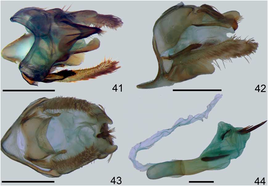
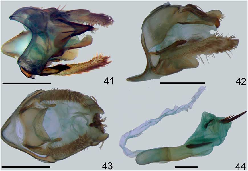
Redescription ♂. Head. Proboscis light brown. Labial palpi dark brown, exceeding the vertex; third palpomere twice longer than wide. Frontoclypeus entirely dark brown, longer than wide, ventrally smooth-scaled, and dorsally rough-scaled. Vertex grey. Postocciput and dorsal cervical area light red. Scales posterior to the antennal socket white. Scape and pedicel predominantly brown, grey anteriorly. Flagellomeres dark brown, except for the proximal third, grey on the inner surface. Thorax. In dorsal view, thorax brown and grey. Posterior portion of mesoscutellum beige. Metascutellum brown. Patagia brown, with anterior and lateral margins beige. Tegulae beige with two brown areas, one on the external margin, rounded, and the other on the internal margin, long. Epimera and episterna with long, brown scales. Procoxae light red with a brown spot anteriorly and brown laterally. Profemora predominantly brown, light red proximally. Protibiae and tarsi brown. Mesolegs and metalegs with the same pattern, except for the outer surface of the coxae, light red instead of brown, and the proximal surface of the tibiae, with a light red stripe. Forewings. Entirely scaled. Dorsal surface with veins predominantly covered with beige scales and dark brown spots of different shapes and sizes forming four distinguishable groups: the most proximal with three spots; the next group with four spots; the third with 12, comprising the discal area and the region immediately distal to it, and all cells of the wing; distalmost group with eight spots, separated in a subdistal group of three spots, and an apical group of five spots. External margin with dark brown scales anteriorly, and white scales posteriorly. Ventral surface predominantly brown, except for four white areas between the discal cell and the apex, and a less conspicuous white area inside the discal cell. Venation: R1 arising near the distal end of the discal cell. R2 arising at the same distance of the transversal vein and R3. M2 and M3 branching from the transversal vein very near each other, without a common stalk. Hindwings. Entirely scaled. Veins and most of the dorsal and ventral surfaces covered with brown scales. Anterior surface of discal cell and costal area immediately above it with white scales. Sometimes the posterior surface of the discal cell also has white scales. Venation: Sc vestigial. M2 arising at a small distance from the stalk of M3 and CuA1. M3 and CuA1 shorter than their stalk. Abdomen. Tergites entirely brown. S2–6 light red; S7–8 brown.T8 and S8 sclerotised. Intersegmental membrane seven to eight of males unmodified, coremata absent. Male genitalia. Aedeagus with approximately the same width across its entire length, smooth and straight, approximately twice longer than the genital capsule when with the vesica everted. Ejaculatory duct positioned dorsally. Coecum rounded. Vesica with two branches; lateral branch with a single spiniform cornutus, dorsal branch with a sclerotised plate proximally, and another large sclerotised plate surrounded by smaller spiniform cornuti distally. Saccus long, wide and acute, symmetrical. Tegumen with few setae, considerably wider than the vinculum, with the ventral part not covering the proximal portion of the valvae. Posterior margin of the tegumen with filiform sclerotised projections, more or less rounded near the apex. Internal surface of the tegumen with two membranous pouches. Uncus symmetrical, not compressed laterally or dorsoventrally, with few setae at its base and long setae at its distal portion, dorsally. Apex sharp. Transtilla membranous. Juxta glabrous, shield-like, wider than high. Valvae subequal, reaching the apex of the uncus. Each sacculus with a small digitiform projection covered with few setae. Distal portion of valvae bifurcate, the dorsal projection bearing spines, shorter and more sclerotised than the ventral projection, which is densely covered by brown scales. Cucullus heavily sclerotised, bearing large spines on its inner surface. Valvula densely scaled, distinct from cucullus and longer than it. Female genitalia. Unknown.
Distribution. Heliura stolli is apparently restricted to the lowlands of the Amazonian Forest and from Guiana Shield.
Additional material examined (16 ♂). BRAZIL: Amazonas, Benjamin Constant, rio Javary, alto Amazonas, coll. Dirings, one male (USNM); Fonte Boa, v.1906, S. M. Klages, one male (BMNH); FRENCH GUIANA: Montagne de Kaw, pk. 37, 29 VII 2000 G. Miral, one male (JAC); GUYANA: Esseq. Wineperu, 18–24.iii.[19]69, Duckworth and Dietz, one male (USNM); Shanklands, 35 m SW Georgetown, Dist. 3, 6°8'N 58°62'W, 23.ix.2006, D. Margua, one male (LACM); Mazarumi River, Maruma Mont. 600 m, IV 2000, P. Bleuzen two males (JAC); PERU: Loreto, Callicebus Res. Station, Mishana, rio Nanay, 25km SW Iquitos, 120 m, 10–17.i.[19]80, J. B. Heppner, tropical wet forest, two males (USNM); Madre de Dios, Rio Tambopata Res., 30 air km SW Puerto Maldonado, 290 m, 6–10.xi.1979, J. B. Heppner, subtropical moist forest, one male (USNM); Rio Tambopata Res., 30 air km SW Puerto Maldonado, 290 m, 11–15.xi.1979, J. B. Heppner, subtropical moist forest, one male (USNM); Tambopata Preserve, Laguna Chica, 12°51'S 69°18'W 200 m, 7.xii.1996, at light, Miller/Snyder/Brower/Rab-Green leg., two males (AMNH); Manu Biosphere Reserve Pakitza, 250 m, 12°07'S 70°58'W, 18.xi.1988, M. G. Pogue, one male (USNM); Manu Biosphere Reserve Pakitza, 250 m, 11°56''S 71°18''W, 250 m, 21.xi.1989, Trail 1, marker 10, UV light #16, D. Adamski and M. Epstein, one male (USNM); Manu Biosphere Reserve Pakitza, 250 m, 11°56''S 71°18''W, 250 m, Pakitza trail 1, marker 8 near Campground 13–14.xi.1989, UV lite, N. E. Adams, one male (USNM); VENEZUELA: Bolivar , Guiaquinima tepui camp I, 5°55'N 63°30'W, 1150 m, 24–28.ii.1990, D. Grimaldi, one male (AMNH).
Etymology. The specific epithet is in honour of Caspar Stoll, Pieter Cramer’s disciple, and responsible for the continuation of the Papillons Exotiques after his death.
Remarks. Heliura stolli was described from an unspecified number of specimens. Two males with label data compatible to those in the original description were found, but only one of them with a second label written “type” with Rothschild’s handwriting. This is here considered the lectotype as designated by Hampson (Reference Hampson1914). It is unclear whether the other specimen is also part of the type series.
Heliura parcipuncta (Hampson, Reference Hampson1914), new combination
Heliura stolli meridionalis Rothschild, Reference Rothschild1912: 170. Lectotype male, by subsequent designation (Hampson Reference Hampson1914: 297), with five labels: “Type” / “Alianca, below S. Antonio, Rio Madeira, Nov. Dec. 1907 (W. Hoffmanns)” / “Heliura stolli meridionalis type Rothsch.” / “Rothschild Bequest B. M. 1939-I” / “Kb-Dia-Nr. 1072 B. Kreusel dok” (BMNH) (examined); Draudt 1915: 170.
Delphyre parcipuncta Hampson, Reference Hampson1914: 296; Hampson Reference Hampson1915: plate 16, fig. 30 (replacement name).
Diagnosis ♂. Heliura parcipuncta can be distinguished from all other species of the H. stolli species group by the red scales covering all the abdominal sternites, except for S8. The only exception is H. stolli, from which it differs by the white (instead of brown) scales near the stalk of the cubital veins, the absence of ventral projections on the tegumen, and the dorsal lobe of the valvae, slightly longer than the ventral lobe, and with a subterminal flat dilatation (as opposed to a much shorter dorsal lobe with small spines in H. stolli).
Redescription ♂. Head. Proboscis light brown. Labial palpi dark brown, exceeding the vertex; third palpomere longer than wide. Frontoclypeus entirely dark brown, slightly longer than wide, ventrally smooth-scaled, and dorsally rough-scaled. Vertex uniformly grey or with darker scales at the centre. Postocciput and dorsal cervical area light red. Scales posterior to the antennal socket white. Scape and pedicel predominantly brown, greyish anteriorly. Flagellomeres dark brown, except for the proximal ones, grey on the inner surface. Thorax. In dorsal view, thorax brown and grey. Posterior portion of mesoscutellum grey. Metascutellum yellow. Patagia brown, with anterior and lateral margins beige. Tegulae predominantly grey with two brown, one on the external margin, rounded, and the other on the internal margin, long. Epimera and episterna with long, brown scales. Procoxae light red with a brown spot anteriorly, brown laterally. Profemora predominantly brown, light red proximally. Protibiae and tarsi brown. Mesolegs and metalegs with the same pattern, except for the outer surface of the coxae, light red instead of brown, and the proximal surface of the tibiae, with a grey stripe. Forewings. Entirely scaled. Dorsal surface with veins predominantly covered with beige scales and dark brown spots of different shapes and sizes forming four distinguishable groups: the most proximal with three spots; the next group with four spots; the third with 12, comprising the discal area and the region immediately distal to it, and all cells of the wing; distalmost group with eight spots, separated in a subdistal group of three spots, and an apical group of five spots. External margin with dark brown scales anteriorly, and white scales posteriorly. Ventral surface predominantly brown, except for four white areas between the discal cell and the apex, and a less conspicuous white area inside the discal cell. Venation as in Figure 13: R1 arising near the distal end of the discal cell. R2 arising at half distance between R1 and R3. M2 and M3 branching from the transversal vein very near each other, without a common stalk. Females with two frenular bristles. Hindwings. Entirely scaled. Veins and most of the dorsal and ventral surfaces covered with brown scales. Discal cell and costal area immediately above it with white scales. Venation shown in Figure 13: Sc absent. M2 arising very close to M3+CuA1, which are fused. Abdomen. Tergites entirely brown. S2–5 light red, S6-8 brown. T8 and S8 sclerotised. Intersegmental membrane seven to eight of males with coremata. Female pre-genital segments unmodified. Male genitalia. Aedeagus with approximately the same width across its entire length smooth and straight, slightly longer than the genital capsule when with the vesica everted. Ejaculatory duct positioned laterally, to the left. Coecum rounded. Vesica with three areas, the anterior with a set of small cornuti, the medial with two large cornuti and a line of small cornuti, and the posterior with a sclerotised plate. Tegumen with few setae and scales, slightly wider than the vinculum, with the ventral part not covering the proximal portion of the valvae. Tegumen without membranous pouches, posterior margin without sclerotised projections. Uncus symmetrical, laterally compressed, with few setae at its base and long setae at the mid portion of its lobe, dorsally and laterally. Apex sharp. Transtilla membranous, with minute setae. Juxta sclerotised, glabrous, long and narrow. Valvae subequal, exceeding the apex of the uncus. Each sacculus with a small digitiform projection covered with few setae at the tip. Distal portion of valvae bifurcate, with both parts having approximately the same length. Dorsal branch of the valvae more heavily sclerotised than the ventral part, with the mid portion convex, and a sharp apex. Ventral branch densely covered by brown scales. Dorsal part non-scaled and with spines. Female genitalia. Pheromone glands undeveloped. Ostium centralised. Antevaginal lamella sclerotised, without setae. Post-vaginal lamella more heavily sclerotised than the antevaginal lamella. Anterior apodemes almost as long as the posterior apodemes. Ductus bursae sclerotised and with approximately the same length as the corpus bursae. Corpus bursae covered by large signa on its anterior half, which is rounded. Posterior half of corpus bursae predominantly membranous, with a sclerotised area continuous with the sclerotisation of ductus bursae, and two small patches of signa on the right side. Ductus seminalis diverging from corpus bursae, dorsally, near its posterior margin.
Additional material examined (10 ♂ and 1♀).BRAZIL: Pará, Faz. Taperinha, prox. Santarém, 19–29.ii.1968 E. Hagmann, one male (USNM); Faz. Taperinha, prox. Santarém, x.1942, Hagmann, one female (LRP539) (MPEG); Rondônia, 62 km S Ariquemes, 165 m, Faz. Rancho Grande, 10°32'S 62°48'W, 27.viii–8.ix.1994, Ron Leuschner, one male (LACM); PERU: Cuzco, Pilcopata, 600 m, 11–14.xii.1979, J. B. Heppner, premontane moist forest, one male (USNM); Loreto, Magnas, Muyun Lodge, 50 m, 8–10.vi.2006, Hannu Saarenmaa, light trap, SMNS-Lep 2006-07, one male (SMNS); Madre de Dios, Manu Biosphere Res. Pakitza, 250 m, 12°7'S 70°58'W, 9.ix.1988, M. G. Pogue, one male (USNM); Manu Biosphere Res. Pakitza, 250 m, 12°7'S 70°58'W, 18.ix.1988, M. G. Pogue, black light trap on fallen tree, one male (USNM); Manu Biosphere Res. Pakitza, 11°56'S 71°18'W, 250 m, 29–31.viii.1989, UV light #1, D. Adamski and M. Epstein, one male (USNM); Manu Biosphere Res. Pakitza, 11°56'S 71°18'W, 250 m, 6–8.ix.1989, mkr. 8 UV light #6, D. Adamski and M. Epstein, two males (USNM); Manu Biosphere Res. Pakitza, 11°56'S 71°18'W, 250 m, trail 1, marker 8 near Campground, 13–14.ix.1989, UV lite, N. E. Adams, one male (USNM).
Distribution. All records for this species are from the Amazonian forest of Brazil and Peru.
Etymology. The specific epithet suggests a sparse concentration of spots (from Latin parco, meaning “to spare”, and punctum, meaning “a small, distinct point”).
Remarks. Heliura stolli meridionalis was described from an unspecified number of specimens, and only one was found with label data matching those provided in the original description. Hampson (Reference Hampson1914) mentioned a male “type”, which, according to articles 74.6 (International Commission on Zoological Nomenclature 1999) makes him the author of the lectotype designation. The name Delphyre parcipuncta Hampson, Reference Hampson1914 was erected by this author as a replacement name for Heliura stolli meridionalis, which became a secondary junior homonym of Heliura klagesi meridionalis Rothschild, Reference Rothschild1912 when Hampson (Reference Hampson1914) placed both in the genus Delphyre.
The association between the sexes here proposed is based on the typical wing venation and the colouration of the metascutellum of H. parcipuncta for both sexes: hindwings with veins M3 and CuA1 fused, and metascutellum yellow-scaled.
Heliura pieroides Rothschild, Reference Rothschild1912, revalidated combination
Heliura pieroides Rothschild, Reference Rothschild1912: 169. Lectotype female, by subsequent designation (Hampson Reference Hampson1914: 297), with five labels: “Type” / “Fonte Boa, Upp. Amazon, August 1907 (S. M. Klages) / “Heliura pieroides type Rothsch.” / “Rothschild Bequest B.M. 1939-1 / “Kb-Dia-Nr. 1073 B. Kreusel dok.” (BMNH) (examined); Draudt 1915: 170.
Delphyre pieroides; Hampson, Reference Hampson1914: 297; Hampson Reference Hampson1915: plate 16, fig. 31.
Diagnosis ♀. Heliura pieroides differs from most of the other species of the H. stolli complex by the yellow, instead of red, surface of the procoxae. This character is shared with H. crameri, from which it differs mainly in the yellow, as opposed to grey, metascutellum, the T8 brown and white, and the abdominal sternites, predominantly yellow, instead of brown.
Redescription ♀. Head. Proboscis light brown. Labial palpi dark brown, exceeding the vertex; Vertex grey. Postocciput and dorsal cervical area light yellow. Scape and shaft black-brown, the inner face white on the basal third. Flagellomeres dark brown. Thorax. In dorsal view, thorax brown and grey. Posterior portion of mesoscutellum beige with a large, black spot. Metascutellum brown with a dorsal yellow spot. Patagia brown, with anterior and lateral margins grey. Tegulae grey with two brown areas, one on the external margin, rounded, and the other on the internal margin, long. Procoxae light yellow. Legs and tarsi black brown, profemora and mesofemora, white on the inner surface, the mesotibia with some white at base. Forewings . Dorsal surface with veins predominantly covered with olive-grey scales and dark brown spots of different shapes and sizes forming four distinguishable groups: the most proximal with three spots; the next group with four spots; the third with 14, comprising the discal area and the region immediately distal to it, and all cells of the wing; distal-most group with eight spots, separated in a subdistal group of three spots, and an apical group of five spots. Ventral surface predominantly brown, except for four white areas between the discal cell and the apex, one inside the discal cell and one under the base of the distal cell between the veins 1A+2A–CuA2. Venation: M2 and M3 branching from the transversal vein very near each other, without a common stalk. Hindwings . Veins and most of the dorsal and ventral surfaces covered with brown scales. Anterior surface of discal cell and costal area immediately above it with white scales. The posterior surface of the discal cell and also a part of the space immediately below, between the veins A2–CuA2 with white scales. Venation: Sc present. M2 arising at the beginning of the stalk M3–CuA1. M3 and CuA1 slightly shorter than their stalk. Abdomen. Tergites entirely brown except for a yellow dorsal line on the first tergite, several yellow dorsal setae on the second tergite and the posterior margin white on the last tergite. S2–6 light yellow; S7–8 brown. Female genitalia. Unknown.
Etymology. The specific epithet of H. pieroides is a reference to H. pierus (Cramer, 1781).
Remarks. Heliura pieroides was described from an unspecified number of specimens. Only one with label data matching the information provided in the original description was found, and it is the one mentioned by Hampson (Reference Hampson1914) as the female “type”. It is so far the only known specimen of this species.
According to Rothschild (Reference Rothschild1912: 169), he had in front of him three very similar species, two unnamed, and another one that he believed to be H. pierus. He did not offer any clue as to why he thought the third species corresponded to H. pierus, which type is apparently lost (see below).
Addendum: Heliura pierus (Cramer, 1781)
Sphinx pierus Cramer, 1781: 148, plate 367, fig. E. SURINAME. Types(s) of unknown sex, probably lost.
Charidea pyerus; Herrich-Schäffer 1854: 23.
Eucereon pierus; Kirby Reference Kirby1892: 200.
Heliura pierus; Hampson Reference Hampson1898: 484; Zerny Reference Zerny1912: 137; Draudt 1915: 170; Draudt 1917: plate 24, row g.
Etymology. Pierus is the name of two characters from the Greek mythology. It is unknown which character the name of the species refers to.
Remarks. Potential types of Sphinx pierus were searched for at the BMNH and RMNH, the two institutions that are known to house Cramer’s types (Horn and Kahle 1935–Reference Horn and Kahle1937, Vane-Wright Reference Vane-Wright1975; Chainey Reference Chainey2005). None could be found, neither the original description nor the original illustration (the original volume of Papillons Exotiques was consulted at the BMNH) are enough to determine the identification of the species.
There are many species that resemble the original illustration of H. pierus, and all collections visited by the first author that have specimens identified as such are, in fact, either H. stolli, H. parcipuncta, H. crameri, or H. pauloi. However, in our opinion, the original illustration corresponds to none of them, but to a species much closer to H. pusilla Butler, 1878 (Butler 1878, fig. 10). The reasons are as follows: the size of the most proximal spots on forewing pattern in the specimen illustrated by Cramer are more similar to those of D. pusilla than to those of the species of the H. stolli species group. The original illustration of H. pierus shows a subproximal spot that exists in specimens of H. pusilla, but not in H. stolli, H. parcipuncta, H. crameri, nor H. pauloi. Moreover, H. pierus does not have a brown spot below the discal cell; the same appears in H. pusilla, while in all species of the H. stolli species group, there is a brown spot below the discal cell.
However, the brief description of H. pierus mentions that the abdominal sternites are grey. The abdominal sternites of H. pusilla are mainly light red. This character makes questionable the synonymy between H. pusilla and H. pierus. Though it is possible that Cramer’s specimen was greasy, the fauna of Suriname is still mainly unexplored. For that reasons, and in the absence of consistent evidence of the identity of H. pierus, this species is here considered a species inquirenda until the Surinamese fauna is more properly explored.
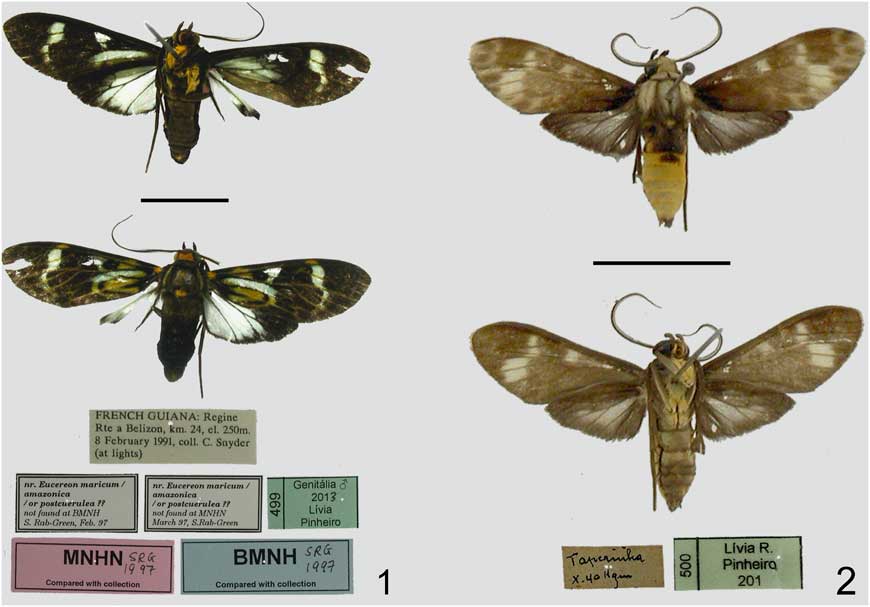
Habitus and labels. 1. Heliura laguerrei, holotype, dorsal and ventral views. 2. Heliura beneluzi, holotype, dorsal and ventral views.
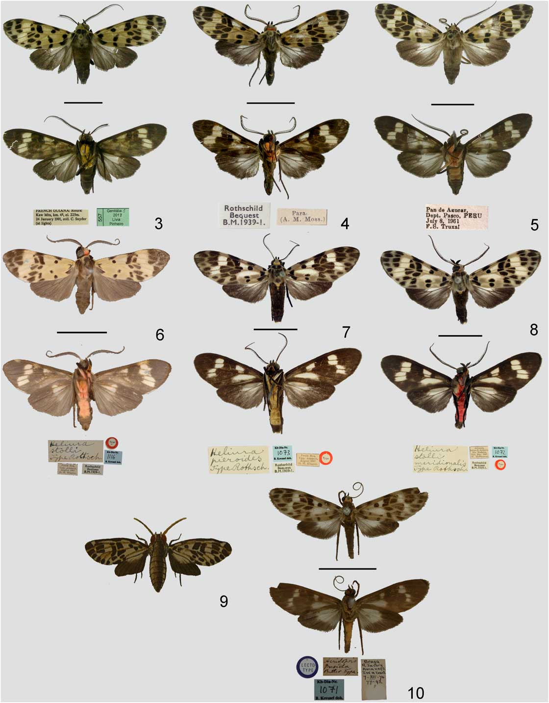
Habitus and labels. 3. Heliura crameri, holotype, dorsal and ventral views. 4. Heliura nigriventris, holotype, dorsal and ventral views. 5. Heliura pauloi, holotype, dorsal and ventral views. 6. Heliura stolli, holotype, dorsal and ventral views. 7. Heliura parcipuncta, lectotype, dorsal and ventral views. 8. Heliura pieroides, lectotype, dorsal and ventral views. 9. Original figure of Sphinx pierus. 10. Lectotype of Delphyre pusilla, dorsal and ventral views.
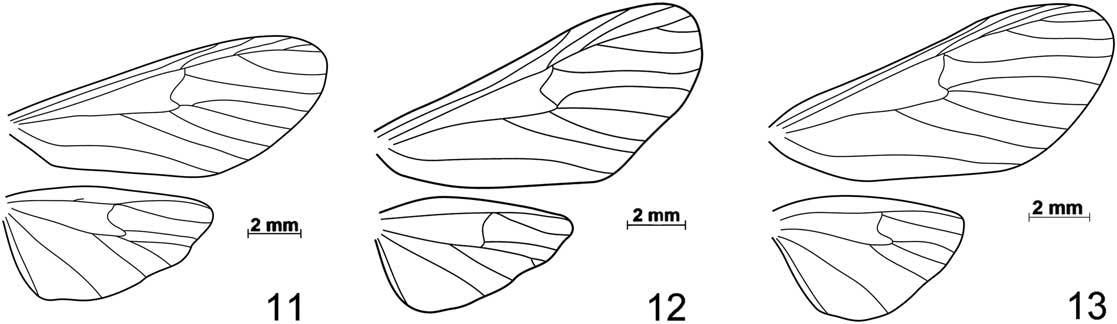
Wing venation patterns. 11. Heliura laguerrei; 12. Heliura beneluzi. 13. Heliura parcipuncta.
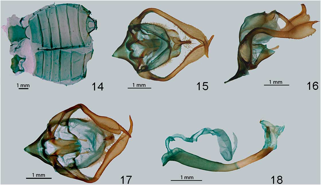
Genitalia of Heliura laguerrei. 14. Abdomen. 15. Male genitalia, dorsal view. 16. Male genitalia, lateral view. 17. Male genitalia, posterior view. 18. Aedeagus.
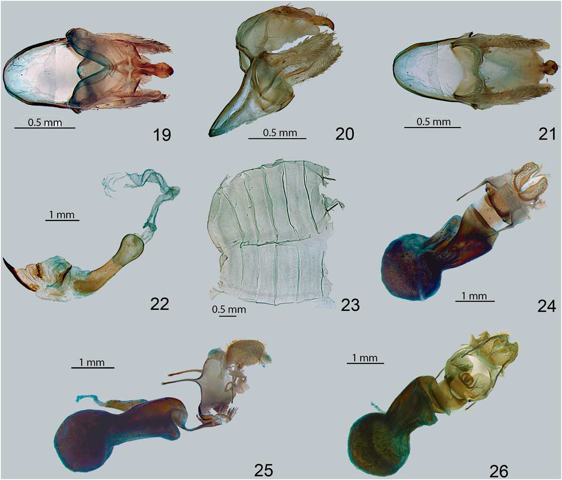
Genitalia of Heliura beneluzi. 19. Male genitalia, dorsal view. 20. Male genitalia, lateral view. 21. Male genitalia, posterior view. 22. Aedeagus. 23. Female abdomen. 24. Female genitalia, dorsal view. 25. Female genitalia, lateral view. 26. Female genitalia, ventral view.
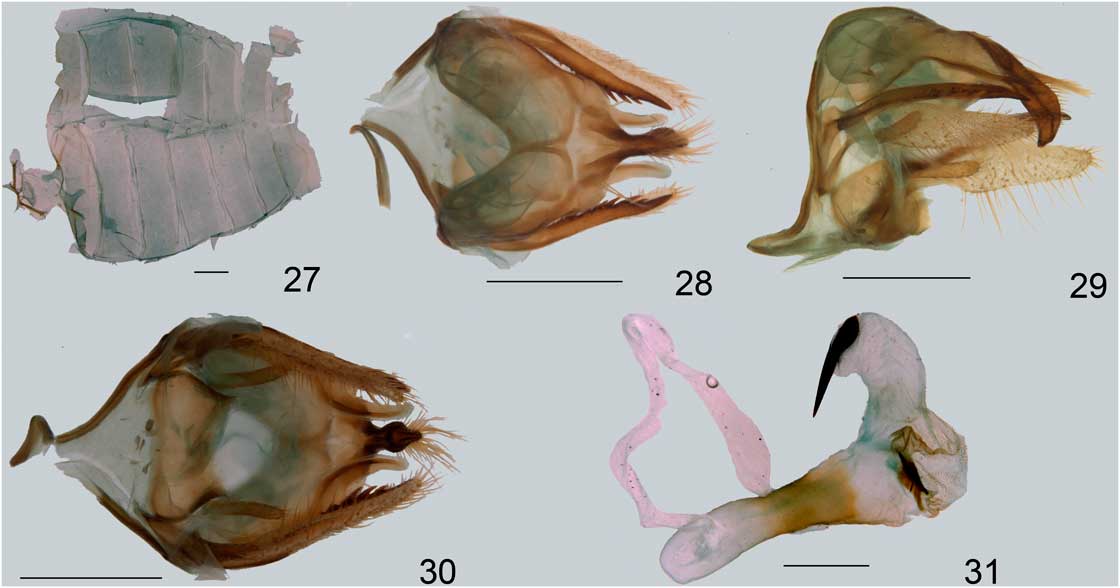
Genitalia of Heliura crameri. 27. Male abdomen. 28. Male genitalia, dorsal view. 29. Male genitalia, lateral view. 30. Same, posterior view. 31. Aedeagus. Scale bars: 1 mm.
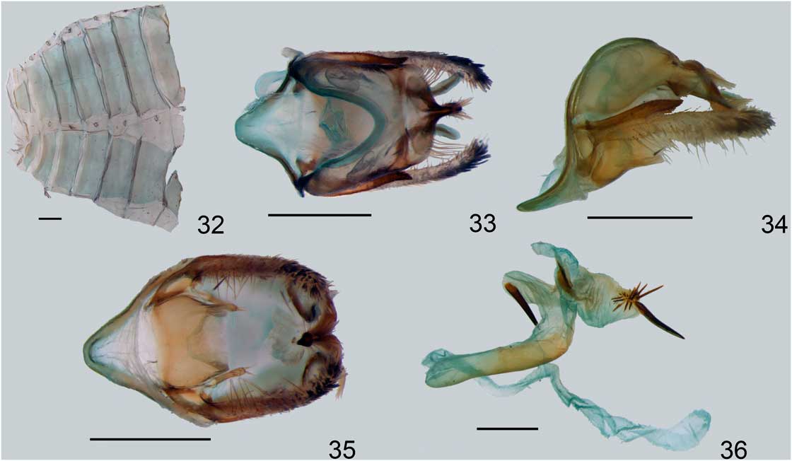
Genitalia of Heliura nigriventris. 32. Male abdomen. 33. Male genitalia, dorsal view. 34. Male genitalia, lateral view. 35. Male genitalia, posterior view. 36. Aedeagus. Scale bars: 1 mm.
Genitalia of Heliura pauloi. 37. Male genitalia, dorsal view. 38. Male genitalia, lateral view. 39. Male genitalia, posterior view. 40. Aedeagus. Scales bars: 1 mm.
Genitalia of Heliura stolli. 41. Male genitalia, dorsal view. 42. Male genitalia, lateral view. 43. Male genitalia, posterior view. 44. Aedeagus. Scales bars=1 mm.
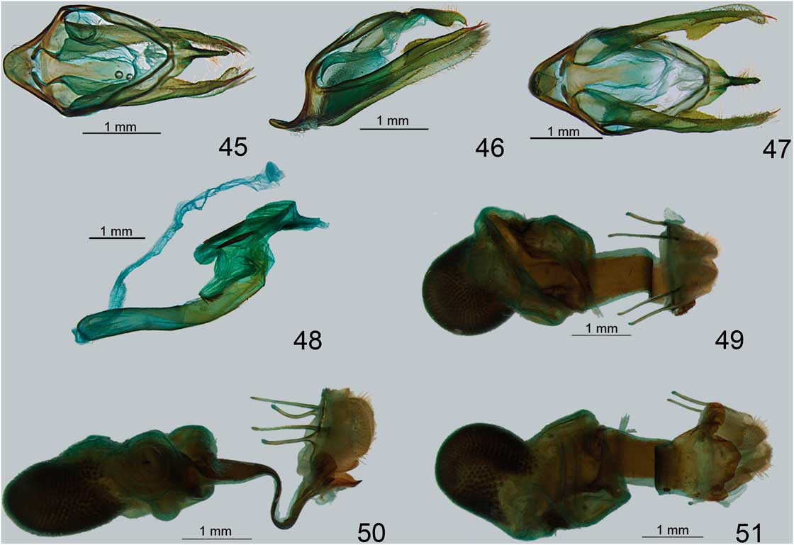
Genitalia of Heliura parcipuncta. 45. Male genitalia, dorsal view. 46. Male genitalia, lateral view. 47. Male genitalia, posterior view. 48. Aedeagus. 49. Female genitalia, dorsal view. 50. Female genitalia, lateral view. 51. Female genitalia, ventral view. Scales bars=1 mm.

Acknowledgements
L.R.P. thanks the curators of the collections visited for their support during her visit: William Overal (MPEG); David Grimaldi and Suzanne Rab-Green (AMNH); and Alessandro Giusti, Geoff Martin, and Martin Honey (BMNH). Thanks also to Rob de Vos for help with the search for potential types at the RMNH. This research was funded by Fundação de Amparo à Pesquisa do Estado de São Paulo (FAPESP grants 2009/11159-5, 2012/02444-0, 2002/13898-0, 2010/14682-8, and 2011/50225-3), Conselho Nacional de Desenvolvimento Científico e Tecnológico (CNPq, as part of the project “National Research Network for Conservation of Lepidoptera/Sistema Nacional de Pesquisa em Biodiversidade SISBIOTA-Brasil”; grant 563332/2010-7), and the Universidade de São Paulo (USP) (Projeto 1). M.D. also thanks CNPq for the Research Productivity Fellowship (process 305905/2012-0).