Small for gestational age is usually defined as birth weight below the 10th percentile for gestational age or more than two standard deviations below the mean for gestational age.Reference Battaglia and Lubchenco 1 Infants whose soft tissue mass is below normal for the stage of skeletal development can be identified by the Ponderal index. Thus, a Ponderal index of below the 10th percentile is the most commonly used parameter to identify small for gestational age infants.Reference Cole, Henson, Tremble and Colley 2 In babies asymmetrically small for gestational age, especially at the third trimester of gestation, maternal reasons and factors related to placenta and uterus would disturb the uteroplacental blood flow.Reference Kliegmann 3 Disturbed placental flow with increased vascular resistance is an important cause of intrauterine growth restriction related to insufficiency of both nutrients and oxygen. The resultant hypoxaemia can affect cardiac functions in growth-restricted foetuses.Reference Bahtiyar and Copel 4 – Reference Larsen, Sloth, Petersen, Pedersen, Sorensen and Uldbjerg 6
Tissue Doppler imaging is a new echocardiographic technique that allows measurement of systolic and diastolic myocardial velocities at early stages.Reference Sutherland, Stewart and Groundstroem 7 , Reference Naqvi 8 Mori et alReference Mori, Nakagawa and Nii 9 and Ekici et alReference Ekici, Atalay, Özçelik, Uçar, Yılmaz and Tutar 10 evaluated myocardial velocities by tissue Doppler echocardiography and reported normal values in term neonates. The few studies evaluating small for gestational age babies included the preterm small for gestational age babies. Decreased stroke volume, increased left ventricular output, and left ventricular diastolic dysfunction have been reported in preterm small for gestational age babies and growth-retarded foetuses.Reference Tsyvian, Malkin, Artemieva and Wladimiroff 5 , Reference Larsen, Sloth, Petersen, Pedersen, Sorensen and Uldbjerg 6 , Reference Robel-Tillig, Knüpfer and Vogtmann 11 – Reference Harada, Suzuki and Takahashi 13 These studies indicate that cardiac abnormalities occur at early stages.
To the best of our knowledge, this is the first study evaluating cardiac systolic and diastolic functions in term small for gestational age babies with conventional and tissue Doppler echocardiography. Our hypothesis was that cardiac functions may be impaired in term small for gestational age babies. Therefore, the present echocardiographic study was designed to evaluate the cardiac systolic and diastolic functions and age-related changes of these functions in full-term small for gestational age infants during the first 3 months of life using the both conventional Doppler and tissue Doppler echocardiography.
Patients and methods
Study population
All infants were examined on the first day after birth; gestational age was determined by Dubowitz score.Reference Dubowitz, Dubowitz and Goldberg 14 A total of 40 term babies between 38 and 42 weeks gestational age according to the Dubowitz score were included in the present study. The exclusion criteria were respiratory and cardiac diseases, congenital malformations, history of drug usage, illness, syndrome, chromosomal abnormality, or intrauterine infection. Moreover, infants whose mothers had diabetes mellitus, eclampsia, preeclampsia, and infants with low Apgar scores were not included.
Of these 40 babies, 20 were asymmetrically small for gestational age and the remaining 20 were appropriate for gestational age. The Ponderal index was used (body weight/lengthReference Kliegmann 3 × 100) for the definition of small for gestational age. The babies having Ponderal index <10% were considered asymmetrically small for gestational age.Reference Cole, Henson, Tremble and Colley 2 A detailed medical history was obtained from the parents, and a thorough physical examination together with routine laboratory tests including haemoglobin, serum electrolytes, total protein, albumin, and additionally troponin I level were performed in all infants on their postnatal day 5.
Cardiac systolic and diastolic functions of all infants in the two groups were prospectively evaluated by conventional echocardiography and tissue Doppler imaging on postnatal day 5 and 1 and 3 months after delivery. The study protocol was approved by the Ethics Committee of Medical School, Pamukkale University. Written informed consents were obtained from the parents of all infants.
Echocardiography examination
All infants underwent M-mode, two-dimensional Doppler, and tissue Doppler evaluation using a vivid 7 model echocardiography and multi-frequency transducer (7 MHz) (GE Vingmed Company, Horten, Norway). Images were taken with infants in the supine position. All infants were evaluated under non-sedated resting state. Three consecutive measurements were recorded. M-mode measurements were made at the level of the tips of the mitral valve leaflets in the parasternal long-axis view of the left ventricle.Reference Sahn, DeMaria, Kisslo and Weyman 15 Standard parameters measured in parasternal long-axis views by M-mode echocardiography were left ventricular dimension at the end of diastole, interventricular septum diastolic thickness, left ventricle posterior wall end-diastolic thickness, and right ventricular dimension at the end of diastole. Left ventricular ejection fraction and fractional shortening were calculated as defined previously.Reference Silverman 16 Left ventricular mass was calculated with a standard formula.Reference Devereux and Reichek 17 , Reference de Simone, Daniels and Devereux 18
Conventional Doppler blood velocities were measured for mitral and tricuspid valves using apical four-chamber view. Early and late inflow velocities and the ratio between early and late velocities were determined.
Tissue Doppler echocardiography
The tissue Doppler mode of the same machine was used by lowering the velocity range (0–30 cm/s) to determine the myocardial velocities and setting the Doppler gain achieve maximum colour information in the myocardium. Pulsed-wave tissue Doppler imaging was used to record myocardial contraction velocities, early diastolic and atrial contraction velocities in basal portions of the left ventricle – at the level of the lateral mitral annulus, the right ventricle – at the level of the lateral tricuspid annulus, and the interventricular septum in the apical four-chamber view. The ratio of peak velocities in early diastolic and atrial contraction was determined for the assessment of diastolic function.
Statistical analysis
All statistical analyses were performed using the Statistical Package for the Social Sciences program (version 17.0, Chicago, Illinois, United States of America). Normality of the continuous data was assessed by the Shapiro–Wilk test. The Student t-test, chi-square test, repeated measures analysis of variance, paired t-test, Pearson's correlation, and linear regression analysis were used for data analysis. The statistical analysis for age-related changes of measured parameters was performed by repeated measures analysis of variance. If statistical significance was found, the paired t-test was used for post-hoc analysis. A p-value <0.05 was considered significant.
Results
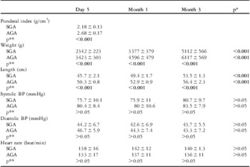
There were 13 girls and 7 boys in both of the small for gestational age and appropriate for gestational age groups. The study groups were similar with regard to conceptional age, sex, Apgar score, heart rate, systolic and diastolic blood pressure (p > 0.05). Apgar scores were 9–10 on minute one and 10 on minute ten. Ponderal index was below 2.32 (10%) in all the small for gestational age babies. The descriptive data including weight, length, blood pressure, and heart rate are presented in Table 1. The weight and length increased with increasing age in all babies (p < 0.001). The weight and length were lower in the small for –gestational age babies (p < 0.001).
Descriptive data of the study population (mean ± standard deviation).

AGA = appropriate for gestational age; BP = blood pressure; SGA = small for gestational age
*Indicates statistical analyses of serial evaluation in each group
**Indicates statistical analyses of two groups
Haemoglobin levels in the small for gestational age and appropriate for gestational age babies were 17.5 ± 2.3 and 16.2 ± 2.1 g/dl, respectively (p = 0.045). Although troponin I was in normal ranges in the two groups, troponin was higher in the small for gestational age babies (0.06 ± 0.005 ng/ml for the small for gestational age babies versus 0.02 ± 0.01 ng/ml for the appropriate for gestational age babies, p < 0.01). The levels of electrolytes, total protein, and albumin were in normal ranges in all subjects, and there was no significant difference between the groups (p > 0.05).
At the time of study, haemodynamic parameters of the patients were normal and none of the babies displayed clinical signs of any cardiovascular disease. All babies were in sinus rhythm and none exhibited any electrocardiographic evidence of cardiac failure or arrhythmia, and standard echocardiographic evaluations were in normal ranges.
Echocardiographic evaluations
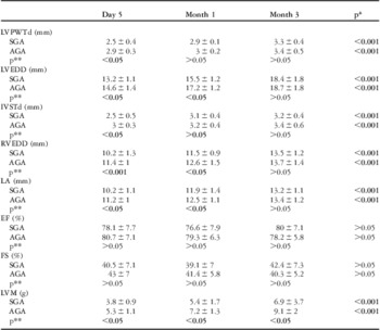
Table 2 presents various conventional echocardiographic parameters for the determination of ventricular function in the study groups. Left ventricular dimension at the end of diastole, interventricular septum diastolic thickness, left ventricle posterior wall end-diastolic thickness, right ventricular dimension at the end of diastole, and left atrium increased with age in all babies (p < 0.001). However, in the small for gestational age babies, the left ventricular dimension at the end of diastole, interventricular septum diastolic thickness, and left ventricle posterior wall end-diastolic thickness were lower than the appropriate for gestational age babies on day 5 (p < 0.001). Left ventricular ejection fraction and fractional shortening were not significantly different in small for gestational age babies compared with controls, suggesting a preserved systolic function (p > 0.05). Although left ventricular mass increased with age in the two groups (p < 0.001), left ventricular mass was lower in the small for gestational age babies compared with the appropriate for gestational age babies at all evaluation time points including day 5 and months 1 and 3 (p < 0.05; Table 2).
Parameters of the M-mode echocardiography in the study population (mean ± standard deviation).

AGA = appropriate for gestational age; EF = ejection fraction; FS = fractional shortening; IVSTd = interventricular septum diastolic thickness; LA = left atrium; LVEDD = left ventricular end-diastolic dimension; LVM = left ventricular mass; LVPWTd = left ventricular posterior wall end-diastolic thickness; RVEDD = right ventricular end-diastolic dimension; SGA = small for gestational age
*Indicates statistical analyses of serial evaluation in each group
**Indicates statistical analyses of two groups
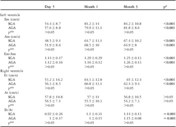
Peak early diastolic (E) and atrial contraction (A) velocities and the ratio of E/A determined by conventional Doppler echocardiography for the left and right ventricles changed with age (p < 0.001), but there was no statistically significant difference between the two groups at any of the evaluation time points (p > 0.05; Table 3).
Parameters of the conventional Doppler echocardiography in the study population (mean ± standard deviation).

AGA = appropriate for gestational age; Am = peak late mitral inflow velocity; At = peak late tricuspid inflow velocity; Em = peak early mitral inflow velocity; Et = peak early tricuspid inflow velocity; SGA = small for gestational age
*Indicates statistical analyses of serial evaluation in each group
**Indicates statistical analyses of two groups
Tissue Doppler imaging results
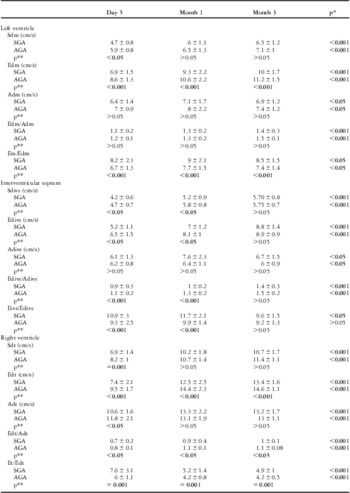
Peak systolic (Sd), peak early diastolic (Ed), and atrial contraction (Ad) velocities together with Ed/Ad and E/Ed ratios changed with increasing age (p < 0.001; Table 4).
Parameters of the tissue Doppler echocardiography in the study population (mean ± standard deviation).

Adivs = peak atrial contraction velocity for the interventricular septum; Adm = peak atrial contraction velocity for the left ventricle; Adt = peak atrial contraction velocity for the right ventricle; AGA = appropriate for gestational age; Edivs = peak early relaxation velocity for the interventricular septum; Edm = peak early relaxation velocity for the left ventricle; Edt = peak early relaxation velocity for the right ventricle; Eivs = peak early mitral inflow velocity; Sdivs = peak systolic velocity for the interventricular septum; Sdm = peak systolic velocity for the left ventricle; Sdt = peak systolic velocity for the right ventricle; SGA = small for gestational age
*Indicates statistical analyses of serial evaluation in each group
**Indicates statistical analyses of two groups
The systolic velocity in the small for gestational age babies was lower than the appropriate for gestational age babies at the first evaluation for the left and right ventricles, and at the first and second evaluation for the interventricular septum (p < 0.05). Peak early diastolic velocity was significantly lower in the small for gestational age babies at all evaluations for the left and right ventricles, and at the first and second evaluations for the interventricular septum (p < 0.05). Atrial contraction velocity was significantly lower in the small for gestational age babies only on the first evaluation for the right ventricle (p < 0.05). We detected that the ratio Ed/Ad was lower, and E/Ed was higher in the small for gestational age babies (p < 0.05). These differences were statistically significant for the Ed/Ad ratio in the right ventricle and interventricular septum. The differences in the E/Ed ratio were significant in the left and right ventricle at all evaluations, in the interventricular septum at the first and second evaluations (p < 0.001; Table 4).
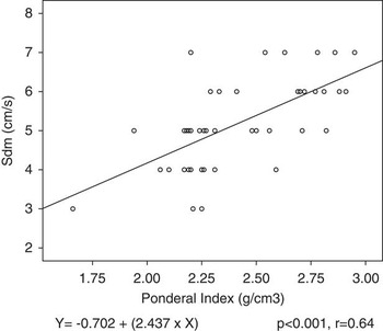
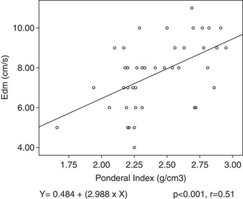
Significant positive correlations were found between the Ponderal index and systolic and early diastolic velocities for the left and right ventricles and interventricular septum (Sdm, Edm, Sdivs, Edivs, Sdt, Edt) with tissue Doppler imaging (r = 0.64, 0.51, 0.38, 0.56, 0.50, 0.51, respectively, p < 0.001). Figures 1 and 2 show the correlations between the Ponderal index and systolic and early diastolic velocities for the left ventricle.
The correlation between the Ponderal index and Sdm. Sdm = peak systolic velocity for the left ventricle.

The correlation between the Ponderal index and Edm. Edm = peak early relaxation velocity for the left ventricle.

Discussion
Small for gestational age is associated with an increased risk of cardiovascular disease in adult life, but it is unclear whether this relationship is present during the infancy period. Small for gestational age infants are at an increased risk for neonatal morbidity and for premature death from cardiovascular disease, hypertension, and diabetes mellitus in adult life.Reference Barker 19 In the present study, a group of 20 term small for gestational age babies were compared with 20 term appropriate for gestational age babies and age-related changes were prospectively evaluated on postnatal day 5, and also months 1 and 3. Our tissue Doppler findings suggest that cardiac systolic and diastolic functions are affected in term small for gestational age infants.
Chronic hypoxaemia, which is likely to occur in the low birth weight foetuses, can cause significant impairment of cardiac relaxation as a result of delayed inactivation of myocardial contraction and impairment of intracellular calcium transporting system.Reference Silverman, Ninomiya and Blank 20 Moreover, subclinical cardiac abnormalities are detected in the intrauterine growth-restricted foetuses.Reference Tsyvian, Malkin, Artemieva and Wladimiroff 5 , Reference Larsen, Sloth, Petersen, Pedersen, Sorensen and Uldbjerg 6 , Reference Naujorks, Zielinsky and Beltrame 21 , Reference Comas, Crispi, Cruz-Martinez, Figueras and Gratacos 22 Tsyvian et alReference Tsyvian, Malkin, Artemieva and Wladimiroff 5 examined left ventricular diastolic functions in growth-restricted foetuses by conventional echocardiography, whereas Naujorks et alReference Naujorks, Zielinsky and Beltrame 21 and Comas et alReference Comas, Crispi, Cruz-Martinez, Figueras and Gratacos 22 investigated the cardiac dysfunction in small for gestational age foetuses using tissue Doppler imaging.
The few published studies in small for gestational age babies comprised the preterm low birth weight babies and conventional echocardiography.Reference Robel-Tillig, Knüpfer and Vogtmann 11 – Reference Harada, Suzuki and Takahashi 13 A higher incidence of patent ductus arteriosus, together with decreased stroke volume, increased left ventricular output and increased left ventricular mass in relation with patent ductus arteriosus in the very low birth weight infants have been reported.Reference Robel-Tillig, Knüpfer and Vogtmann 11 , Reference Leipala, Boldt, Turpeinen, Vuolteenaho and Fellman 12 Left ventricular diastolic dysfunction has also been reported in preterm small for gestational age babies.Reference Harada, Suzuki and Takahashi 13 Our findings indicated decreased left ventricular mass in term small for gestational age babies, but conventional echocardiography did not reveal differences in systolic and diastolic functions between the study groups.
Chaiworapongsa et alReference Chaiworapongsa, Espinoza and Yoshimatsu 23 found higher troponin I levels in umbilical cord blood of the term small for gestational age babies. The present finding of higher troponin I levels in the small for gestational age babies provides further support for the myocardial affection detected on day 5 in term small for gestational age babies.
To the best of our knowledge, this is the first study to evaluate the postnatal cardiac functions and age-related changes in term small for gestational age babies by tissue Doppler echocardiography. Experimental studies have shown that subendocardial fibres are the earliest to be affected by the hypoxia.Reference Bijnens, Claus, Weidemann, Strotmann and Sutherland 24 Peak velocities by tissue Doppler imaging mainly reflect the motion of longitudinal myocardial fibres, which are located mostly in the subendocardial layer.Reference Anderson, Smerup, Sanchez-Quintana, Loukas and Lunkenheimer 25 Thus, the present data suggest that tissue Doppler imaging may be a sensitive technique to detect subtle forms of hypoxia. In this study, we found lower systolic velocity in the small for gestational age babies for all myocardial regions on day 5 by tissue Doppler imaging. Furthermore, early diastolic velocity and the ratio between early diastolic and atrial contraction velocities were significantly lower in the small for gestational age babies. Although there were statistically significant differences in Doppler parameters between the study groups, these differences may not indicate a clinical significance, as the numerical differences were too small. These results demonstrate that systolic and diastolic functions were affected in small for gestational age babies, and affection of the diastolic functions was continuous during the first 3 months of life. In addition, our findings indicate a relation between the Ponderal index and tissue Doppler parameters as systolic and diastolic dysfunction severity was related with Ponderal index. Therefore, it may be suggested that there is a positive correlation between the severity of malnutrition and the severity of the intrinsic myocardial affection. The present data suggest that indexes of systolic and diastolic function, including tissue Doppler imaging, are significantly impaired in the small for gestational age babies. Conventional echocardiographic techniques have failed to distinguish subclinical cardiac dysfunctions when global functions were examined.
Tissue Doppler technique has been reported to have greater sensitivity than conventional Doppler in detecting subclinical cardiac dysfunction without the limitations of transvalvar mitral and tricuspid flow analysis by conventional Doppler, which is influenced by a high cardiac rate and preload and afterload conditions.Reference Sutherland, Stewart and Groundstroem 7 , Reference Naqvi 8 , Reference Ommen, Nishimura and Appleton 26 Normal values of myocardial tissue Doppler velocities in term neonates have been demonstrated in a few previous studies.Reference Mori, Nakagawa and Nii 9 , Reference Ekici, Atalay, Özçelik, Uçar, Yılmaz and Tutar 10 , Reference Ciccone, Scicchitano and Zito 27 – Reference Kozak-Barany, Jokinen and Rantonen 30 Ekici et alReference Ekici, Atalay, Özçelik, Uçar, Yılmaz and Tutar 10 investigated left ventricular myocardial tissue velocities in 1- to 5-day-old neonates. Our findings in term appropriate for gestational age babies on day 5 for the left ventricle are in accordance with the data reported by Ekici et alReference Ekici, Atalay, Özçelik, Uçar, Yılmaz and Tutar 10 and Ciccone et al.Reference Ciccone, Scicchitano and Zito 27
Furthermore, we investigated the cardiac systolic and diastolic functions with regard to postnatal age during the first 3 months of life using both the conventional and tissue Doppler echocardiography. The neonatal myocardium had a relatively higher collagen content in relation to myocytes and larger amount of type I related to type III collagen and this resulted in a more rigid and less compliant heart in the neonate than in older infants. Parallel to postnatal age, both types of collagen decrease progressively, reaching the normal levels of collagen content during the late infancy. These features suggest that, owing to myocardial maturation, developmental changes occur in the left and right ventricular diastolic functions.Reference Marijianowski, Van der Loos, Mohrschlandt and Becker 31 Kozak-Barany et alReference Kozak-Barany, Jokinen and Rantonen 30 evaluated postnatal changes in left ventricular diastolic functions by serial conventional echocardiographic evaluation from days 3–5 up to 6 months in 20 term infants. The authors reported age-related changes in the left ventricular diastolic functions and increased left ventricular mass during the first 6 months.Reference Kozak-Barany, Jokinen and Rantonen 30 Our present prospective study indicated postnatal-age related changes in the diastolic functions for the left and right ventricles during the first 3 months of life. We found that peak early diastolic velocity, the ratio of E/A, and left ventricular mass increased with postnatal age in both groups, but no age-related changes were found with the conventional echocardiography in the fractional shortening, which represents systolic function. However, tissue Doppler imaging revealed that both systolic and diastolic functions changed with postnatal age in both groups. Therefore, the present age-related findings provide further support for the postnatal myocardial maturation. Our data seem to point more towards a delayed maturation of the myocardium in small for gestational age babies.
As a conclusion, the present findings suggest that tissue Doppler imaging may be a more sensitive technique than conventional echocardiography for detection of regional systolic and diastolic myocardial dysfunctions in small for gestational age babies. Our data provide reference values of conventional and tissue Doppler-derived global cardiac functions in term small for gestational age babies, and also provide insight to the maturational process in these babies. The present findings suggest that cardiac systolic and diastolic functions are affected, and myocardial maturation is delayed in term small for gestational age babies as evaluated by tissue Doppler echocardiography and this effect is continuous during the first 3 months of age. Cardiac dysfunction severity seems to be related with Ponderal index. Long-term prospective studies using echocardiographic assessment for systolic and diastolic ventricular functions in term small for gestational age babies are warranted to better clarify this issue.
Acknowledgements
The authors would like to thank Dr Timur Köse (Research Assistant, Department of Biostatistics and Medical Informatics, School of Medicine, Ege University, İzmir, Turkey) for performing all the statistical analysis of the data presented in this study.