Implications
This paper identifies commercially important hair fibre products and animals, which produce them. It reviews the biology of fibre production in the context of similar tissues in the body and regulation of activity internally within the follicle and by external environment. It considers how follicles develop in skin and produce fibre in a cyclical manner. Important physical properties of fibres are recognised. Attention is given to identifying genes and their specialist products, which form fibre structures and regulating molecules, which direct their production. Reference is made to the potential use of such molecules as markers for selection of genetically superior animals.
Introduction and important production characteristics
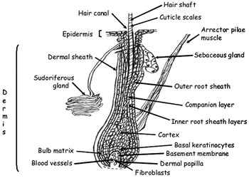
The contribution of the hair coat to environmental protection and behavioural display of animals is well recognised (Gerken, Reference Gerken2010). Important properties of hair include thermal insulation and visual appearance, which affect the value of fibre harvested for use by human commerce. Hair is the product of synthetic processes by hair follicles (Figure 1), which are embedded in the skin, uniquely in mammals. The follicles occur in two main anatomical structures in ‘primary’ or ‘secondary’ forms and in a range of subtypes.
Diagramatic representation of a non-medullated mammalian primary anagen hair follicle showing basal and suprabasal keratinocytes, central cortex (hair shaft) and cuticle and increasingly peripheral inner root sheath layers, companion layer, outer root sheath (all epidermal) and dermal sheath. The dermal papilla, arrector pilae muscle and sebaceous and sudoriferous glands are also shown.

The most commercially important animal fibres are those produced by secondary hair follicles. Commercial value of fibres is dependent on numbers and density of follicles, which affect the quantity of fibre produced, and the smallness of diameter that determines ‘fineness’. Typical values of fineness and annual raw fibre yield for the major fibre-producing animal species are: South American camelids: Vicuña (<15 μm: 0.5 to 1.5 kg), Guanaco (15 to 18 μm: 0.5 to 1.5 kg), Alpaca (18 to 30 μm: 1.5 to 5.5 kg) and Llama (>20 μm: 1.5 to 2.0 kg). Alpaca have a predominantly secondary follicle single coat and are domesticated as the double-coated Llama while the two essentially undomesticated Vicuna and Guanaco are also double-coated. Approximate diameters and annual yields for Cashmere (from double-coated goats) and mohair (from predominantly single-coated goats) are 12 to 18 and 22 to 30 μm and 0.06 to 1.0 kg and 2.0 to 5.0 kg, respectively. For sheep, yields typically up to 6 kg and in excess of 15 kg at production extremity, may be obtained with average diameters varying, for example, from 17 μm for certain predominantly single-coated fine wool Merino genotypes to >30 μm for double-coated Scottish Blackface. Annual production of fibre by Angora rabbits is typically up to 1.5 kg at 10 to 13 μm. Other important properties of fibre are medullation (poorly formed or hollow central core of fibres), cellularity of external cuticular surface and presence of pigment-producing melanosomes. Greater quantities of fibre, with generally reduced fineness, are normally associated with increasing age and production by male animals. Additional factors that determine value of harvested secondary fibres include uniformity, crimp, staple length and the presence of primary fibres (e.g. guard hair and kemp), which require separation during processing. Commercial value is generally optimised from animals, which exhibit high numbers and ratios and activity of secondary to primary follicles and genetic potential for rapid and prolonged growth while maximising fineness of fibres. There are therefore a number of properties of hair follicles, which contribute to the value of their fibre products and understanding of which offers potential for their improvement. This review addresses current knowledge, derived from a range of species and follicle types, at the level of (a) whole animal (integumental tissue: follicular v. non-follicular regions in skin), (b) skin (pre-natal development; post-natal type, numbers and densities of follicles) and (c) follicle level (cycling; changes in anatomy and synthetic capacity; cellularity and extracellular structures; genomic and proteomic expression; pigmentation). Brief consideration is given to nutrition and to the relationships between regulatory processes and expression of phenotype as tools to improve selection of animals of superior genotype.
General properties of integumental tissues
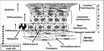
Hair follicles are important components of the mammalian integument which has many tissues which share common physical and compositional properties (Goldsmith, Reference Goldsmith1991; Galbraith and Scaife, Reference Galbraith and Scaife2008). Integumental tissues are composed largely of dermis and epidermis, which interact to synthesise the keratinised cellular end products of the epidermis, which include skin, hair, hoof and head horn. These end products are typically produced by proliferation and differentiation of specialized epidermal (epithelial) cells located on the basement membrane adjacent to the underlying dermis such as in a general model (Figure 2) or specifically in the matrix of the hair follicle (Figure 1). The dermis is composed principally of an extracellular matrix of adhesion and connective tissue molecules, which are produced by a population of fibroblast cells specialised to anatomical location and physiological function. The dermis is vascularised and supplies regulatory molecules and nutrients to the non-vascularised epidermis. Regulatory signalling between epidermis and dermis is recognised to have a major influence on epidermal gene expression, development, and rates of synthesis of end-products. Typically, also positioned on the basal layer are melanocytes. These produce melanic pigments which are transferred to adjacent keratinocytes in melanosome organelles and which give colour to the epidermal tissues produced. Proliferation of basal epithelial cells occurs by mitosis with one ‘daughter’ cell normally remaining to divide further and the other committing to differentiation and suprabasal migration. Included in the differentiating process is the development of a keratinised cytoskeleton with expression of ‘hard’ keratins or ‘softer’ cytoskeletal keratins, which form intermediate filaments (IFs), and keratin-associated proteins (KAPS). Adhesion between keratinocytes includes desmosomal junctions, which also attach internally to cytoskeletal keratin-containing IFs, and adherens junctions involving actin-containing microfilaments. Adhesion to the basement membrane involves hemi-desmosomes and adhesion plaques with connections to IFs and microfilaments, respectively (e.g. Odland, Reference Odland1991; Lodish et al., Reference Lodish, Berk, Zipursky, Matsudaira, Baltimore and Darnell2000). Protein molecules with important roles in adhesion include members of the cadherin and integrin families in epidermis and fibronectins in dermis. The dermal extracellular matrix typically contains connective tissue molecules such as collagens and elastins and hydrophilic negatively charged proteoglycans such as hyalurans and sulphated keratans (Malgouries et al., Reference Malgouries, Thibaut and Bernard2008). Remodelling of dermal matrix typically involves activation of catabolic enzymes including matrix metalloproteinsases and synthesis of macromolecules by fibroblasts. The proliferation and migration of epidermal cells and modelling of structures such as the hair follicle involve changes in presence and activity of adhesion molecules to permit detachment and re-attachment of cells. Terminal events occur at variable distance and layers of cells from the basal layer and typically result in specialised apoptosis and denucleiation and partial dehydration of cornified cells. The physical properties of integumental tissue end-products such as hair fibre may be attributed to the presence of a chemically resistant fibrous cytoskeleton and effective adhesion between keratinocytes. Integumental tissues are also populated by stem/progenitor cells, which support long-term production and renewal of cells of the epidermis.
Diagramatic representation of typical anatomy of mammalian integumental tissue showing outer ectodermally derived epidermis in close contact, via a contiguous basement membrane, with mesenchymally derived dermis. The epidermis is non-vascular and contains basal keratinocytes, which on division, differentiation and movement, form outermost layers of cornified keratinocyte end products such as skin, hoof horn or hair fibre. Note the presence of a neuro-ectodermally derived melanocyte that contains melanosomes. These synthesise and transfer pigment to keratinocytes. The dermis is composed of connective tissue molecules of the extracellular matrix, which is synthesised by specialised fibroblasts. These also secrete a range of signalling molecules important in determining proliferation and differentiation of keratinocytes. Additional signalling affecting both fibroblasts and epidermal cells derives from systemic provision via the dermal vascular capillary system. (see text for details).

Functional anatomy of the hair follicle
The main anatomical features of a representative anagen, non-medullated, primary hair follicle are shown in Figure 1. The structure of the follicle is embedded in the skin and constitutes a hair shaft surrounded by concentric cylinders of ectodermally derived root sheaths and a mesenchymally derived outermost dermal sheath. The dermal sheath contains progenitor mesenchymal cells that contribute to maintenance and regeneration of the distally located dermal papilla (DP) (Jahoda and Reynolds, Reference Jahoda and Reynolds2001). The DP has an essential role in regulating the activity of basal epidermal cells positioned in close proximity on the basement membrane and suprabasally. The volume of the DP is important in determining the numbers of basal epidermal keratinocytes positioned on the basement membrane and consequently diameter of the hair cortex and fibre product. Hair is synthesised by proliferation and differentiation of cells in the matrix region of the bulb which produce the most centrally positioned medulla (if present), cortex and cuticle and the increasingly peripheral cuticle and Huxley’s and Henle’s layers of the inner root sheath (IRS). The outer root sheath (ORS)-supporting epidermal layer is known to contain progenitor cells which can contribute both to regeneration of follicle matrix and to replacement of skin epidermis in, for example, response to wounding (Jahoda and Reynolds, Reference Jahoda and Reynolds2001).
During fibre growth, while the hair-forming cells migrate towards the skin surface along with cells of the IRS, the latter separate and slough into the piliary canal typically at the level of the sebaceous gland. Although requiring a supply of nutrients, IRS cells do not contribute directly to synthesis of hair shaft product and so reduce efficiency of utilisation of nutrients by the follicle (Hynd, Reference Hynd1989). The cuticle of the IRS forms adjacently to the hair shaft cuticle and contributes to the patterns characteristic of individual fibre types (e.g. Antonini, Reference Antonini2010). The single companion layer of cells separating IRS from ORS has an important role in facilitating separation of these supporting structures in processes involving disconnection and reconnection of intercellular adhesion (Orwin, Reference Orwin1989). The hair fibres that are present at birth and postnatally are the product of keratinocyte proliferation, with progressive expression of cytoskeletal and other genes associated with differentiation. Genes expressed in keratinocytes basally and during suprabasal movement include those for filamentous keratins (low to intermediate cysteine-containing), keratin-associated proteins (KAP: non-filamentous, (a) high cysteine content or (b) high glycine and tyrosine (HGT) content) in the cytoskeleton, and adhesion proteins. The latter proteins are important for basement membrane-cell and cell-cell adhesion. They also contribute to physical properties of fibre in forming connections with cytoskeleton and have central roles in hair follicle development and movement of cells during the hair follicle cycle (see below). Additional proteins that contribute to cytoskeleton of keratinocytes are actins and tubulins, which form microfilaments and microtubules, respectively.
There is major interest in keratin proteins as components of IFs and which have been characterised as contributing ca. 0.85 of total protein in cornified epidermal cells (Fuchs and Marchuk, Reference Fuchs and Marchuk1983). Keratins have been identified according to possession of either acidic (Type 1) or neutral/basic basic (Type II) properties. These form obligate hetero-polymers (i.e. each Type 1 combining with an associated type II molecule) and which polymerise further to provide the 8 to 10 nm diameter IFs. These combine with the non-filamentous KAPs in formation of the cytoskeleton of the hair shaft keratinocytes. Keratins have been characterised according to electrophoretic profiling and more recently by proteomic analysis for individual molecular species (Plowman, Reference Plowman2003) in wool. Proteomic analysis has more recently described the presence of hair type keratins in claw horn of cattle (Galbraith et al., Reference Galbraith, Flannigan, Swan and Cash2006; Galbraith and Scaife, Reference Galbraith and Scaife2008) with two dimensional electrophoretic profiles similar to those described for hair by Plowman (Reference Plowman2003).
The origins of keratins have been the subject of investigation in the evolutionary development of integumental structure and function (Fuchs and Marchuk, Reference Fuchs and Marchuk1983). There has been particular interest in the role of hair and its insulating properties in the change from poikilothermic (e.g. reptilian) to endothermic (e.g. mammalian) systems of regulating body temperature (Gerken, Reference Gerken2010). There has thus been interest in genomic comparisons across a range of species.
Results from the human genome project have been particularly valuable in, for example, identifying a total of 54 functional keratin genes. These encode for 28 type I and 26 type II keratins, clustered on chromosomes 17q21.2 and 12q13.13, respectively, with the type I keratin K18 gene also sited on the type II keratin gene domain (Schweizer et al., Reference Schweizer, Bowden, Coulombe, Langbein, Lane, Magin, Maltais, Omary, Parry, Rogers and Wright2006). Among these are epithelial cytokeratins (17 type I and 20 type II) and hair-type ‘hard’ α-keratins (11 type 1 and 6 type II) which have been investigated to determine structural properties in IFs by techniques such as X-ray diffraction (e.g. Rafik et al., Reference Rafik, Doucet and Briki2004). The location of human KAP genes in at least 21 molecular families and grouped together on five chromosomes has also been identified (e.g. Rogers and Schweizer, Reference Rogers and Schweizer2005; Wu et al., Reference Wu, Irwin and Zhang2008) with reference made to knowledge and nomenclature available for application to other species. Previous reports in the sheep have described mapping of type 1 and type II keratin genes to chromosome 11 and 3, respectively (Powell, Reference Powell1997) with the high glycine-tyrosine KAPs ascribed to Chromosome 3 (McLaren et al., Reference McLaren, Rogers, Davies, Maddox and Montgomery1997). The deposition of specific keratins and KAPs in the follicle has also been described for particular hair follicle cell lineages. For example, follicles of human anagen hair were dissected and analysed for the presence of keratins as differentiation markers by Limat et al. (Reference Limat, Breitkreutz, Stark, Hunziker, Thikoetter, Noser and Fusenig1991). These workers described the exclusive presence of acidic and basic ‘hard’ alpha-keratins in cortex with peptides derived from, or related to, K (keratin) 1 and K10 in IRS and cuticle and the ‘soft’ (cyto)keratins K5, 6, 14, 16, and 17 present in ORS. These results indicate site-specific regulation by mechanisms that have not been described in detail.
Hair follicle cycling
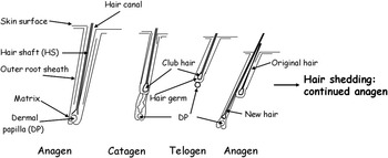
A characteristic property of the post-natal hair follicle is a cyclical pattern of activity (Figure 3). Active growth (anagen) typically involves proliferation and differentiation of matrix cells to produce hair shaft and IRS with incorporation of a small number in the ORS (Ebling et al., Reference Ebling, Hale and Randall1991; Schneider et al., Reference Schneider, Werner, Paus and Wolf2008). The length of anagen varies between follicles differing in anatomical type (i.e. primary v. secondary), body locations and species of animal. For example, pelage secondary follicles of Merino sheep or Angora goats, which are essentially single-coated, may retain activity for 2 years or longer which contrasts with 6 months or less for the double-coated cashmere-bearing animal (e.g. Allain and Renieri, Reference Allain and Renieri2010). Anagen is followed by a short catagen phase which features cessation of proliferation, apoptosis of the lower portion and shortening of the follicle and retention of the hair in a keratinised ‘club end’. The follicle shortens further in the telogen resting phase, which includes loss of the inner root sheath while the DP remains in contact with epidermal germ cells, extending beyond the club end. Changes in intracellular volume of fibroblasts and composition of the extracellular matrix of the DP occur concomitant with changes in physiological state of the hair follicle. New anagen involves proliferation and movement of stem cells from the bulge area (located below the sebaceous gland) in the mouse or from ORS in the sheep (Rogers, Reference Rogers2006) to form a new matrix of progenitor cells which surround the dermal papilla. These cells then proliferate and differentiate according to anatomical position to form the hair shaft, medulla, if present, and other concentric epithelial layers of the renewing follicle. The timing of shedding or moulting ‘exogen’of the club hair during telogen or frequently at the beginning of a new anagen is important in harvesting of fibre. This is particularly so where there is synchronisation of follicle activity, which frequently occurs in double-coated animals such as cashmere-bearing goats.
Diagramatic representation of the major events in the cycle of a secondary follicle (sebaceous gland not shown) exhibiting the morphology of anagen growth phase, regression into catagen and quiescence in telogen, followed by regeneration and resumption of anagen hair growth.

How do follicles get there? Embryonic and post-natal development
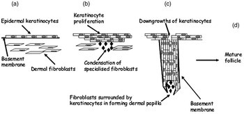
Typically, across species, hair follicles develop prenatally from cells in the externally positioned embryonic ectoderm and underlying mesenchyme, which ultimately form, in the skin, epidermal and dermal tissues, respectively (e.g. Stenn and Paus, Reference Stenn and Paus2001; Schmidt-Ullrich and Paus, Reference Schmidt-Ullrich and Paus2005). Follicle numbers are considered to be genetically determined and fixed around birth, although changes in activity of individual follicles occur during the lifetime of the animal. Cells from the neural crest provide pigment-producing melanocytes. Development of hair follicles has been described in eight stages with major features depicted in Figure 4. The initial inductive events result in the clustering together of epidermal keratinocytes and formation of a ‘hair placode’ and formation, in the underlying mesenchyme, of a condensate containing specialist fibroblasts. Molecular signalling inter-communication gives rise to proliferation of keratinocytes and fibroblasts and stimulation of downgrowths of keratinocytes into the dermis. Subsequent organogenic development produces a ‘bulbous peg’ structure, which has, at its base, a mesenchmally derived DP in close contact, across a basement membrane, with surrounding basal epidermal cells.
Diagramatic representation of hair follicle development under the influence of ectodermal-dermal signalling (see text). (a) undifferentiated ectodermal keratinocytes and mesenchymal fibroblasts separated by basement membrane. (b) proliferation of keratinocytes and condensation of fibroblasts to form a dermal condensate. (c) keratinocytes grow down into dermis and surround the dermal papilla at the base of the follicle. (d) subsequent development includes commitment of basal keratinocytes to inner root sheath and other cell lineages, formation of accessory structures, recruitment of melanocytes and vascularisation of dermal papilla to form the mature follicle (see Figure 1). Modified from Millar (Reference Millar2002) and Jamora et al. (Reference Jamora, Lee, Kocieniewski, Azhar, Hosokawa, Chai and Fuchs2005).

Mesenchymally derived cells also form the outer connective tissue sheath that surrounds the follicle. Subsequent development involves cytodifferentiation of the follicular epidermis and formation of cylindrical structures from cells with defined lineages, of first the IRS and then ORS and the differentiation of cells, which form the central hair shaft (see Figure 1). Subsequent recruitment of melanocytes and initial synthesis of melanins occurs and individual keratinocytes commit to formation of associated apocrine and/or sebaceous glands. The DP also become vascularised and innervated at this later stage. Recruitment of Langerhans and T cells of the immune system also occurs in the follicular epidermis concomitant with attraction of mast cells and macrophages into surrounding mesenchyme.
The mouse (murine) pelage provides a useful general model to describe the synchronised sequence of events in the formation of primary and secondary hair follicles. Regulatory mechanisms which are becoming increasingly recognised for mice and which have parallels in development and activity of other multicellular organisms provide pointers for investigation in commercial fibre-producing animals. For example, mice have four major pelage hair types (e.g. Schmidt-Ullrich and Paus, Reference Schmidt-Ullrich and Paus2005; Schlake, Reference Schlake2007). These are produced by primary (‘guard hair’) follicles, which are first to initiate development at embryonic (E) day 14 and produce associated structures, the arrector pili muscle, the sebaceous and apocrine glands and hair canal (e.g. Figure 1). Secondary follicles with associated sebaceous gland and hair canal only, develop next. The latter follicles produce different hair types including awls (E 15.5: straight fibre), auchene (E 15.5: single bend in fibre) and zig-zags (E17: more than one bend).
The development of primary, secondary and derived follicles in the pelage of Merino sheep has been described by Rogers (Reference Rogers2006). Primary follicles with arrector pilae muscle, sebaceous and apocrine glands commence development first (E70) in the form of a central follicle surrounded by two lateral primaries. Secondary follicles (So) associated with primaries appear next (E85). Additional (SD) follicles develop by branching from So follicles and are apparent by E105. These share common hair canals and the latter follicles contribute up to 0.80 of the mature follicle population. Although induction of all follicles and maturity of primaries is considered to be complete by birth at E145 to 150, growth of hair by secondaries continues to develop for a further 4 to 5 months. Major features of genetic selection in Merino sheep are therefore large numbers of secondary follicles producing high yields of uniformly small-diametered wool fibres. A similar pattern for follicle activity has been described for Angora goats (e.g. Dryer and Marincowitz, Reference Dryer and Marincowitz1967). In these animals, reductions in the activity of primary follicles occurs by months 3 to 4 with secondary follicle maturation completed by 6 months postnatally. Subsequent changes may occur during the lifetime of the animal according to the characteristics of, for example, the hair follicle cycle (see below).
Regulation of development: principles of morphogenesis and mitogenesis, signalling and genetic variation
The development of specialised structures in animals occurs by the process of morphogenesis in which cells and extracellular products are synthesised to form characteristic functional anatomical structures (Figure 5). Such growth of tissues both prenatally and postnatally typically occurs in a genetically programmed sequence and during ‘windows’ of development under the influence of signalling molecules and adequacy of nutrient supply (Godfrey, Reference Godfrey1998). Such signalling is conventionally thought to be induced by gradients of morphogens where effects are produced as a result of direct interaction with target cells and according to the concentration present. Similarly, increase in cells numbers arising from mitotic events in the process of cell division are subject to regulation by signalling molecules with mitogenic activity. Normal development requires the presence of, and conformity to, regulatory processes in order to prevent the uncontrolled growth associated with neoplasic lesions.
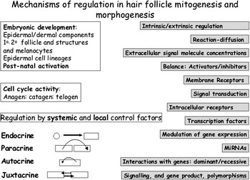
Summary of mechanisms involved in regulating mitogenesis (cell proliferation) and morphogenesis (development of anatomical features) in hair follicle. Major events are associated with gene expression, which is affected by composition of DNA at individual loci in polymorphic forms, and composition and activity of expressed proteins (e.g. ligands, and their antagonists, receptors and enzymes) in producing particular phenotypes.

There are a number of the mechanisms by which chemical signalling ‘messenger’ molecules interact with tissues in mammalian systems and regulate behaviour and activity of constituent cells (e.g. Figure 5). These include the production of molecules distant from site of action by non-endocrine tissues and ‘classical’ hormones produced by cells in endocrine glands (e.g. Lodish et al., Reference Lodish, Berk, Zipursky, Matsudaira, Baltimore and Darnell2000). Regulation produced by molecules close to target cells is generally classified as ‘paracrine’ in action with ‘autocrine’ activity defined as occurring in which cells both synthesise, and are acted on by, the secreted molecule. ‘Juxtacrine’ signalling arises from the interaction of molecules extending from external membranes of adjacent cells.
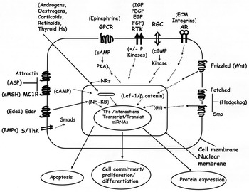
Signalling molecules may be classified chemically and by the system of interaction with target cells. A range of such interactions is shown diagrammatically in Figure 6. For example, proteins and peptides, catecholamines, prostaglandins and eicosanoids interact as ‘ligands’, with receptors positioned in the cell membrane. For these, transduction (transfer) of signal may activate transmembrane G protein-coupled receptors in a process involving exchange of guanosine diphosphate (GDP) with guanosine triphosphate (GTP) in associated binding proteins. Subsequent activation of downstream enzymes within the cell produces second messengers including cyclic adenosine monophosphate (cAMP), inositol 1,4,5 triphospate (IP3), diacylglycerol (DAG) and release of sequestered (Ca2+) calcium ions. Increases in concentration of, for example, cAMP may then activate protein kinase A (PKA) and phosphorylation of other proteins involved in interaction with DNA and affecting expression of certain genes. Smaller molecules such as Ras protein also express activity when bound to GTP and loss of activity on hydrolysis of GTP to GDP. The receptor guanyl cyclase responds to ligand binding by synthesis of cyclic guanosine monophosphate. Other membrane receptors exhibit receptor tyrosine kinase (RTK) activity, which on reacting with ligand on extracellular surface, activate intracellular auto-phosphorylating activity on tyrosine residues in addition to phosphorylating and activating proteins in other signalling pathways. Certain receptors (S/ThK), on activation, also target phosphorylation of serine/threonine residues. It is evident that the addition by kinases, or removal of phosphate groups, by phosphatase enzymes, may alter the functional activity of proteins by changing enzyme activity or interactions with other proteins in signalling networks and pathways including those regulating gene expression. Adhesion receptor systems typically involving integrins and binding molecules in extracellular matrix may alter conformation and signal via cytoskeletal interactions.
Diagram showing signalling systems in an epidermal cell. Receptors (general transmembrane structure shown and not to scale) located in membranes typically have an external ligand recognition site, variable transmembrane components and intracellular domain, which is activated on occupancy by ligand. ‘Message’ transmission occurs via synthesis of molecules such as c(cyclic) AMP, cGMP, IIP3 and DAG or by phosphorylation (kinases) and dephosphorylation (phosphatases) of intracellular proteins in complex signalling pathways. Certain ligands (steroids, retinoids, thyroid hormones) interact directly with receptors located in cytoplasm or nucleus. Typically, these pathways terminate in activation or inhibition of expression of transcription factors (TFs), which along with accessory molecules, interact with nuclear DNA binding sequences and promote gene expression. Outcomes include synthesis of proteins, cell proliferation and differentiation and apoptosis. Abbreviations: AR, adhesion receptors; GPCR, G-protein-coupled receptors; NR, nuclear receptors; RGC, receptor guanyl cyclases; RTK, receptor tyrosine kinases; S/ThK, serine/threonine kinases. Individual systems include EDa/EDAr (Dickkof) and NFκB; BMPs (Noggin), Smad proteins; Wnt/Frizzled, Lef/β-catenin; Shh (hedgehog)/Patched/Smo, Gli, αMSH/Asp/MC1R/attractin; IGFs/Igfbps; EGFs; FGFs. (See text for details).

The second mechanism of signalling involves the movement of ligand into the cell, and interaction with specific cytoplasmic or nuclear receptors (NRs). The majority of these receptors require the presence of the ligand for activation and expression which can involve dimerisation, movement to the nucleus, binding to the hormone response element of the genetic DNA, activation of the transcription process and synthesis of messenger RNA followed by translation into protein. Signalling molecules that regulate gene expression via such intracellular receptors/transcription factors include steroids, thyroid hormones and retinoids. Productive signalling requires both the presence of a secreted ligand and its receptor in a receptive form. Receptors in membranes are synthesised by gene expression, typically transported by endocytotic process (Seto et al., Reference Seto, Bellen and Lloyd2002) and subjected to recycling by internalisation and repositioning. Molecular outcomes are regulated by concentrations of ligand, affinity to receptor, numbers of receptors and concentrations and affinities of inhibitors. Evolutionary changes in genes encoding for signalling proteins have given rise to ‘polymorphic’ variants which have changes in their composition, structure and affinity for receptors. Similar genetic changes in expressing genes have given rise to variants in protein structure and signalling properties of receptors. Taken together, these changes in ligands and receptors have been related to recognised variation in hair fibre phenotypes.
As indicated, modulation of signalling pathways may be achieved at the level of receptor where competing molecules (antagonists) may block interaction with primary ligand, or where alternative pathways are activated by the presence of other ligands acting on the same receptor. Induction of other ligand-receptor-activated pathways may induce expression of different genes and promotion of morphogenic and/or mitogenic events. Variation in response may be introduced by expression of poymorphisms in genes affecting composition of protein/peptide products acting within pathways or as factors in transcription, epigenetic modification (e.g. changes in histone proteins in chromatin (methylation, acetylation, phosphorylation or ubiquitination) or methylation or acetylation of cytosine bases in DNA without changing its primary sequence (Mathers, Reference Mathers2008)) or by post-translational modification of expressed proteins. There is also increasing evidence, in multicellular organisms, of regulation of translation into protein products by interactions of messenger RNA with short length microRNA species. These are single stranded, typically contain 21 to 23 nucleotides and appear to act by repressing translation activity and/or increasing degradation of mRNA (Nilsen, Reference Nilsen2007).
In the context of developmental signalling, a range of common molecules and pathways are well recognised to determine, in multicellular organisms, pre- and post-natal development in organs and tissues, including the hair follicle, (Schmidt-Ullrich and Paus, Reference Schmidt-Ullrich and Paus2005; Schneider et al., Reference Schneider, Schmidt-Ullrich and Paus2009) There is also a requirement for closely controlled signalling in the regulation of cycling of the hair follicle. This process, uniquely in mammals, involves breakdown and regeneration of the hair-synthesising structures in a process similar to events occurring during embryonic development. The mechanism by which such events occur are becoming better understood and may have a basis in the concept of gradients of concentrations of activators and inhibitors in a ‘reaction-diffusion’ system (e.g. Nagorcka and Mooney, Reference Nagorcka and Mooney1989). Such a system may be applied to the explanation of the interactions between epidermis and dermis and the differences in gene expression between for example, basal and increasing layers of suprabasal keratinocytes and differentiation into the defined cell lineages of hair shaft and root sheaths.
Molecular and gene biology and phenotypic expression
How do molecular and gene biology of hair follicles relate to issues of importance in the phenotypic expression of fibre production of animals of commercial importance (e.g. Figure 7)? Other papers in the symposium have described the presence or absence of hair follicles in different areas of the skin (Gerken, Reference Gerken2010) and important roles in physiology of thermal regulation. The numbers, types and activities of hair follicles and physical characteristics of fibres in different genotypes of South American Camelids have been described (Antonini, Reference Antonini2010). In addition, the paper of Allain and Renieri (Reference Allain and Renieri2010) has evaluated aspects of quantitative and molecular genetics relating to properties of follicles and fibre. Traits of economic importance considered included yield, quality and colour. The importance of applying this information in breeding programmes has been established and may be advanced further by a deeper analysis of the underlying mechanisms regulating hair follicle development, cycling and production of fibre and identification, for example, of important molecular markers to assist in genetic selection. The following section provides a contemporary review on aspects of such mechanisms with information derived from a range of investigative approaches.
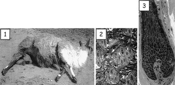
Images of phenotypic expression of hair follicle characteristics at levels of: 1, whole animal (Llama) afollicular and follicular skin (with variation in hair fibre pigment) according to anatomical site (Gerken, Reference Gerken2010); 2, bundles of primary and secondary follicles in histological horizontal skin section of Q’aras llama (Sacpic staining (Antonini, Reference Antonini2010)); 3, vertical section of a cashmere follicle (Toluidine blue; ×128 magnification; Lee and Galbraith (unpublished).

Regulation of pre- and post-natal development of the hair follicle
In terms of biological characteristics that determine the morphogenesis of the different types of hair follicle in individual animals, the major events are initiation, anatomical development, differentiation of keratinocytes into specific cell lineages and positioning in skin of hair follicles (e.g. Schmidt-Ullrich and Paus, Reference Schmidt-Ullrich and Paus2005; Schneider et al., Reference Schneider, Schmidt-Ullrich and Paus2009). Information on molecular regulation of these developmental processes and subsequent fibre production is limited in the major fibre-producing species. Consequently, much of the following information is derived from studies on small animals and particularly genetically modified mice. Such studies have particularly illuminated developmental regulation prenatally although they have contributed less in describing post-natal control processes where the gene modifications produce lethal mutations. It is evident that regulation is complex and tightly regulated by processes based on signalling pathways involving chemically distinct molecules. These molecules, or their homologues, have been implicated in controlling interactions, within and between, epithelia and mesenchyme in organisms across a long evolutionary time frame and include tissues such as scales and feathers. Such apparent conservation of regulatory mechanisms does encourage a common approach to explanation across species. However, caution should be exercised particularly where specialised species-specific hair follicle phenotypes are produced. Aspects of regulation of hair follicle development and fibre production have been considered in detail and extensive bibliographies provided by authors including Millar (Reference Millar2002), Schmidt-Ullrich and Paus (Reference Schmidt-Ullrich and Paus2005) and Schneider et al. (Reference Schneider, Schmidt-Ullrich and Paus2009). The review of Galbraith (Reference Galbraith2006) has context in biology of fibre production by South American camelids and those of Rogers (Reference Rogers2006) and Purvis and Jeffery (Reference Purvis and Jeffery2007) provide focus to production by sheep and goats.
Regulatory pathways and signalling molecules in tissue and follicle development
Hair follicles are uniquely found in mammals, and are formed in the context of growth of specialised structures and tissues, which develop from a single egg cell following fertilisation in utero. Studies on regulation of pre-implantation development from morula to blastocyst stages suggest the presence of intracellularly driven autopoietic mechanisms and the absence of a requirement for externally derived chemical signalling (e.g. O’Neill, Reference O’Neill2008). This process involves the synthesis of signalling ligands within cells and their secretion and action by autocrine action on specific receptors on the membranes of the producing-cells (e.g. Figures 5 and 6). Included in the receptor-mediated responses to these ligands are 1-o-phosphatidinyl-3-kinase and phosphatidylinositol-3,4,5-trisphosphate. This latter molecule is involved in signalling for insulin and IGF 1 and acts in the cell membrane to provide a docking site for regulatory proteins containing the pleckstrin homology domain. These include the kinases that activate the Wnt/β-catenin and NF-κB (NF kappa B) pathways and which are essential for normal blastocyst implantation and development. Members of the Wnt gene family of secreted ligands activate the Frizzled receptor-mediated pathway, which inhibits phosphorylation and resulting inactivation of β-catenin which is then available to translocate to the nucleus (Figure 6). It then forms complexes with members of the lymphoid enhancer binding factor/T cell factor (Lef/Tcf) family and activates transcription of target genes. Other authors (e.g. Mohamed et al., Reference Mohamed, Dufort and Clarke2004) have indicated the importance of the expression of Wnt ligands (particularly Wnt 7a with antagonism by its antagonist, secreted Frizzled related protein 2 (sFRP 2)) in mediating implantation-related embryo-uterine communication in mice. Following implantation, the cells in the embryo proliferate and reposition during gastrulation to form the cell layers of endoderm in addition to ectoderm and mesoderm from which epidermis, dermis and ultimately hair follicles are derived. Secreted Wnt proteins have been ascribed importance in posterior-anterior positioning of cells with modulation of their activity induced by antagonists to which they bind directly and interfere with binding to the (Wnt) receptor Frizzled (Caneparo et al., Reference Caneparo, Huang, Staudt, Tada, Ahrendt, Kazanskaya, Niehrs and Houart2007). Similarly, Dickkof 1 (DKK 1), a recognised inhibitor of the Wnt/β-catenin pathway, has been suggested to modulate gastrulation in pathways which include those independent of β-catenin signalling.
These pathways are examples of systems, which regulate a diverse range of physiologically essential processes in development of tissues and organs in animals. They are of particular relevance because they also have specific importance in hair follicle morphogenesis, which follows initial differentiation of embryonic cells into ectoderm and underlying mesoderm and in the epithelial–dermal interactions indicated below.
For example, post-implantation data from murine studies implicates Wnt/β-catenin signalling as a possible first dermal signal that is essential for initiation of placode formation with subsequent expression in both epithelial and mesenchymal cells (Figure 4). Placode proliferation is promoted by a range of other activators including fibroblast growth factors (FGFs), transforming growth factors (TGFs), and the juxtacrine-acting molecules, Delta 1 and its receptor Notch 1. Mesenchymal cells, under the influence of epithelial signals, congregate to form condensates that develop into the dermal papilla. Activators which promote proliferation of epithelial cells, downgrowths into dermis and formation of the germ, peg and bulbous peg include platelet derived growth factor and sonic hedgehog (Shh). Shh has a central role in embryogenesis and organogenesis in mammalian systems as a member of the hedgehog family of secreted molecules. Binding to its receptor Patched 1 (Ptch 1) removes the inhibition otherwise exerted by Ptch 1 on the associated receptor complex with smoothened protein and permits signalling via the transcription factors Gli 1 and Gli 2 on target genes. Although Shh protein is expressed only in epidermis, its receptors are present in both epithelium and mesenchyme. One important effect of Shh signalling is stimulation of proliferation of epithelial cells needed for anatomical development of the maturing hair follicle. Jamora et al. (Reference Jamora, Lee, Kocieniewski, Azhar, Hosokawa, Chai and Fuchs2005) have described a TGF β2 signalling system that transiently induces the transcription factor Snail causing down-regulation of E-cadherin activation of the Ras-mitogen activated protein kinase pathway and enhanced keratinocyte proliferation. There is also recent evidence for an essential role for certain microRNA species produced by the ribonuclease enzyme Dicer in the formation of epidermal downgrowths (Yi et al., Reference Yi, O’Carroll, Pasolli, Zhang, Dietrich, Tarakhovsky and Fuchs2006). Other work (Wenguang et al., Reference Wenguang, Jianghong, Jinquan and Yashizawa2007) has described the presence, in skin of adult sheep and goats, of microRNAs associated with murine hair follicles. Such microRNAs have been ascribed important roles in regulation of gene expression by interfering with translation of messenger RNA (Nilsen, Reference Nilsen2007).
Although regulation of follicular initiation and growth is important, interest remains in the properties of afollicular epidermis such as described by Gerken (Reference Gerken2010) or epidermis with variable concentrations of hair follicles such as described for South American camelids by Antonini (Reference Antonini2010) (Figure 7). Current information suggests that hair follicle development is inhibited unless constitutively expressed placode inhibitors such as bone morphogenic proteins (BMPs) 2 and 4, or Activin βA are adequately suppressed by Noggin or Follistatin, respectively (Schmidt-Ullrich and Paus, Reference Schmidt-Ullrich and Paus2005). Noggin forms a complex with BMP 4 and prevents binding to its receptor in the early dermal condensate and facilitates Lef 1 expression. Similarly, as indicated above, DKK 1 is an effective inhibitor of the Wnt signalling needed to stabilise β-catenin and permit its interaction with the Tcf/Lef DNA binding proteins and so induce hair follicle initiation. In addition, Wnt/β-catenin and Noggin/Lef 1 may also inhibit the expression of the inter-cell adhesion molecule E-cadherin that is required to permit placode downgrowth. The expression of other adhesion molecules such as α-catenin and β-integrin is thought to affect exchange of signals and adhesive interaction between and within epidermal and dermal components.
In addition to issues concerning commitment to hair follicle morphogenesis or interfollicular epidermal and dermal fates, questions may be directed to mechanisms governing the production of the range of follicle phenotypes in mammalian skin. This question is particularly relevant for animals which express follicle phenotypes that produce fibre of yield and quality of high economic value and, for example, numbers and activities of primary and secondary hair follicles such as described for Merino sheep (Rogers, Reference Rogers2006). Results from mice do indicate differences in mechanisms that include time points and molecular signals. For example, initiation of primary follicle at E13/E14 requires Wnt and Ectodysplasin (Eda) A1/Eda receptor (Edar) and NF-κB regulation (Schmidt-Ullrich and Paus, Reference Schmidt-Ullrich and Paus2005) with patterning and interfollicular spacing influenced by the repression of Edar by dermal BMPs 4 and 7 and responsiveness, rather than localisation of ligand, determining outcome (Mou et al., Reference Mou, Jackson, Schneider, Overbeek and Headon2006). Initiation of secondary follicles (awl, auchene) at E16/E17 is dependent on Wnt/β-catenin and Noggin/Lef 1 and independent of EdaA1/Edar and NF-κB regulation. Regulation of zig-zag follicle type development is less well characterised although Eda A1/Edar and NF-κB regulation appears to be necessary for production of hair. This information is valuable since it provides targets for investigation of signalling pathways and gene expression governing the formation, location and numbers of hair follicles and the properties of their fibres in commercial species.
The close association of the different layers of epithelial cells formed in the maturing hair follicle during the stage of cytodifferentiation indicates concise commitment to individual cell phenotypes and over short spatial distances (Figure 1). The inhibition, by the gene product Noggin, of effects induced by BMP 4 has been shown to impair differentiation of hair shaft and cuticle while permitting proliferation of epithelial cells. The transcription factor Foxn 1 has been shown to suppress differentiation while promoting expression of certain hair keratin genes (Schlake, Reference Schlake2001). Signalling molecules involved in the formation of hair shaft cortex and cuticle include Notch 1 (and ligands Serrate 1 and 2) and BMPs (BMP 4) and Wnt paracrine signallers (with Wnt 10b specifically identified by Ouji et al. (Reference Ouji, Yoshikawa, Moriya, Nishiofuku, Matsuda and Ishizaka2008)). An essential role for the transcriptional repressor Cutl 1 has been indicated in the development of IRS (Ellis et al., Reference Ellis, Gambardella, Horcher, Tschanz, Capol, Bertram, Jochum, Barrandon and Busslinger2001). Similarly, the Hox family of transcription factors, which are usually expressed sequentially according to spatial position and time with respect to their positions in the Hox complex has been ascribed a role in regulation of patterning and developmental morphology of hair follicles (and other tissues of the integument) (Potter et al., Reference Potter, Peterson, Barth, Pruett, Jacobs, Kern, Argraves, Sundberg and Awgulewitsch2006). Expression of Hox C13 was detected in medulla, cortex, IRS, companion layer and upper inner ORS. Defects in Hox C13 expression have been shown to give rise to loss of hair in mutant mice. Potter et al. (Reference Potter, Peterson, Barth, Pruett, Jacobs, Kern, Argraves, Sundberg and Awgulewitsch2006) also showed that the regulatory transcription factor-encoding gene Foxq, is a downstream target for Hox C13 and acting through a common regulatory pathway controls differentiation of hair follicle medulla. Other transcriptional regulatory molecules include (a) the Sox 9 gene product, which has been ascribed an essential role in development of ORS and stem cell compartment of murine hair follicles in which it has continuous expression (Vidal et al., Reference Vidal, Chaboissier, Lützkendorf, Cotsarellis, Mill, Hui, Ortonne, Ortonne and Schedl2005), (b) Foxe 1, detected in the lower undifferentiated region of the HF and involved in hair follicle morphogenesis, as an apparent downstream target of the Shh/Gli pathway (Brancaccio et al., Reference Brancaccio, Minichiello, Grachtchouk, Antonini, Sheng, Parlato, Dathan, Dlugosz and Missero2004) and (c) Gata 3 (Kaufman et al., Reference Kaufman, Zhou, Pasolli, Rendl, Bolotin, Lim, Dai, Alegre and Fuchs2003) which has been ascribed a role in early specification of basal cells towards IRS as opposed to hair shaft lineage. The development of cell lineages has been associated with expression of keratins and other proteins according to cell type in differentiating hair shaft or supporting sheath structures (Limat et al., Reference Limat, Breitkreutz, Stark, Hunziker, Thikoetter, Noser and Fusenig1991).
Regulation of hair follicle cycling
Mechanisms governing the behaviour of the hair follicle cycle are gradually becoming understood (Figures 3 and 5). Control is applied at three inter-relating levels with extrinsic influences arising from response to environment (e.g. photoperiod and melatonin-prolactin axis (Lincoln, Reference Lincoln2002; Lincoln et al., Reference Lincoln, Andersson and Loudon2003); thermoregulation (Gerken, Reference Gerken2010); systemic endogenous physiological (e.g. androgens, oestrogens) and metabolic hormones (e.g. insulin, somatotrophin, thyroid hormones, corticosteroids (Messenger, Reference Messenger1993; Stenn and Paus, Reference Stenn and Paus2001) and intrinsic paracrine/autocrine interactions between and within epidermal and dermal components. These interactions are characteristic of the individual follicle type, its location and rhythmic expression of cellular components (Millar, Reference Millar2002; Schneider et al., Reference Schneider, Schmidt-Ullrich and Paus2009). For the latter category, there is considerable evidence to suggest that paracrine/autocrine ‘intrinsic’ signalling systems, which regulate post-fertilisation development and initial hair follicle morphogenesis as described above, also have roles in regulating cycling in post-natal hair follicles. These include Wnts (such as Wnt 10b)/β-catenin and Ptch 1 involved in early anagen. Although Shh appears not to be required for initiation of anagen, it appears essential for proliferation of epithelial cells and downgrowths of the regenerating follicle into the dermis. It contributes to maintenance of hair growth and its expression ceases on onset of catagen. The upregulation of expression of Noggin has been ascribed a role in initiating anagen, in which the inhibitory action of BMP 4, which is downregulated, is neutralised (Botchkarev et al., Reference Botchkarev, Botchkareva, Nakamura, Huber, Funa, Lauster, Paus and Gilchrest2001). BMP 4 has also been shown to inhibit anagen development in secondary hair follicles. Noggin increased Shh gene expression, which was downregulated by BMP 4. These results suggested mediation of its effects, at least partially, by Shh. Similarly, Fessing et al. (Reference Fessing, Sharova, Sharov, Atoyan and Botchkarev2006) have demonstrated a role for Eda and Edar signalling, particularly in the transition from active anagen to catagen with main expression in outer and inner root sheaths and secondary hair germ, and associated with apoptosis in ORS. The transcription factor p53 has also been demonstrated as being upregulated during apoptotic events in catagen (Botchkarev et al., Reference Botchkarev, Botchkareva, Nakamura, Huber, Funa, Lauster, Paus and Gilchrest2001).
Roles in hair follicle biology for insulin-like growth factors (IGF) 1 and II expressed predominantly in dermal papillae, and certain IGF binding proteins (Igfbps) have been considered by Weger and Schlake (Reference Weger and Schlake2005). IGF 1 has been ascribed a stimulatory role in epidermal proliferation and has been shown to contribute to cycle regulation and hair shaft differentiation in IGF 1-expressing transgenic mice. The binding protein Igfbp 3 has been shown to be increased in expression in DP cells during regression and early telogen, with Igfbp 5 evident during anagen. Similarly, Kawano et al. (Reference Kawano, Komi-Kuramochi, Asada, Suzuki, Oki, Jiang and Imamura2005), analysed murine skin samples for expression of the 22 genes encoding for members of the FGF family of secreted growth factors and for the four FGF RTK genes. Attention was drawn to previous reports of the effect of FGF 5, expressed in the ORS, in the induction of catagen (Hébert et al., Reference Hébert, Rosenquist, Götz and Martin1994) and the antagonistic effect of FGF 5S. Expression of FGFs varied throughout the cycle and subcutaneous administration of FGF 18 induced anagen in telogen follicles. These results may be considered in the context of the role of FGFs in regulatory and developmental processes which include patterning and morphogenesis and proliferation, differentiation and migration of cells (Thisse and Thisse, Reference Thisse and Thisse2005). Factors of importance include the proteoglycan heparan sulphate that facilitates ligand binding and the tight regulation in signal transduction, which modulates FGF signalling and is effected by feedback inhibitors such as the Sprouty, Sef and MAP kinase phosphatase 3 gene products. Epidermal growth factor (EGF) and other ligands of the EGF receptor (EGFR) have also been attributed roles in hair follicle regulation (Schneider et al., Reference Schneider, Schmidt-Ullrich and Paus2009). For example, studies with mice have suggested that EGF receptor signalling is essential for initiation of hair growth and if EGF is continuously expressed, entry to catagen is prevented (Mak and Chan, Reference Mak and Chan2003). This work supplements earlier observations of the depilatory action of EGF infusions in adult sheep (Moore et al., Reference Moore, Panaretto and Carter1985) and responses in vitro in stimulation of synthesis in ORS and inhibition in hair shaft of human (Philpott and Kealey, Reference Philpott M and Kealey1994) and caprine (Souri et al., Reference Souri, Galbraith and Scaife1997) hair follicles in inducing a catagen-like state.
Other systems signalling from greater distances from the skin and contributing to ‘extrinsic’ influences include androgens (Messenger, Reference Messenger1993). Recent work (Naito et al., Reference Naito, Sato, Matsumoto, Takeyama, Yoshino, Kato and Ohdera2008) demonstrated the importance of androgen receptors in mediating the inhibition of hair growth in mice by dihydrotestosterone. Ohnemus et al. (Reference Ohnemus, Uenalan, Inzunza, Gustafsson and Paus2006) have addressed the roles for oestrogens. They reported, for example, that topically applied oestradiol 17β induced premature catagen via oestradiol 17β-receptor (ER)α mediation in murine mouse skin and that ERβ-knockout mice exhibited accelerated catagen and greater numbers of apoptotic follicular keratinocytes. Their results suggested that ERβ may antagonise induction of catagen via the ERα signalling pathway. Certain corticosteroids have also been suggested to influence hair follicle cycling and relevant work includes the observation that elevations in blood plasma cortisol were associated with reductions in the ratio of secondary to primary follicles in perinatal twin Merino lambs (Thompson et al., Reference Thompson, Hebart, Penno and Hynd2007).
The duration of secretion of melatonin from the pineal gland during darkness is well recognised to signal perception, via hypothalamic-pituitary pathways, of photoperiod and season by certain species of animals. Recent studies have indicated the importance of hypothalamic triiodothyronione (T3) concentrations (via related melatonin, TSH and deiodinase actions), in mediating daylength-related changes in Siberian hamsters (Barrett et al., Reference Barrett, Ebling, Schuhler, Wilson, Ross, Warner, Jethwa, Boelen, Visser, Ozanne, Archer, Mercer and Morgan2007). These workers showed that replacement of T3 in the hypothalamus of hamsters exposed to short day length prevented the development of short day reproductive physiology and body weight although not the appearance of winter pelage. Elevated hypothalamic T3 concentrations caused by melatonin-induced increases in thyroid stimulating hormone and altered deiodinase activities have been ascribed an important role in the summer pattern of reproductive physiology in Soay sheep (Hanon et al., Reference Hanon, Lincoln, Fustin, Dardente, Masson-Pevet, Morgan and Hazlerigg2008). Other hormones which vary in concentration according to photoperiod include alpha melanocyte stimulating hormone (αMSH) with systemic concentrations lowest during long days and which maximised during short days (Lincoln and Baker, Reference Lincoln and Baker1995). Alpha MSH has an important role in melanogenesis in hair-forming keratinocytes (see Figures 6 and 8). Changes in systemic prolactin concentrations are also well recognised to signal perception of photoperiod in peripheral tissues including hair follicles (Lincoln and Baker, Reference Lincoln and Baker1995). Reduced secretion of melatonin in increasing day length has been associated with elevated systemic secretion of pituitary-derived prolactin and moulting of telogen follicles in Spring (Allain et al., Reference Allain, Thebault, Rougeot and Martinet1994) in certain animal species. Effects of prolactin are mediated by receptors in hair follicles including DP (Choy et al.,Reference Choy, Nixon and Pearson1995; Nixon et al., Reference Nixon, Ford, Wildermoth, Craven, Ashby and Pearson2002). Craven et al. (Reference Craven, Nixon, Ashby, Ormandy, Blazek, Wilkins and Pearson2006) have identified, in skin and hair follicles of transgenic mice, local production of prolactin which is considered to interact with that present systemically. The addition of prolactin (200 μg/l) to culture medium been shown to stimulate both hair shaft elongation and protein synthesis in isolated cashmere and mohair anagen hair follicles (Galbraith, Reference Galbraith2010). However, downstream effects of prolactin and potential interaction with, for example, paracrine regulators and pathways appear not to have been described.
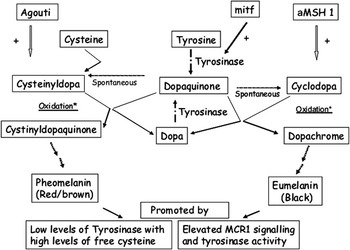
Diagrammatic representation of pathways for synthesis of pheomelanin and eumelanin involving initial conversion of l-tyrosine to dopaquinone catalysed by tyrosinase enzyme. Subsequent events typically include either interaction with l-cysteine under oxidative conditions to form pheomelanin via cystinyldopaquinone and stimulated by Agouti signalling protein (ASP) or conversion to dopachrome and synthesis of eumelanin under the influence of αMSH 1(αmelanocyte stimulating hormone). Expression of tyrosinase is regulated by Mitf (Micropthalmia-associated transcription factor).

As regards melatonin, early reports indicated responsiveness to exogenous application in isolated cashmere follicles in vitro (Ibraheem et al., Reference Ibraheem, Galbraith, Scaife and Ewen1994). More recent work (Fischer et al., Reference Fischer, Slominski, Tobin and Paus2008) has reported extra-pineal synthesis of melatonin and expression of melatonin membrane binding (MT2) receptor transcripts in integumental tissues, which provides evidence for a direct role for melatonin in regulation of the murine hair cycle. These authors also suggest that melatonin may directly modulate hair cycle activity via activities, which include downregulation of apoptosis and ERα expression and modulation of its binding receptors according to position in the follicle cycle.
These latter mechanisms may be expected to contribute to the maintenance of endogenous cycles of hair follicle activity in the absence of photoperiodic stimulation, which occurs following, for example, surgical ablation of pineal signalling (Allain et al., Reference Allain, Thebault, Rougeot and Martinet1994) in animal species, which normally respond to photoperiodic change. Intrinsic rhythms that regulate activity of each individual follicle are also identified as important by these authors. In this regard, parallels may be drawn with the expression of clock genes which have been identified in tissues such as the pars tuberalis of the pituitary gland (Lincoln et al., Reference Lincoln, Andersson and Loudon2003) and in human skin cells, cultured keratinocytes, melanocytes and dermal fibroblasts (expression of Clock and Period 1: Zanello et al., Reference Zanello, Jackson and Holick2000). Lin et al. (Reference Lin, Geyfman, Kumar, Chudova, Ihler, Smyth, Paus, Takahashi and Andersen2009) have described the control of Clock-regulated genes in mouse hair follicle cycling. Products of these genes appear to have a role in mammalian biological time-measurement with the additional possibility of signalling of photoperiod via reception in skin. Recently Bull et al. (Reference Bull, Pelengaris, Hendrix, Chronnell, Khan and Philpott2005) have implicated the transcriptional regulator c-Myc in the control of hair follicle cycling. Suggested effects, in contributing to the presumed ‘hair cycle clock’ include activation of follicular stem cells and maintenance of epidermis and sebaceous gland.
However, despite information on expression of a range of ligands and extracellular and intracellular receptors, the precise mechanisms regulating the onset of the different phases of the hair growth cycle remain to be fully elucidated. There is a particular gap in understanding of the molecular mechanisms responsible for the economically important differences in developmental properties and duration of anagen in pelage secondary hair follicles of commercially important animal genotypes. This relates to fibre-producing animals such as Merino sheep and Angora goats (long anagen) and Cashmere-bearing goats (short anagen) and is likely to include differences in transduction of photoperiodic signalling.
Molecular basis for phenotypic properties of hair fibre
Additional features that are important expressions of the many types of phenotype of fibre in animals include the structure and shape of the hair product (Schlake, Reference Schlake2007). Such properties of commercial importance include the length, diameter and degree of ‘bend’ of the fibre, presence, or not, of medullation and the degree of overlap and flatness of cuticle scales. These characteristics affect thermal insulation, surface smoothness and light-reflection and refraction in addition to response of fibre to post-harvest processing. Information from mouse models supplements that from other species such as sheep and goats (Allain et al., Reference Allain, Thebault, Rougeot and Martinet1994; Rogers, Reference Rogers2006), South American camelids (Antonini, Reference Antonini2010) and rabbits (Allain and Renieri, Reference Allain and Renieri2010) and confirms the importance of the bulb matrix in providing epithelial follicle components (hair shaft, medulla and cortex and layers of the IRS (cuticle, Henle’s, Huxley’s). Chemical signals regulating proliferation and differentiation of epithelial cells derive from the underlying DP, and expression in cells committed to the structural lineages of shaft and sheaths. The physical size of the DP affects the length of the contiguous basement membrane, and hence number of basal cells it can accommodate. Factors that influence follicle cycle behaviour by increasing rates of epithelial cell proliferation and duration of anagen will increase volume of fibre produced.
An analysis (e.g. Schlake, Reference Schlake2007) of factors that influence physical dimensions include regulators of keratinocyte proliferation such as IGF 1, which increases both length and diameter of fibre with effects antagonised by its binding proteins Igfbp 3 and Igfbp 5. Keratinocyte growth factor (FGF 7), derived from the DP, and certain BMPs such as BMP 4 expressed in both matrix and DP, have been ascribed roles in regulation of keratinocyte proliferation. A role for the secreted signalling molecule Shh and its receptor Ptch have also been described particularly in the proliferation of keratinocytes in post-telogen follicle regeneration. Certain Wnts have also been shown to reduce commitment to proliferation while promoting commitment to differentiation and expression of associated keratin genes (Kulessa et al., Reference Kulessa, Turk and Hogan2000). The orderly expression of keratin genes is important since their products contribute to the formation of cytoskeleton in keratinocytes which in turn constitute the bulk of the hair fibre and so affect its tensile strength. The expression of adhesion molecules such as desmoglein in intercellular junctions has also shown to be essential in maintaining integrity of hair shaft (Koch et al., Reference Koch, Mahoney, Ishikawa, Pulkkinen, Uitto, Shultz, Murphy, Whitaker-Menezes and Stanley1997). In addition to signalling from DP and cortical keratinocytes, IRS and ORS also express gene products, which influence hair shaft formation and activity. These include important signalling molecules affecting hair shaft formation and activity such as Cutl 1and Gata 3 (IRS) and FGF 5 (ORS); the latter influencing the time-length of anagen. Similarly, FGF 7 and FGF 10 secreted by the DP are important in the regulation of keratinocyte proliferation and differentiation in the mature murine hair follicle (Schlake, Reference Schlake2005). Down-regulation of their receptor, FGFR2 IIIb, an effect associated with elevated expression of Igfbp 5, was shown to decrease thickness of hair shaft and to reduce cellularity of the hair shaft medulla. Precise signals which determine the presence, or not, of medullae in hair shafts derived from follicles varying in anatomical location and across species including South American Camelids (e.g. Antonini, Reference Antonini2010) and rabbits (e.g. Allain and Renieri, Reference Allain and Renieri2010) appear not to have been established. Schlake (Reference Schlake2007) describes the importance of the medulla in the light reflective (sheen) properties of murine hairs and effects of fat deposits or air spaces and refers to the role of the cuticle and limited information on the regulation of its production. Greater information is available describing the importance of cuticle cell structure and frequency in the light reflective and felting characteristics of, for example, mohair fibres (Vassis et al., Reference Vassis, Butcher, Lee and Holt2003; Council for Scientific and Industrial Research (CSIR), 2007).
Another important property of the hair shaft is the degree of bend or crimp associated with individual hair types. Such effects on axial polarity may arise as a consequence of differences in rates of proliferation of keratinocytes located on opposite locations in the hair matrix, or, alternatively, differences in size or shape of cells. Genetic mutations associated with wavy hair in mice include defects in the receptor for EGFR and expression of TGF α (Schlake, Reference Schlake2007). Examples of natural bending include the zig-zag hairs which are secondary follicle products of the mouse skin epidermis and for which Igfbp 5a has been recently identified as a molecular marker in DP and shaft medulla. Schlake (Reference Schlake2007) describes the interaction between ‘bursts’ of the transcription factor Krox 20 and the induction of Igfbp 5 activity in the hair matrix, which stimulates movement of differentiating cells along the proximo-distal axis. Subsequent suppression of Igfbp 5 expression in the DP is described as taking place. Ensuing events include suppression of Krox 20 expression and its re-expression to produce a new ‘burst’ with continuation of the oscillatory cycle. An essential role for Wnt and Eda signalling in positioning and angling of the hair follicle in skin and bending of shaft in zig-zag hair formation has also been described by Hammerschmidt and Schlake (Reference Hammerschmidt and Schlake2007). These authors indicate that both signalling pathways direct the expression of Hedgehog, which acts to promote asymmetry both in the mature hair shaft follicle and during its morphological development. Loss of zig-zag hair production is reported to occur in the absence of this endogenous signalling.
The appearance of crimped fibres (short changes in direction of fibre growth) in sheep fine wool has been associated with the production of a bilateral structure of the hair into orthocortex (outer aspect) and paracortex (inner aspect) (e.g. Rogers, Reference Rogers2006). These differ in composition of KAPs where the former have a greater ratio of HGT KAPs and the latter exhibits more high cysteine (sulphur) KAPS. Molecular mechanisms appear not to have been described. Interestingly, loss of crimp has been associated with an increase in lustre in the ‘Felting lustre mutation in Merino sheep’ and loss of expression of certain HGT KAPs and orthocortex in wool fibres (e.g. Li et al., Reference Li, Ouyang, Rogers and Bawden2009). Similarly, a transgenic sheep mutation exhibiting high levels of hair shaft cortical expression of the Keratin type II gene K2.10 (Bawden et al., Reference Bawden, Powell, Walker and Rogers1998) was shown to produce a more lustrous fleece with fibres, which had disrupted ortho- and para-cortex. The results from these studies provide pointers towards explanation of the genetic basis for differences in fibre phenotype. Of particular interest are differences in the characteristics of fibre lustre and curl with similarities, within and across, caprine and SAC genotypes. In this context, while differences between mohair and cashmere fibre characteristics have an apparently multi-gene aetiology, the expression of Mendelian inheritance in comparisons of Suri and Huacaya genotypes of SAC suggest an as yet unexplained and simpler genetic basis (Allain and Renieri, Reference Allain and Renieri2010).
Biology of pigment production
The presence or absence of pigmentation and its distribution is another important property of the integument (e.g. Hepburn et al., Reference Hepburn, Kinnimonth and Galbraith2007) with implications for processing and commercial value of animal fibres (Allain and Renieri, Reference Allain and Renieri2010). The biology of hair follicle pigmentation in human, murine and other species has been the subject of recent detailed consideration (e.g. Nordlund et al., Reference Nordlund, Boissy, Hearing, King, Oetting and Ortonne2006; Kauser et al., Reference Kauser, Slominski, Wei and Tobin2006 and Renieri et al., Reference Renieri, Valbonesi, La Manna, Antonini and Lauvergne2008) to which reference should be made for details to supplement the following brief summary. Reference is made to the involvement of 150 alleles at over 90 loci, which support and regulate production of coat colour in the mouse. Similarly, Renieri et al. (Reference Renieri, Valbonesi, La Manna, Antonini and Lauvergne2008) refer to regulation of coat colour in mammals as under regulation of more than 800 alleles at 127 identified loci, with 11 loci associated with variation in pigmentation in sheep. Expressed protein products of these loci, which vary in composition and activity, target a range of functions in cells. These include ligands and their receptors, enzymes, structural proteins, transcription factors and membrane transporters. Control is exerted at the levels of (a) developmental migration and localisation in skin and melanocyte differentiation and proliferation, (b) hair follicle according to cycle dynamics, apoptosis and recruitment and (c) melanocyte and melanosome in regulating synthesis of pigments and transfer to keratinocytes.
Pigmentation is produced in melancoytes, located in the epidermis of the hair follicle, by enzymatic pathways which convert the amino acid l-tyrosine to either eumelanin or pheomelanin which confer black and red/yellow colours, respectively (Figure 8). The sites of synthesis are melanosomes that are produced from endosomal intermediates within melanocytes, and transferred, via dendrite structures, to cortical and medullary keratinocytes in the bulb region of the follicle. Melanosomes are specialised lysosomes, which contain filamentous protein lamellae and which, following transfer, interact with lysosomes of keratinocytes and form pigment-containing vesicles.
How do melanocytes colonise hair follicles? Melanocytes are known to originate during embryogenesis from neural crest precursor melanoblasts. Studies in mice indicate that these are released from the cell mass under the influence of BMPs 4 and 7 which induce gene products including Slug and Rhob which break tight junctions connecting the cells (Slug) and promote cell mobility (Rhob) (Quevedo and Holstein, Reference Quevedo and Holstein2006). Paracrine factors such as endothelin (ET) three direct melanoblasts towards the epidermis and commitment to differentiation as melanocytes in a process that is supported by Wnt proteins of mesenchymal origin from underlying dermis (Quevedo and Holstein, Reference Quevedo and Holstein2006). These melanoblasts migrate from neural crest, colonising first, the basal epidermis before entering the hair follicle placode during early follicular development. Other important gene products required for entry and normal development within the bulb matrix and function include the tyrosine kinase receptor c-Kit, in melanocytes. Its ligand, stem cell factor (SCF), is produced at the Steel locus in mesenchymal fibroblasts. Certain mutant alleles of both ligand and receptor intefere with normal proliferation and differentiation of melanocytes. These give rise to recognised phenotypes varying in pigment production. SCF/c-Kit signalling in melanocytes is required for expression of the Micropthalmia transcription factor (Mitf) encoded by the micropthalmia gene. Activated Mitf and cofactors are centrally involved in expression of genes involved in synthesis of melanins. Although there is a requirement postnatally for c-Kit during the hair growth cycle for activation of melanocytes, their stem cells are apparently not dependent on SCF/c-Kit signalling. In the mouse follicle under hair-growing conditions, melanocytes are present in at least three locations. These are basally sited pigment-producing cells in matrix; differentiating cells in ORS and stem cells in bulge region. Melanocytes exhibit activity according to the hair follicle cycle. Regression occurs towards late anagen and the apoptosis of most, if not all, of mature melanocytes takes place by occurrence of telogen. Some less differentiated melanocytes are thought to survive catagen to contribute to regeneration during new anagen along with recruitment from the stem cell reservoir in the ORS (Chintala et al., Reference Chintala, Li, Lamoreux, Ito, Sviderskaya, Bennett, Park, Gahl, Huizing, Spritz, Ben, Novak, Tan and Swank2005). As anagen progresses, these cells increase in volume, proliferate and increase dendrite connections with, and transfer melanosomes to, hair-shaft forming keratinocytes. Melanososmes have been characterised according to their eumelanic or pheomelanic products to give morphologically distinct eumelanosomes or pheomelanosomes, respectively. The biogenesis of melanosomes, synthesis of melanins and their transfer to keratinocytes are complex processes influenced by fibroblasts of the DP and which contribute to hair shaft phenotypes. These processes have been the subject of studies targeted at understanding genetic and other mechanisms of regulation.
Synthesis of melanin pigments
l-tyrosine is the major precursor for synthesis of melanins in reactions involving the enzyme tyrosinase protein (monophenol monooxygenase) which catalyses hydroxylation to l-dopa (l-3, 4-dihydroxyphenylalanine) with subsequent oxidation to dopaquinone (Figure 8). Dopaquinone is a common substrate for both eumelanin and pheomelanogenic pathways (e.g. Figure 8, Slominski et al., Reference Slominski, Wortsman, Plonka, Schallreuter, Paus and Tobin2005). The following reactions involve oxidation-reduction and intramolecular conversions, which occur spontaneously according to local conditions affected by concentrations of oxidising and reducing compounds and facilitation by, for example, metal cations. For synthesis of eumelanin, the specificity and rate of the reactions are principally regulated by Tyrosinase. For synthesis of pheomelanin, dopaquinone conjugates with cysteine (Figure 8) or glutathione to produce cysteinyldopa or glutathionyldopa. Under oxidative conditions, cysteinyldopa undergoes ring closure to yield 4-benzothiazinylalanines that may undergo peroxidase/H2O2-promoted or Tyrosinase-catalyzed oxidation reactions in a multistep sequence, which results in synthesis of pheomelanin. Alternatively, cysteinyldopa may be formed by the conjugation of dopaquinone with glutathione to generate glutathionyldopa with subsequent hydrolysis by glutamyltranspeptidase. Dopaquinone appears as a key molecule, which controls switching of eumelanogenesis to pheomelanogenesis by processes, which regulate activity of glutathione reductase and inhibit pheomelanogenesis under conditions of active eumelanin production. Steps of pheomelanogenesis that occur after the production of cysteinyldopa are stimulated by peroxidase and tyrosinase reactions, which are involved in the synthesis of benzothiazinylalanines.
What regulates pigment production in skin and hair follicle?
Interestingly, there is increasing evidence of the presence of locally produced and activated signalling systems, which mirror those of the classical hypothalamic-pituitary-adrenal (HPA) axis (Kauser et al., Reference Kauser, Slominski, Wei and Tobin2006) in addition to systemic and locally produced influences, which act via paracrine and autocrine mechanisms (Slominski et al., Reference Slominski, Wortsman, Plonka, Schallreuter, Paus and Tobin2005). Included among these are the seasonal changes and prolactin-mediated production of melanins in the post-natal hair follicle in some species (e.g. Nixon et al., Reference Nixon, Ford, Wildermoth, Craven, Ashby and Pearson2002). It is also interesting to note the involvement of the HPA axis and similar signalling proteins in appetite regulation and control of body composition at sites outwith the hair follicle (Mastorakos and Zapanti, Reference Mastorakos and Zapanti2004).
Stimulators of melanogenesis include, principally αMSH, in addition to adrenocorticotrophic hormone and β-endorphin (Kauser et al., Reference Kauser, Slominski, Wei and Tobin2006). These proteins are ‘melanocortins’ that are derived by action of prohormone convertase enzymes, from the locally synthesised parent molecule pro-opiomelanocortin and under control of corticotrophin releasing hormone. Additional positive influences on melanogenesis include certain prostaglandins, leukotrienes, endothelins (ET 1, ET 3, ET A and ET B), histamine, SCF, estrogens, androgens, cortisol, vitamin D3, BMPs and substrates l-tyrosine, l-dopa and l-cysteine. These act in parallel or sequentially, through pathways involving G-protein or kinase-coupled receptors, or NRs and involving SCF/c-Kit and ET 1, ET 3/ET A and ET B.
The action of αMSH and other melanocortins in skin and hair follicle adds to constitutive production of pigment and is mediated by binding to the melanocortin (MC) 1 receptor (MC 1R), produced at the extension locus, and which is a member of the MC family of transmembrane receptors (Figure 6). The major modifier and inhibitor of melanin synthesis in animal hair follicle epidermis, is Agouti (signalling) protein (ASP) produced in the DP with responses dependent on duration of signal and genetic and local intracellular environment. Differences in activity of ASP expression have been associated with different isoforms, variations in mRNA transcripts and untranslated first exons (coding regions in RNA), particularly during the hair follicle cycle. Outcomes from ASP signalling are typically a switch from eumelanogenesis to pheomelanogenesis in a pathway associated with reduced MC 1R-mediated adenyl cyclase activity and downsteam Tyrosinase activity. The mechanism of action of ASP appears to involve (i) direct competition with melanocortins as a consequence of high affinity binding of its c-terminal domain to MC 1R and (ii) interaction of its N-terminal domain to the closely located transmembrane gene product Attractin (Figure 6). Similarly, the gene product Mahoganoid (Mgrn 1) which presents as an intracellular protein with E3 ubiquitin ligase activity, appears essential for normal expression of Agouti signalling (He et al., Reference He, Eldridge, Jackson, Gunn and Barsh2003; Barsh, Reference Barsh2006). Genomic interference with attractin or Mgrn1 function has been shown to reduce production of pheomelanin and to produce large amounts of eumelanin particularly in hair coats of mice homozygous for the mutations (Barsh, Reference Barsh2006). In addition, expression of the transmembrane serine protease (proteolytic enzyme) corin, produced by fibroblasts in the DP, has been shown to inhibit ASP signalling and contribute to maintenance of eumelanin production during anagen in pelage hair follicles of mice (Enshell-Seijffers et al., Reference Enshell-Seijffers, Lindon and Morgan2008).
The presence of cysteine has been recognised as an important component of regulation in melanocytes and the Slc7a11 gene, which encodes for the cystine/glutamate exchanger xCT has been implicated as having a direct regulatory effect on pheomelanin production with little effect on eumelanin production. Mutation in this gene has been shown to cause the subtle gray (sut) mouse pigmentation phenotype (Chintala et al., Reference Chintala, Li, Lamoreux, Ito, Sviderskaya, Bennett, Park, Gahl, Huizing, Spritz, Ben, Novak, Tan and Swank2005). Negative regulators of melanic pigment production include melatonin, tumor necrosis factor α, TGF β, interleukins (IL)1, IL 6, and, interferon-γ, triiodothyronine, glucocorticoids and dopaminergic and cholinergic agonists.
The major tyrosinase proteins have been characterised as having a high degree of conservation between species and include tyrosinase, TRP 1, expressed by TYRP 1 (human) or TYRP b (mouse: Brown) loci and TRP 2 expressed by TYRP 2/DCT (dopachrome tautomerase) (human), and slaty (mouse) loci, respectively. TRP 1 appears to be particularly important for eumelanogenesis. Additional regulators of melanogenesis include PMEL 17 protein, catechol-o-methyltransferase and macrophage migration inhibitory factor. Signalling by Wnts has been ascribed a role in regulating the activity of bulge-derived melanocyte stem cells in pigment production in new hair follicles formed after wounding or UV radiation of murine skin (Ito et al., Reference Ito, Yang, Andl, Cui, Kim, Millar and Cotsarelis2007). The Mitf is a well-recognised ‘master’ regulator of melanocytes and has been used as a marker for development and presence of melanocytes in embryonic and foetal human skin tissue (Gleason et al, Reference Gleason, Crum and Murphy2008). Mitf has also been shown to regulate expression of tyrosinase proteins (Smith et al., Reference Smith, Zhou and Orlow1998).
A recent example of work in ruminants (Norris and Whan, Reference Norris and Whan2008) has described the molecular mechanisms regulating white and black fleece colour in dominant white/tan (A(Wt)) Agouti sheep. These workers observed that the dominance of the white phenotype was caused by the expression of a duplicated coding sequence in ASP and AHCY (s-adenosylhomocysteine hydrolase) coding regions located immediately downstream of a duplicated ITCH (E3 ubiquitin-protein ligase) gene promoter region. In contrast, the black sheep recessive phenotype exhibited a single ASP copy with a silenced ASP promoter, which was indicative of limited ASP expression. They also reported the expression of a single copy gene at the ASP locus in the ancient breed of Barbary sheep. This was concluded to regulate production of a tan body fleece coat in keeping with observations in other species of animals such as the mouse. Similarly, differences in pelage coat colour (light v. dark) in feral Soay St Kilda sheep has been shown to be regulated by a single autosomal locus at which the dark allele is dominant to the light allele (Gratten et al., Reference Gratten, Wilson, McRae, Beraldi, Visscher, Pemberton and Slate2008). The light-fleeced phenotype occurs due to homozygous expression of an allele exhibiting, at coding position 869, a single nucleotide substitution in Tyrosinase related protein TYRP 1G locus (causing loss of a cysteine residue) and altered activity of the enzyme associated with observed colour polymorphism. Further consideration of factors regulating pigment production in wool of sheep and utilisation of information to inform genetic selection, has been provided by Renieri et al. (Reference Renieri, Valbonesi, La Manna, Antonini and Lauvergne2008) and Allain and Renieri (Reference Allain and Renieri2010).
Additional considerations in patterning
An important feature of pigmentation, which occurs frequently in animals is the presence of recognisable patterns in the hair coat. These have origins in the positioning of melanocytes in ectodermal skin precursor cells during embryogenesis and recruitment into developing follicles and subsequent dynamic response to signalling systems regulating melanogenesis (Chang, Reference Chang2007). The absence of pigmentation in white-wooled merino genotypes has been ascribed to the failure of recruitment of melanocytes during developments of follicles (Fleet et al., Reference Fleet, Forrest, Walker and Rogers2004). Recent work (Chang, Reference Chang2007; Yamaguchi et al., Reference Yamaguchi, Passeron, Watabe, Yasumoto, Rouzaud, Hoashi and Hearing2007) has described differences in melanocyte numbers and pigmentation in murine skin at non-hair-bearing palmoplantar (lower) and trunk (greater) sites and demonstrated greater expression of the Wnt signalling inhibitor DKK 1. Addition of DKK 1 to cultured melanocytes in vitro repressed proliferation. This was associated with inhibited expression of Mitf. It was concluded that epithelial-mesenchymal interactions regulate melanocyte expression according to programming of fibroblasts along with three developmental axes with important positional features subsequently retained by expression of Hox genes in fibroblasts according to original site. Other work, relating regional positioning of melanocytes in murine hair follicles, has been summarised by Chang (Reference Chang2007). Differences in expression of pigmentation in hairs in ventral (lighter) and dorsal (darker) locations have been attributed to ventral-specific expression of an isoform of ASP in dermal papillae and affecting hair follicle melanocytes. These cells switch synthesis from black eumelanins to red/yellow pheomelanins. ASP is, in turn, subject to suppression by the T-box transcription factor, Tbx 15, synthesised by dermal papillae of the dorsally positioned hair follicles (Candille et al., Reference Candille, Van Raamsdonk, Chen, Kuijper, Chen-Tsai, Russ, Meijlink and Barsh2004). Further studies will be required to advance knowledge of expression of such signalling molecules and their use as potential molecular markers for selection of improved animal genotypes.
Nutrients
The importance of nutrients in supporting hair follicle function has been the subject of a number of reviews (e.g. Galbraith, Reference Galbraith2000; Hynd, Reference Hynd2000; Rogers, Reference Rogers2006). Briefly, competition for, and availability of, nutrients at follicle level are influenced by physiological state of the animal with changes in requirements of tissues supporting somatic growth, pregnancy, lactation and thermal regulation. Individual nutrients are required directly to provide energy (ATP), substrates for synthesis of cellular and extracellular structures, catalysis (enzymes) and molecular regulation (ligands, inhibitors, receptors, co-factors). Indirect effects may be produced from systemic responses producing signalling molecules that act locally on individual follicles. Reported outcomes are modulation of hair follicle cycle and altered fibre production arising from changes in matrix cell proliferation and/or differentiation. Important nutrients include amino acids, carbohydrates, lipids, minerals and vitamins. Studies in goats have shown the relationships between protein/energy supply, genetic potential for fibre production and physiological state with, for example, reductions in fleece growth during pregnancy and lactation (Galbraith, Reference Galbraith2000). In addition, pre-natal development of hair follicles and other tissues, in embryo and foetus depends on supply from maternal circulation. The availability of amino acids from dietary protein sources for absorption at the small intestine depends on a number of factors which include, (a) for the dietary supply, ruminal degradability and digestibility of the undegraded fraction and (b) for ruminal microbial protein, its synthesis and post-ruminal digestibility with uptake at the small intestine (Galbraith, Reference Galbraith2000). Sheep wool has a much higher concentration of cysteine (ca. 9.0 residues/total amino acid residues) than (i) skeletal muscle (1.1) as a competing tissue within the body, (ii) dietary supply from either rumen microbial protein (1.0) or (iii) typical dietary protein sources or supplements of extracted heat-treated soya bean meal (1.0), or white fish meal (0.9). The disparity between the concentrations of cysteine in hair fibre and that in sulphur amino acid supply are factors in the relatively low value (0.3) relating conversion of metabolisable protein to hair follicle protein in animals such as sheep (Agricultural and Food Research Council (AFRC), 1993). Additional factors of importance are transport in blood and uptake into follicles by mechanisms which are now becoming understood in integumental tissues including the hair follicle (Thomas et al., Reference Thomas, Tivey, Penno, Nattrass and Hynd2007; Hepburn et al., Reference Hepburn, Knight, Wilde, Hendry and Galbraith2008; Galbraith, Reference Galbraith2010). Post-absorptive utilisation depends on supply at the follicle level and synthetic activities of structures (particularly medulla: cortex, cuticle and IRS) according to position in the follicular cycle. Cysteine has a particular role in di-sulphide bonding in cytoskeletal keratins and the sulphur-containing KAPS. It is also a required substrate for production of pheomelanins in melanocytes, where its concentration may influence switching from eumelanin synthesis. Methionine is essential to provide the sulphur moiety for cysteine synthesis in transulphuration, in tissues for synthesis of polyamines, molecular methylation and initiation of polypeptide synthesis important in cell proliferation, differentiation and nuclear epigenetic modifications (Galbraith et al., Reference Galbraith, Norton and Sahlu2000). Tyrosine supply is important for synthesis of the HGT KAPS and in synthesis of proteins important in cell signalling via phosphorylation of tyrosine moieties. Similarly, phosphorylation or dephosphorylation of serine and threonine in signalling proteins also provides important cellular regulation.
A number of vitamins and minerals are recognised as essential for normal function in skin and for hair follicles also (Sherertz and Goldsmith, Reference Sherertz and Goldsmith1991; Galbraith, Reference Galbraith1998; Rogers, Reference Rogers2006). Deficiency symptoms have been described for inadequacy of vitamin supply, which relate to generally well-understood roles in cell biochemistry. These include ribloflavin, niacin, pantothenic acid, folic acid and biotin (general effects on integumental tissues including caprine and sheep hair follicles (Tahmasbi et al., Reference Tahmasbi, Galbraith and Scaife2007a and Reference Tahmasbi, Galbraith and Scaife2007b; Galbraith Reference Galbraith2010)), vitamin A and retinoids, which are essential for cell proliferation and vitamin D that influences TGF β2 upregulation and follicle development (Inoue et al., Reference Inoue, Aoi and Yoshimura2009). Important cations include calcium that provides signal transduction in keratinocytes (Pruche et al., Reference Pruche, Boyera and Bernard1996) and magnesium, which has a role in energy-providing phosphate transfer reactions and DNA degradation and synthesis. Potassium deficiency in the rat causes hair loss and, in mice, production of a hair coat lacking in lustre. Manganese is essential for the synthesis of glycoproteins such as chondroitin, dermatan sulfate and collagen in the dermal extracellular matrix and in stabilising metabolic phosphate transfer. Selenium has been shown to be essential for the synthesis of seleno-cysteine. It is incorporated in the active site of enzymes such the deiodinases which convert thyroxine to its active form, triiodothyronine, which is essential for development and activity of hair follicles (Rhind and Kyle, Reference Rhind and Kyle2004; Tiede et al., Reference Tiede, Klatte, Wenzel, Lynaugh and Paus2009). Iodine is also essential for synthesis of such thyroid hormones, which have recently been shown to modulate cycle behaviour and pigment production in cultured human scalp hair follicles (van Beek et al., Reference Van Beek, Bodó, Kromminga, Gáspár, Meyer, Zmijewski, Slominski, Wenzel and Paus2008) Copper also has an important enzymatic role (Danks, Reference Danks1991), for example, in the production of cross-links in elastin and collagen fibres in the dermis, melanin pigment production in melanocytes (as a component of tyrosinase), post-translational formation of di-suphide bonds between cysteine molecules in cytoskeletal proteins of cortical and cuticle proteins and a role in the formation of crimp in wool. Zinc has an important function in ‘zinc finger’ structures, which stabilise proteins or peptides and provide DNA recognition sites in regulatory molecules such as transcription factors. Zinc also has a role in function of metallo-enzymes, which mediate metabolism of protein (includes collagenase and gelatinase zinc-metalloproteinase enzymes involved in tissue remodelling in dermal matrix), fat and carbohydrate (Neldner, Reference Neldner1991).
Acknowledgements
This review is based on an invited presentation at the 59th Annual Meeting of the European Association for Animal Production held in Vilnius, Lithuania during August 2008. The input of the many co-workers and support of national and international funding agencies, which contributed to work on hair follicle and other integumental tissues at the University of Aberdeen is gratefully acknowledged. Thanks are extended to the School of Biological Sciences, Directorate of Information Technology and Library for providing research, and literature-access facilities, respectively. The assistance of Ms Jane Pool in the preparation of illustrations is appreciated. Grateful acknowledgement is also made to fellow members of the European Working Group on Animal fibres and to Professor Carlo Renieri and colleagues at the University of Camerino, Italy for support and opportunities for collaboration.