Echocardiographic assessment of right ventricular function is difficult due to its complex geometric shape and regional differences in wall motion and contractility.Reference Haddad, Doyle, Murphy and Hunt1,Reference Rudski, Lai and Afilalo2 This is particularly true in patients with repaired tetralogy of Fallot because the normal pattern of right ventricular contractility is disrupted.Reference Lopez, Cohen and Anderson3,Reference Srinivasan, Sachdeva and Murrow4 Placement of a transannular patch during initial surgical repair of tetralogy of Fallot can cause dyskinesia and dilatation of the right ventricular outflow tract. Recent studies have suggested that right ventricular outflow tract dysfunction has a significant impact on the occurrence of ventricular arrhythmias in this population, which is a risk factor for sudden death.Reference Li, Luo, Liu and An5,Reference Luo, Li, Yang, Zhou, An and Chen6
Cardiac MRI is the gold standard measure of right ventricular function, and important clinical decisions for these patients are largely based on cardiac MRI data.Reference Valente, Ko and Cook7 Several recent studies have shown that there is significant regional variation in right ventricular function in this patient population.Reference Kutty, Zhou and Gauvreau8,Reference Greutmann, Tobler and Biaggi9 However, cardiac MRI remains expensive and often requires sedation in the paediatric population. Therefore, accurate echocardiographic assessment of right ventricular function continues to be an important goal.
Traditional echocardiographic measures of right ventricular function are not always reliable. There are many proposed quantitative measures of right ventricular function, but their correlation with global right ventricular ejection fraction by MRI continues to be suboptimal.Reference Hamilton-Craig, Stedman and Maxwell10,Reference Koestenberger, Nagel and Ravekes11 This may be in part due to the fact that regional differences in systolic function are not often taken into account with these measurements.
In adult patients, right ventricular outflow tract fractional shortening correlates with other echo measures of right ventricular function and with right ventricular ejection fraction measured by cardiac MRI.Reference Greutmann, Tobler and Biaggi9,Reference Davlouros, Kilner and Hornung12,Reference Morcos, Vick, Sahn, Jerosch-Herold, Shurman and Sheehan13 To date, no studies have been performed in children with repaired tetralogy of Fallot to assess the validity and utility of right ventricular outflow tract fractional shortening in the assessment of right ventricular function. The goals of this study therefore are to compare regional systolic right ventricular function in children with structurally normal hearts and those with repaired tetralogy of Fallot, compare measures of regional right ventricular systolic function with global systolic right ventricular function measured by cardiac MRI, and to develop a quantitative model that incorporates echo measures of regional right ventricular systolic function to predict global right ventricular ejection fraction by cardiac MRI.
Materials and methods
A retrospective chart review was performed to identify children with repaired tetralogy of Fallot who had undergone both a transthoracic echocardiogram and cardiac MRI within 6 months of one another during the course of routine clinical care between January 2010 and January 2016. The control population of patients included those with structurally normal hearts undergoing echocardiogram and cardiac MRI for a variety of indications including evaluation for myocarditis, sickle cell disease, and vascular anomalies. Patients with repaired tetralogy of Fallot who had undergone right ventricle to pulmonary artery conduit placement were excluded from analysis. If patients with repaired tetralogy of Fallot had undergone pulmonary valve replacement, they were included only if their echocardiogram and cardiac MRI were both performed before or after valve implantation. Patients with repaired tetralogy of Fallot were separated into those with impaired versus preserved right ventricular ejection fraction by cardiac MRI using published normative data.Reference Kawel-Boehm, Maceira and Valsangiacomo-Buechel14 Impaired right ventricular ejection fraction was defined as cardiac MRI right ventricular ejection fraction more than two standard deviations below the mean reference value for age. Using these values, further distinction was made between normal right ventricular ejection fraction (within two standard deviations of the mean), moderately depressed and severely depressed (<35%) right ventricular ejection fraction. Demographic and clinical information was collected about patients and controls by review of the medical record. The study was approved by the institutional review board at the University of Pittsburgh.
Transthoracic echocardiograms were performed following a standardised institutional protocol using iE33 ultrasound systems (Philips Medical Systems, Andover, Massachusetts, United States of America). All study echocardiograms were analysed using Xcelera (Philips Medical Systems, Andover, Massachusetts, United States of America). Measurements were made by a single physician and verified by a second reader blinded to CMR data.
Echocardiographic measures of right ventricular function were performed as follows: Right ventricular outflow tract fractional shortening was calculated by measuring the dimension of the right ventricular outflow tract on M-Mode in the parasternal short axis view at the level of the aortic valve during end-systole and end-diastole. Right ventricular outflow tract systolic excursion was performed on the same M-mode frame by measuring the linear displacement of the anterior wall of right ventricular outflow tract from diastole to systole. Right ventricular area at end-diastole and end-systole was measured in the apical 4 chamber view to calculate the right ventricular fractional area change. Tricuspid annular plane systolic excursion was measured off of the M-mode through the lateral tricuspid valve annulus to the right ventricular apex. The right ventricular myocardial performance index was calculated, and S’ was measured on tissue Doppler imaging traces through the tricuspid valve annulus as previously described. Left ventricular ejection fraction was performed using Simpson’s biplane method from the apical 4 and apical 2 chamber views.
Cardiac MRI studies were performed on a 1.5 T scanner (Signa HDX, GE Medical Systems, Waukesha, Wisconsin, United States of America). Ventricular dimensions and function were assessed with an electrocardiographically gated steady-state free-precession cine magnetic resonance pulse sequence. Measurements of right ventricular end-diastolic and end-systolic volumes were obtained on axial views with the manual tracing of endocardial contours by one of four experienced readers. Right ventricular ejection fraction was calculated as follows: [(end-diastolic volume − end-systolic volume)/end-diastolic volume × 100].
Statistical analysis
The statistical analysis was performed using SPSS version 25 (SPSS Inc., Chicago, Illinois, United States of America). Descriptive statistics for continuous variables is presented as means ± standard deviations. Measures of regional right ventricular systolic function in patients with repaired tetralogy of Fallot were compared to controls and between groups with one-way ANOVAs.
To derive a quantitative model of global right ventricular function, Pearson correlations were performed between regional measures of right ventricular systolic function and cardiac MRI right ventricular ejection fraction. Echo measures with a significant correlation (p < 0.05) were then included in the multi-variate regression model to identify independent predictors of cardiac MRI right ventricular ejection fraction.
Results
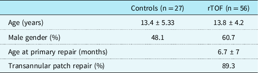
Twenty-seven patients with structurally normal hearts (13.4 ± 5.33 years) and 56 patients with repaired tetralogy of Fallot (13.8 ± 4.2 years) were analysed. There were no significant differences in demographic characteristics between the control and patient groups (Table 1). There was significantly more time between echo and MRI in patients with repaired tetralogy of Fallot (71.2 ± 52 days) compared to those with structurally normal hearts (39.2 ± 49 days, p = 0.009).
Baseline characteristics of control and patient populations

rTOF, repaired tetralogy of Fallot.
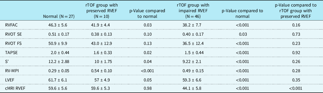
Table 2 compares echocardiographic measures of right ventricular systolic function with cardiac MRI data between patients with structurally normal hearts, patients with repaired tetralogy of Fallot with preserved right ventricular function, and those with impaired right ventricular function. Despite having the same right ventricular ejection fraction by cardiac MRI as normal controls, patients with repaired tetralogy of Fallot and preserved right ventricular function had abnormal echocardiographic measures including right ventricular fractional area change, tricuspid annular systolic excursion, tricuspid valve S’, and right ventricular myocardial performance index. This group showed no difference in right ventricular outflow tract systolic excursion and right ventricular outflow tract fractional shortening compared to normal controls. Patients with repaired tetralogy of Fallot with impaired MRI right ventricular ejection fraction were significantly different than patients with structurally normal hearts on all measures of right ventricular function. No echocardiographic measure of right ventricular systolic function differentiated between patients with repaired tetralogy of Fallot with preserved versus impaired MRI right ventricular ejection fraction.
Comparison of echocardiographic and MRI measures in repaired tetralogy of Fallot

cMRI RVEF = cardiac MRI right ventricular ejection fraction; LVEF = left ventricular ejection fraction; rTOF = repaired tetralogy of Fallot; RVFAC = right ventricular fractional area change; RV-MPI = right ventricular myocardial performance index; RVOT FS = right ventricular outflow tract fractional shortening; RVOT SE = right ventricular outflow tract systolic excursion; TAPSE = tricuspid annular plane systolic excursion.
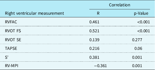
Table 3 shows Pearson correlation statistics comparing echo measures of right ventricular systolic function with cardiac MRI right ventricular ejection fraction. Right ventricular fractional area change and M-mode right ventricular outflow tract fractional shortening correlated most strongly with right ventricular ejection fraction by MRI.
Pearson correlations between echocardiographic and MRI measures of right ventricular function

RVFAC = right ventricular fractional area change; RV-MPI = right ventricular myocardial performance index; RVOT FS = right ventricular outflow tract fractional shortening; RVOT SE = right ventricular outflow tract systolic excursion; TAPSE = tricuspid annular plane systolic excursion.
A multi-variate regression model was created to create a quantitative echocardiographic model to predict cardiac MRI right ventricular ejection fraction. The final model included the two most reproducible echo measures that were most strongly correlated with MRI right ventricular ejection fraction and were defined by the regression equation MRI right ventricular ejection fraction = 21.7 + (0.3 × RVOT FS) + (0.41 × RVFAC).
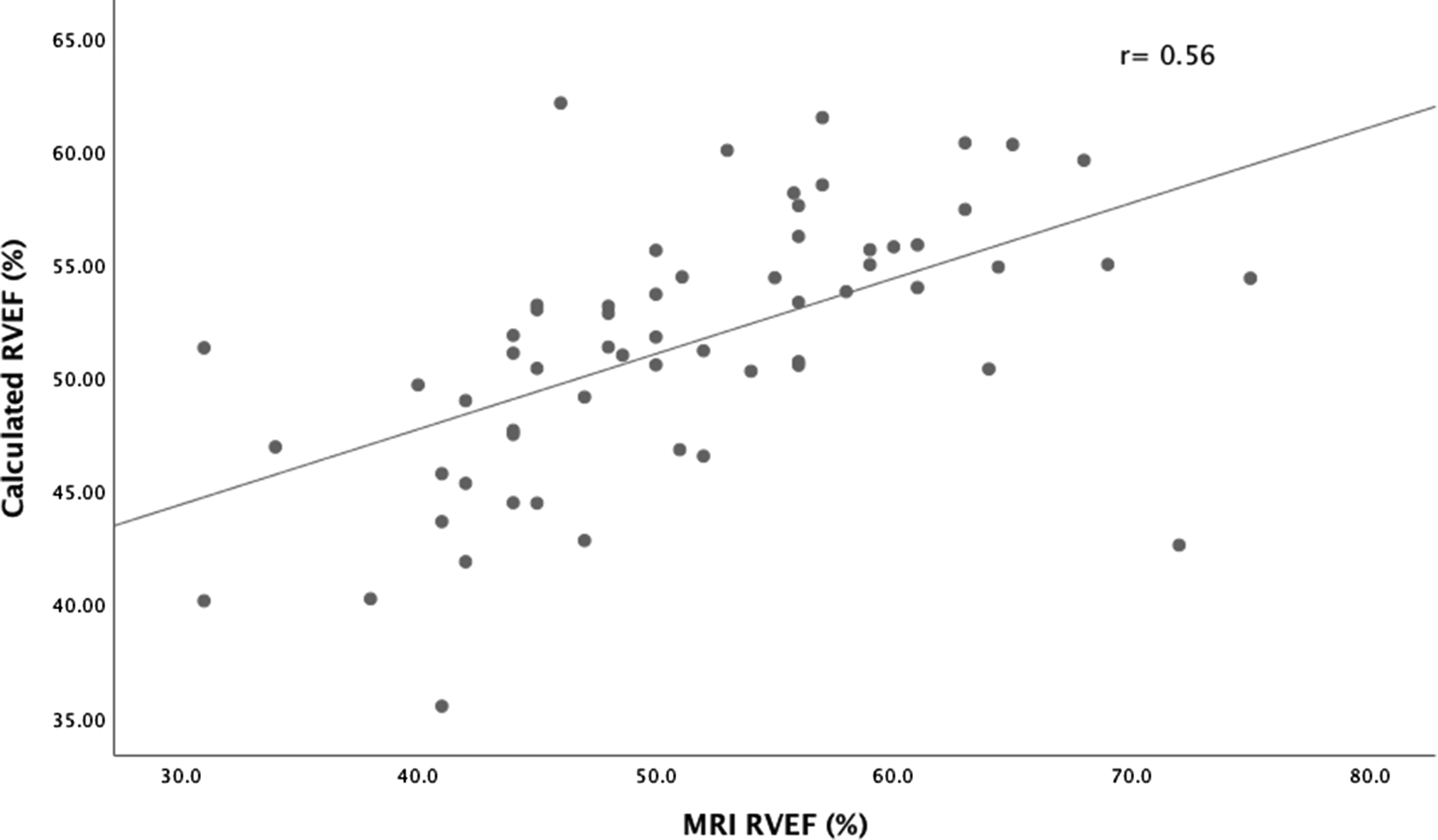
There was no significant difference between the cardiac MRI right ventricular ejection fraction and the right ventricular ejection fraction calculated by the model (51.3% ± 9.6 versus 51.5% ± 5.7; p = 0.9). Figure 1 demonstrates the Pearson correlation between calculated and measured right ventricular ejection fraction.
Linear regression plot comparing calculated right ventricular ejection fraction to cardiac MRI right ventricular ejection fraction (n = 61).

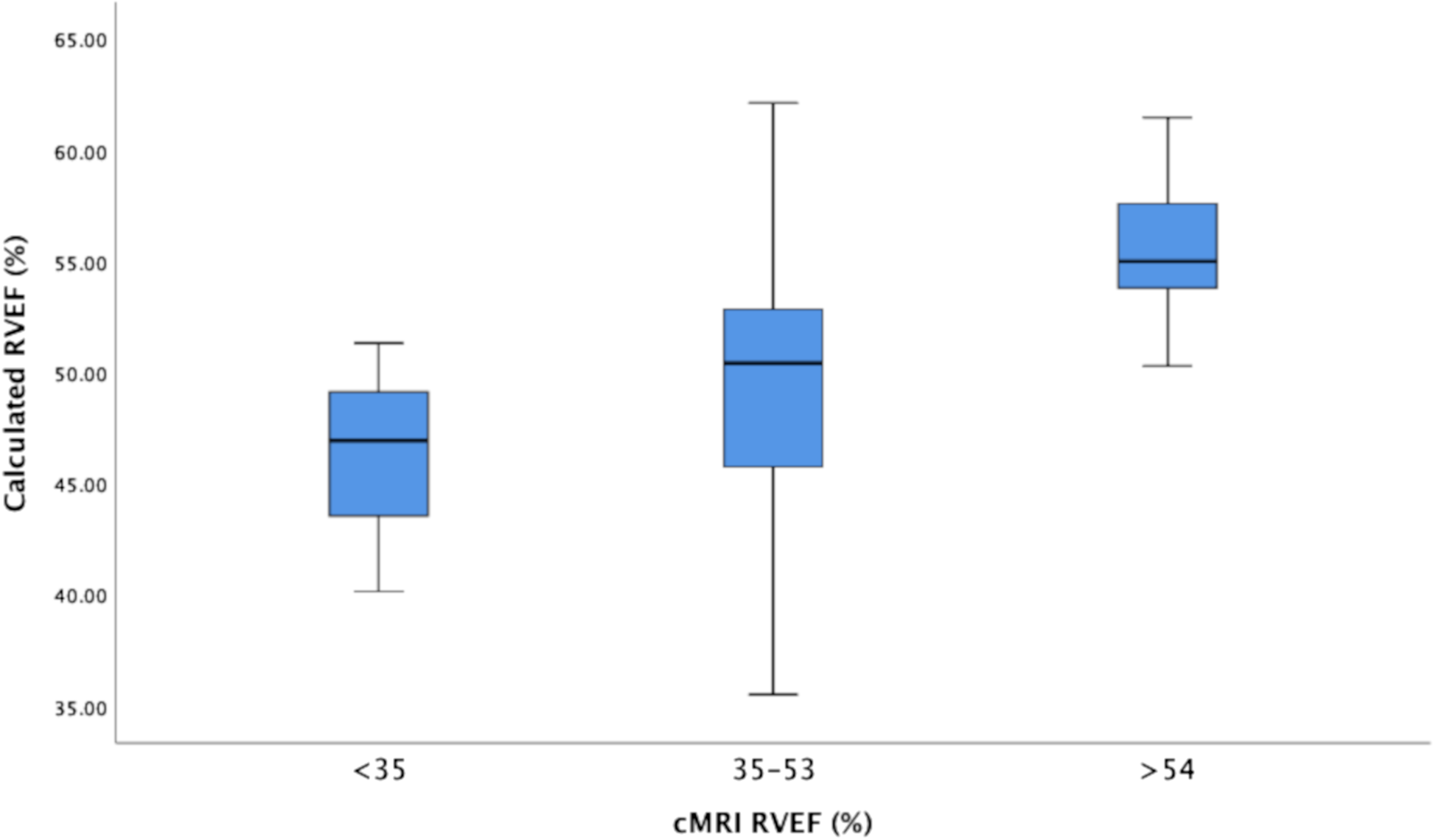
The model had good discrimination between patients with normal, mild-moderately impaired, and severely impaired right ventricular function (Fig 2).
Box plot showing separation of model derived right ventricular ejection fraction (RVEF) for patients with normal, mild/moderate, and severely impaired global RVEF on CMR (n = 61).

Discussion
Patients with repaired tetralogy of Fallot demonstrate significant differences in regional systolic right ventricular function compared to patients with structurally normal hearts.Reference Greutmann, Tobler and Biaggi9,Reference Davlouros, Kilner and Hornung12 Interestingly, patients with repaired tetralogy of Fallot and preserved right ventricular ejection fraction by MRI had preserved function of the right ventricular outflow tract, but lower longitudinal contraction of the right ventricular free wall and global right ventricular systolic and diastolic function compared to patients with structurally normal hearts. While these parameters of right ventricular function in patients with repaired tetralogy of Fallot were still within the accepted normal ranges, they were significantly different than individuals with normal hearts. These subtle differences in regional systolic function in the tetralogy of Fallot group can likely be attributed to their prior cardiac surgery, scarring, fibrosis, and/or the presence of patch material. In this particular group, these small differences in regional function were not enough to effect global systolic function assessed by the gold standard cardiac MRI. Patients with impaired right ventricular ejection fraction by cardiac MRI showed deterioration in the function of the right ventricular outflow tract, which contributes to worse global right ventricular systolic function.
This is in contrast to a study by Greutman et alReference Greutmann, Tobler and Biaggi9 which found that adult patients with repaired tetralogy of Fallot had impaired function of the right ventricular outflow tract regardless of their global ejection fraction and tended to compensate for this by increased contraction of the right ventricular body as measured by right ventricular fractional area change. This difference in findings may be secondary to our younger patient population, suggesting that deterioration in right ventricular outflow tract function may happen later after repair of tetralogy of Fallot. There may be some effect of surgical era as well, as more recent surgical techniques emphasise having smaller right ventriculotomies and smaller transannular patches compared to repairs performed 20–30 years ago.
In this cohort, no echocardiographic measure of global or regional right ventricular systolic function differentiated between patients with repaired tetralogy of Fallot with preserved versus impaired right ventricular ejection fraction measured by cardiac MRI. This may reflect the small sample size of the repaired tetralogy of Fallot with preserved right ventricular function. This also highlights the challenges inherent in using echocardiography for screening and clinical decision-making in this patient population. The linear regression model created to predict cardiac MRI right ventricular ejection fraction has additional value in this situation because it would allow more precise determination of the need for and timing of MRI imaging.
Examination of the data as whole demonstrated that the regional differences in right ventricular systolic function contribute significantly to global right ventricular ejection fraction by cardiac MRI. Specifically, radial contraction of the right ventricular outflow tract and longitudinal and transverse contraction of the right ventricular body were most strongly correlated with cardiac MRI by right ventricular ejection fraction. The regression model incorporated regional measure of the longitudinal and transverse contraction of the right ventricular body as well as the outflow tract as the strongest predictors of cardiac MRI right ventricular ejection fraction. These echocardiographic measures are easily reproducible and have high inter-rater reliability. This model could be used to follow patients over time and help determine timing and need for advanced imaging.
Contribution of the right ventricular outflow tract to global right ventricular ejection fraction by cardiac MRI in repaired tetralogy of Fallot is well described and appears to correlate with clinical and functional outcomes in adult patients.Reference Kutty, Zhou and Gauvreau8,Reference Morcos, Vick, Sahn, Jerosch-Herold, Shurman and Sheehan13,Reference Geva, Powell, Crawford, Chung and Colan15 This is the first study to date that has specifically assessed the radial contraction of the right ventricular outflow tract in children with repaired tetralogy of Fallot. The echocardiographic measurement of right ventricular outflow tract fractional shortening was straightforward, simple to perform from standard views, and easily reproducible. Our data suggest that when used in a model that incorporates different echocardiographic measures of right ventricular systolic function, the right ventricular outflow tract fractional shortening may be an important predictor of global cardiac MRI right ventricular ejection fraction in children.
Our study is limited by its retrospective nature, small sample size for the group with repaired tetralogy of Fallot and preserved right ventricular function as well as the 6-year study period which introduces surgical technique differences as well and changing surveillance practices. Our data did not include additional clinical or echocardiographic information such as degree of pulmonary regurgitation or stenosis, other valvular lesions, QRS duration or other rhythm issues, all of which may also affect ventricular function. Furthermore, while our control group did have structurally normal hearts, the indications for obtaining echo and cardiac MRI studies were heterogenous and ranged from a history of myocarditis to vascular anomalies and sickle cell disease screening for iron deposition. While all studies selected did have normal function both by echo and MRI, there may be subtle differences in systolic and diastolic function that may have clouded results.
In conclusion, children with repaired tetralogy of Fallot have abnormal regional and global right ventricular systolic function compared to patients with structurally normal hearts. Despite the presence of patch material, the right ventricular outflow tract does significantly contribute to global right ventricular ejection fraction by cardiac MRI in this patient population. When incorporated into a model that includes regional and global echocardiographic measures of right ventricular function, right ventricular outflow tract fractional shortening can help predict global right ventricular ejection fraction by cardiac MRI. Further prospective studies are needed to further validate this model and determine its relationship to clinical outcomes.
Acknowledgements
The authors would like to thank the statisticians at University of Pittsburgh Clinical and Translational Science Institute for their input on study design.
Financial support
This research received no specific grant from any funding agency, commercial or not-for-profit sectors.
Conflicts of interest
None.
Ethical standards
The authors assert that all procedures contributing to this work comply with the ethical standards of the Helsinki Declaration of 1975, as revised in 2008, and has been approved by the institutional committee of the University of Pittsburgh.