Introduction
Kawasaki disease is an acute, self-limited systemic vasculitis predominantly affecting children younger than five years and is the leading cause of acquired heart disease in developed countries.Reference McCrindle, Rowley and Newburger1 Up to one-quarter of untreated patients develop coronary artery aneurysms; timely intravenous immunoglobulin reduces this risk substantially, but 10–20% of patients are immunoglobulin-resistant and at markedly higher risk of severe cardiovascular complications.Reference McCrindle, Rowley and Newburger1,Reference Tremoulet, Best and Song2 Giant coronary artery aneurysms (≥8 mm or Z-score ≥10) confer the highest risk of thrombosis, myocardial infarction, and sudden death.Reference McCrindle, Rowley and Newburger1 Spontaneous rupture is exceptionally rare but uniformly catastrophic.Reference Suzuki, Kuroko, Kotani, Sakoda and Kasahara3,Reference Zhang, Tuokan and Shi4 Infants frequently present with incomplete criteria, compounding diagnostic delay and increasing the risk of sudden, unexpected death.Reference Pucci, Martino, Tibaldi and Bartoloni5 We describe an 18-month-old boy with immunoglobulin-resistant incomplete Kawasaki disease whose giant coronary aneurysms ruptured despite interleukin-1 blockade, triple antithrombotic therapy, and aggressive mechanical support. To our knowledge, this represents a rare example of fatal rupture occurring despite step-up immunomodulation and veno-arterial extracorporeal membrane oxygenation in infants beyond the first year of life.
Case presentation
An 18-month-old boy was referred after seven days of unremitting fever and clinical features suggestive of Kawasaki disease. Examination revealed dry, erythematous lips, periungual desquamation, and oedema of the dorsal hands and feet; bilateral nonexudative bulbar conjunctival injection and cervical lymphadenopathy were absent. At the referring centre, erythema and induration of the bacille Calmette–Guérin vaccination scar had been documented during the febrile period, transiently appearing and then regressing. Laboratory studies showed marked inflammation (C-reactive protein 177.5 mg/L), anaemia, and hypoalbuminaemia; echocardiography revealed no pericardial effusion. Incomplete Kawasaki disease was diagnosed; intravenous immunoglobulin (2 g/kg) and acetylsalicylic acid were administered.
Because the fever persisted, immunoglobulin-refractory disease was diagnosed, and intravenous methylprednisolone was started, followed by pulse methylprednisolone when inflammation relapsed. Coronary computed tomography angiography demonstrated a 7.5 mm fusiform left main coronary artery aneurysm, an 8.8 mm giant aneurysm of the proximal-to-mid left anterior descending artery, a 5.2 mm proximal circumflex aneurysm, and right coronary artery dilation. Enoxaparin, acetylsalicylic acid and clopidogrel were initiated. Follow-up echocardiography five days later showed further aneurysmal progression (left coronary artery 8–11 mm; right coronary artery 6.6 mm) and re-elevated inflammatory markers; pericardial effusion remained absent. Anakinra was added. On the day of transfer, new-onset pericardial effusion was detected—interpreted in retrospect as the earliest sign of impending rupture—and the patient was urgently referred to our centre.
On admission to our centre the patient remained haemodynamically stable. Admission studies revealed leukocytosis, thrombocytosis, and striking hyperinflammation (ferritin 1023 ng/mL; interleukin-6 5009 pg/mL) with coagulation activation (D-dimer 18.87 µg/mL). Two hours after admission, sudden cardiopulmonary arrest developed; cardiopulmonary resuscitation was initiated. Bedside echocardiography disclosed cardiac tamponade with massive pericardial effusion (Figure 1( a )); emergency pericardiocentesis and bedside sternotomy were performed, with evacuation of approximately 200 mL of blood. Cardiac activity was restored, and the patient was transferred to the ICU.
Echocardiographic and intraoperative findings. (a) Subcostal echocardiographic view at the time of cardiopulmonary arrest, demonstrating massive pericardial effusion with cardiac tamponade physiology. (b) Intraoperative view at surgical re-exploration showing a markedly dilated, friable and inflamed right coronary artery (arrow). (c) Intraoperative view of the dilated and grossly inflamed left anterior descending coronary artery (arrow), at the site of rupture where suture haemostasis ultimately failed. LV = left ventricle.

Figure 1. Long description
Panel A: An echocardiographic image showing a subcostal view during cardiopulmonary arrest. The image highlights a massive pericardial effusion causing cardiac tamponade. The left ventricle (LV) is labeled. Panel B: An intraoperative photo showing a markedly dilated, friable, and inflamed right coronary artery (RCA), indicated by an arrow. Panel C: Another intraoperative photo displaying a dilated and grossly inflamed left anterior descending coronary artery (LAD), marked by an arrow, at the site of rupture where suture hemostasis ultimately failed.
Ongoing low-volume haemorrhagic drainage required transfusion. Three hours later, progressive haemodynamic deterioration developed with worsening contractility and hypotension despite inotropic therapy. In view of refractory cardiogenic shock, veno-arterial extracorporeal membrane oxygenation was initiated. Shortly thereafter, cardiac arrest occurred. Concurrent surgical re-exploration revealed severely dilated, friable, and inflamed coronary arteries (Figures 1( b ) and ( c )), with frank rupture of the left coronary artery. Surgical haemostasis was attempted, but bleeding could not be controlled, as the friable inflamed arterial wall did not hold the sutures. Despite mechanical circulatory support and further resuscitation, cardiac activity could not be restored, and the patient died. Findings were consistent with massive cardiac tamponade from rupture of a giant coronary artery aneurysm on a background of active necrotising arteritis.
Discussion and review of the literature
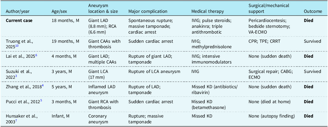
Spontaneous rupture of a giant coronary artery aneurysm is the rarest and most lethal complication of Kawasaki disease.Reference Suzuki, Kuroko, Kotani, Sakoda and Kasahara3 Table 1 compares the present case with previously reported paediatric cases of Kawasaki-related giant aneurysm rupture, massive cardiac tamponade, or sudden cardiac arrest. Ruptured cases cluster in two phenotypes: undiagnosed (“missed”) disease and diagnosed but immunoglobulin-refractory disease.Reference Pucci, Martino, Tibaldi and Bartoloni5,Reference Hunsaker, Hunsaker, Adams, Noonan and Ackermann7 Lai and colleagues recently reported rupture in a four-month-old infant despite intensive immunomodulation.Reference Lai, López-Guillén, Yeung, Ayusawa, McCrindle and Nagy6 The present case extends that observation to infants beyond the first year of life and illustrates that rupture can occur despite step-up immunomodulation, including interleukin-1 blockade, followed by sternotomy and veno-arterial extracorporeal membrane oxygenation.
Summary of reported paediatric cases of severe Kawasaki disease complicated by giant coronary artery aneurysm rupture, cardiac tamponade, or sudden cardiac arrest

CABG = coronary artery bypass grafting; CAA = coronary artery aneurysm; CPR = cardiopulmonary resuscitation; CRRT = continuous renal replacement therapy; ECMO = extracorporeal membrane oxygenation; IVIG = intravenous immunoglobulin; KD = Kawasaki disease; LAD = left anterior descending coronary artery; LCA = left coronary artery; M = male; RCA = right coronary artery; TPE = therapeutic plasma exchange; VA-ECMO = veno-arterial extracorporeal membrane oxygenation.
The rapid expansion and rupture observed here reflect the underlying vasculopathy. Orenstein and colleagues showed that Kawasaki vasculopathy comprises distinct processes, most notably acute necrotising arteritis: a synchronised, neutrophil-dominant destruction of the endothelium, medial smooth muscle, and adventitia during the first weeks of illness.Reference Orenstein, Shulman and Fox8 This transmural destruction strips the vessel of mechanical integrity and produces saccular or giant fusiform aneurysms. Our intraoperative description is the macroscopic correlate. Because autopsy consent was not obtained, histological confirmation is unavailable. Alternative contributors—acute coronary thrombosis with infarction, fulminant myocarditis, or a pre-existing undiagnosed arteriopathy—cannot be formally excluded, although the intraoperative findings and massive haemopericardium make spontaneous rupture on a background of necrotising arteritis the most plausible explanation.
Several management questions are raised. First, anakinra and other interleukin-1 antagonists have yielded encouraging results in refractory disease, including aneurysm regression,Reference Ferrara, Giani, Caparello, Farella, Gamalero and Cimaz9 but efficacy appears contingent on early initiation before irreversible medial destruction. In our patient anakinra was started after imaging confirmed giant aneurysms, by which point structural failure may have been committed. Second, Truong and colleagues reported survival of a catastrophic presentation after therapeutic plasma exchange and continuous renal replacement therapy;Reference Truong, Bui and Nguyen10 whether earlier plasmapheresis modifies outcomes warrants a prospective study. Third, surgical repair during the acute inflammatory phase is notoriously difficult because inflamed vascular tissue does not hold sutures, accounting for the dismal survival rate once rupture occursReference Lai, López-Guillén, Yeung, Ayusawa, McCrindle and Nagy6—directly corroborated by the failure of suture haemostasis in our patient.
Conclusion
Spontaneous rupture of a giant coronary artery aneurysm is a catastrophic and frequently fatal complication of Kawasaki disease, concentrated in infants with incomplete or immunoglobulin-refractory disease. Aggressive step-up immunomodulation, including interleukin-1 blockade, may not reverse vessel-wall fragility once necrotising arteritis has caused structural destruction. Outcomes following rupture remain dismal despite extracorporeal support, and surgical haemostasis may fail as inflamed arterial tissue does not hold sutures.
Acknowledgements
None.
Author contributions
All authors contributed substantially to the conception and design of the work, the acquisition and interpretation of clinical data, drafting and critical revision of the manuscript, and final approval of the version submitted for publication. All authors agree to be accountable for all aspects of the work. Specifically, BK and UP were responsible for the clinical evaluation, echocardiographic imaging, and paediatric cardiology management and led the drafting of the manuscript. AÖ and AKİ performed the bedside sternotomy and surgical re-exploration and provided the intraoperative findings and photographs. SÖ was responsible for the paediatric intensive care management, including extracorporeal membrane oxygenation support. All authors reviewed and approved the final manuscript.
Financial support
This research received no specific grant from any funding agency, commercial or not-for-profit sectors.
Competing interests
None.
Ethical standard
The authors assert that all procedures contributing to this work comply with the ethical standards of the relevant national guidelines on human experimentation and with the Helsinki Declaration of 1975, as revised in 2008. Written informed consent for publication of anonymised clinical details, including intraoperative photographs and echocardiographic images, was obtained from the patient’s parents.