Introduction
Cerebrospinal fluid (CSF) is produced by the choroid plexus of the ventricles, and plays an important role in maintaining the normal homeostasis of the cerebral cortex. It has nutritive and protective effects on the brain, and plays an important role in the maintenance of normal pressures within the cranium.
Cerebrospinal fluid leaks are potentially life-threatening, and result from disruption of the dura and arachnoid, along with bony defects in the skull base. Cerebrospinal fluid can leak into the ear (causing CSF otorrhoea) or the nose (causing CSF rhinorrhoea).
Cerebrospinal fluid rhinorrhoea can occur either spontaneously or following trauma. In recent times, the occurrence of iatrogenic CSF rhinorrhoea following endoscopic surgery has been well documented. The first report of surgical management of CSF rhinorrhoea was published by Dandy in 1926; an external approach was used. The first report of endoscopic management of CSF rhinorrhoea was published by Wigand in 1981. Since then, transnasal endoscopic management has been the mainstay of CSF rhinorrhoea treatment. Various materials have been used for closure of the defect, including fat, fascia, muscle, cartilage, bone, dura and other allografts.
The purpose of this paper is to share our experience of the endoscopic management of CSF rhinorrhoea using the cartilage inlay (underlay) technique.
Materials and methods
We identified retrospectively a total of 223 patients who had been treated for CSF rhinorrhoea in our hospital between 1994 and 2008. The majority of these patients had been referred to us with this diagnosis.
Diagnostic investigation included demonstration of CSF drip, CSF biochemical analysis (including glucose and β2 transferrin (in doubtful cases)) and computed tomography. The demonstration of crystal clear, watery fluid dripping from one nostril was the most important criterion when deciding to undertake surgery. All patients underwent a diagnostic nasal endoscopy; however, this usually did not identify the site of the leak, except in the case of a large meningocoele, an encephalocoele or copious CSF flow. Fluorescein studies were not undertaken in any of the patients.
The study inclusion criterion was CSF repair using a cartilage inlay technique, irrespective of patient age, sex or aetiology. Patients undergoing CSF repair using other materials were excluded.
The medical records and surgical procedure notes of all included patients were reviewed and analysed.
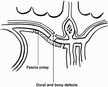
All patients underwent endoscopic repair under general anaesthesia. Marked probes were used to ascertain the size of the defect. Tragal cartilage was used when the defect was larger than 4 mm. (For defects larger than 1 cm, septal cartilage was used, while for defects smaller than 4 mm, temporalis fascia or fascia lata was used as an onlay graft (Figure 1); these patients were not included in this series.)
Diagrammatic representation of fascia onlay graft.

We always performed an endoscopic spheno-fronto-maxillo-ethmoidectomy on the side of the leak. The middle turbinate was removed in cases in which the defect was in the cribriform plate or lateral lamella.
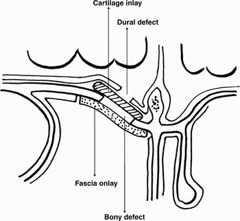
The site of the defect was identified and the margins freshened. In cases in which an obvious leak was not identified, an anesthetic valsalva maneuver was employed (still under general anaesthesia) to identify the leak. Soft tissue in the vicinity of the bony defect was meticulously removed using a diamond burr and diathermy. The size of the bony defect ranged from 4 to 20 mm in its maximum diameter, with a mean of 10 mm. Once all the edges of the bony defect had been delineated, the dura was carefully elevated with blunt instruments to create a pocket so as to accommodate the cartilage in an inlay fashion (Figure 2). Cartilage (either tragal or septal, depending upon the size of the defect) was harvested and shaped according to the size of the defect. The cartilage graft needed to be a few millimetres larger than the size of the bony defect. Although the cartilage graft was larger than the bony defect, its flexibility and malleability allowed it to be manoeuvred into the target pocket. Final adjustments were then made to settle the cartilage graft, before covering it with an appropriately sized piece of temporalis fascia. Care was taken that the fascial graft did not overlap any of the mucosal edges (Figure 2). The fascial graft was stabilised by placing a layer of Surgicel® or Gelfoam®, which was supported by many pieces of Merocel® (Medtronic Xomed, Jacksonville), one below the other, until the ipsilateral nasal cavity was completely filled with Merocel.
Diagrammatic representation of cartilage inlay graft with fascia onlay.

Patients were made ambulant from the first post-operative day. They were prescribed post-operative antibiotics and advised against straining. The CSF leak was expected to continue for a few days after repair, before finally stopping once the grafts had adhered to the graft bed; thus, patients were warned about immediate post-operative seepage of CSF and reassured. We did not use a lumbar drain in any of our patients. Patients were discharged after 72 hours, and their packs removed after one week.
Results
We identified a total of 62 patients who had undergone endoscopic CSF repair using a cartilage inlay technique: 42 women and 20 men. Patients were aged between 23 and 56 years.
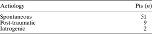
Table I shows the aetiology of patients' CSF leaks. While the aetiology was mixed, the majority of our cases were idiopathic.
Aetiology of cerebrospinal fluid leak
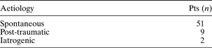
Pts = patients
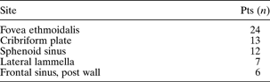
The sites of patients' CSF leaks are shown in Table II.
Site of cerebrospinal fluid leak
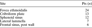
Pts = patients; post = posterior
The size of the offending bony defect ranged from 4 to 20 mm in its maximum diameter, with a mean of 10 mm. The largest defect encountered was approximately 2 × 1.5 cm, in the fovea ethmoidalis.
Defect repair was successful in all our patients, with no recurrence of CSF rhinorrhoea or major complications. Three patients developed post-operative sinus infection, which was promptly controlled by broad spectrum antibiotics.
The longest patient follow-up period was 16 years and the shortest six months; the median follow-up period was 15 months.
Discussion
Cerebrospinal fluid rhinorrhoea results from an abnormal communication between the subarachnoid space and the nasal cavity. This abnormal communication is brought about by trauma, prior surgery or tumours. More recently, endoscopic sinus surgery has been recognised as a major aetiological factor in the formation of CSF fistulae.Reference Mirza, Thaper, McClelland and Jones1, Reference Banks, Palmer, Chiu, O'Malley, Woodworth and Kennedy2 However, in some cases there is no identifiable cause; these cases are thus termed idiopathic or spontaneous. Cases of spontaneous CSF rhinorrhoea have been found to be associated with an abnormal body mass index and increased intracranial pressure, and also with an increased risk of failure following surgery due to increased CSF pressure.Reference Dunn, Alaani and Johnson3
Endoscopic repair of CSF fistulae is an accepted, safe procedure, with a success rate of between 87 and 98 per cent in recent reports.Reference Banks, Palmer, Chiu, O'Malley, Woodworth and Kennedy2, Reference Kirtane, Gautham and Upadhyaya4, Reference Lee, Huang, Chaung and Huang5
A variety of materials have been used to repair the offending skull base defects. Either free or flap grafts may be used. Free graft materials include fascia, fat, cartilage, bone, perichondrium and pericranium. Pedicled grafts commonly consist of middle turbinate or septal flaps. Flap grafts have the advantage of being vascularised, and this is theoretically associated with a reduced incidence of failure. However, studies and meta-analysis have found no statistically significant difference in outcomes, comparing different graft materials.Reference Zweig, Carrau, Celin, Schaitkin, Pollice and Snyderman6, Reference Hegazy, Carrau, Snyderman, Kassam and Zweig7
Grafts may be placed using either an inlay (underlay) or an onlay (overlay) technique. The majority of authors prefer the onlay technique, but there appears to be no difference in outcomes between the two techniques.Reference Zweig, Carrau, Celin, Schaitkin, Pollice and Snyderman6 The choice of graft depends upon the expertise and preference of the surgeon. No clear criteria have been established with regards to the size or site of the fistula and the type of graft best used. One group of authors has recommended the use of free mucosa grafts for defects smaller than 0.5 cm2; for defects larger than 0.5 cm2, a composite graft with rigid support from turbinate bone or septal cartilage is advised.Reference Burns, Dodson and Gross8
It is our observation that almost all cases of chronic CSF rhinorrhoea are associated with encephalocoeles, pseudomeningocoeles, or arachnoid cysts; their presence prevents the closure of the CSF fistula by conservative measures, since CSF lacks the cell population for self-repair. Pseudomeningocoeles and arachnoid cysts may be present some distance away from the bony defect, and they must be meticulously dissected and followed to the bony defect. It is the bony defect and dural tear that require reconstruction.
We are of the opinion that the size of the bony defect should determine the size of the reconstruction. Defects smaller than 4 mm in diameter require only an onlay graft on a well prepared graft bed. From recent experience, we are inclined to believe that simple preparation of the graft bed is adequate to provoke sufficient post-operative fibroblastic activity to close small leaks. Defects of 5 mm and larger require reconstruction of the skeleton, and we use cartilage graft as an inlay and fascia as an onlay. The cartilage graft should be 3–4 mm larger than the bony defect in at least two opposing directions. The flexibility of the cartilage is important, as it enables it to be manoeuvred into the intracranial pocket. In fact, this insertion of cartilage through the bony defect into the intracranial pocket is the trickiest and most important part of the procedure. After inserting the cartilage graft halfway into the pocket, cautious, gentle pressure is applied to the cartilage using a straight curette, until the cartilage buckles a little and passes through the bony defect, before finally ‘clicking’ into place in its ultimate destination.
We use just enough tissue to reconstruct the defect. A well prepared graft bed consists of bare bone all around the defect, devoid of soft tissue. Furthermore, all soft tissue and mucosa in the vicinity should be meticulously removed on the nasal side. We are of the opinion that multilayer closure, in which different materials are heaped one on top of the other, is unnecessary. On the cranial side, the dura is lifted off the bone by blunt instruments and angled elevators. Microscopic ear surgery instruments are useful for this purpose. The bony defect should be large enough to accomplish this step. Sometimes, when the dural defect is large, the cartilage may slip into the subarachnoid space. The surgeon must recognise this and replace it in the proper plane. The tragal cartilage is harvested together with its perichondrium. The perichondrium goes to reconstruct the dura, and the cartilage goes to reconstruct the bony defect. The fascial onlay acts as scaffolding on which the nasal epithelium can grow. When septal cartilage is used, a small piece of fascia is placed between the cartilage and the dura to reconstruct the dural defect. In large defects, placement of cartilage prevents future prolapse of brain tissue and meninges; it also dampens transmission of the strong pulsations of the dura, and improves graft adherence.
Watertight closure of CSF leaks from day one is every surgeon's dream. However, we believe this to be impossible, whatever technique one uses. Some amount of CSF seepage is inevitable until the graft adheres to the graft bed. We suspect that, irrespective of the type of repair performed, all patients will have some amount of CSF seepage in the immediate post-operative period, which goes unnoticed when the patient is confined to strict bed rest. When we first began to perform CSF defect repairs, tissue glue was not available commercially in India; however, lack of tissue glue has not interfered with our results in any way.
We believe that the crucial elements of successful CSF fistula repair are: precise identification of the bony defect; meticulous preparation of the graft bed; careful elevation of the dura; judicious use of just enough graft tissue; and adequate graft support. Some amount of CSF seepage in the immediate post-operative period does not in any way affect the final outcome.
Conclusion
Extradural cartilage inlay is an effective technique in the management of CSF rhinorrhoea, when just enough tissue is used for repair. It creates a stable repair which reduces transmission of dural pulsations, prevents future dural prolapse and enhances onlay fascia graft adherence. Cartilage is particularly useful in repairing defects larger than 4 mm. To the best of our knowledge, the current study represents the largest reported series of CSF fistula repair procedures involving a cartilage inlay technique, with no failures. We believe that the crucial factors in our high success rate for CSF fistula repair are: precise identification of the bony defect; meticulous preparation of the graft bed; careful elevation of the dura; judicious use of just enough graft tissue; and adequate graft support.