Introduction
The goals of cholesteatoma surgery are the complete eradication of pathology, the prevention of recurrent disease, the restoration of the hygiene status of the ear, and the preservation of hearing loss or improvement of hearing.Reference Shelton and Sheehy1
Our preferred surgical procedure for the treatment of middle-ear cholesteatoma has been the closed technique, or canal wall up approach, with bony obliteration of the mastoid and epitympanic cells, the so-called canal wall up bony obliteration technique.Reference Offeciers, Vercruysse, De Foer, Casselman, Somers and Ars2 This technique dramatically lowers the incidence of recurrent disease after primary cholesteatoma surgery, and achieves excellent ear hygiene and acceptable functional results.Reference Offeciers, Vercruysse, De Foer, Casselman, Somers and Ars2–Reference Gantz, Wilkinson and Hansen5 It can also be successfully applied to reconstruct unstable and troublesome cavities caused by canal wall down surgery for cholesteatoma.Reference Offeciers, Vercruysse, De Foer, Casselman, Somers and Ars2, Reference Vercruysse, De Foer, Somers, Casselman and Offeciers3, Reference Roberson, Mason and Stidham6
When performing bony obliteration, the main concern is the risk of burying residual cholesteatoma beneath the obliteration material. Follow up of obliterated mastoids using accurate and reliable imaging is mandatory to prevent late complications, which may take many years to appear.
Several authors have reported on the value of high resolution computed tomography (CT) and magnetic resonance imaging (MRI) for the detection of residual cholesteatoma in mastoids which have undergone bony obliteration.Reference De Foer, Vercruysse, Pouillon, Somers, Casselman and Offeciers7, Reference Yung and Karia8 High resolution CT is characterised by a low sensitivity and specificity for cholesteatoma, and cannot differentiate cholesteatoma from other soft tissues. Recently, non-echo-planar, diffusion-weighted MRI sequences have proven their value in detecting acquired, congenital and residual cholesteatoma down to a size of 2 mm.Reference De Foer, Vercruysse, Pilet, Michiels, Vertriest and Pouillon9–Reference De Foer, Vercruysse, Bernaerts, Deckers, Michiels and Pouillon11
The aim of this study was to report on the long term imaging follow up of bony mastoid and epitympanic obliteration, including CT and MRI modalities.
Materials and methods
We retrospectively evaluated a series of 51 patients who had undergone eradication of cholesteatoma using the bony obliteration technique at the St Augustine Hospital, Antwerp, between 21 September 1998 and 9 December 2002. All surgery had been performed by the senior author (EO).
The indication for bony obliteration of the mastoid and epitympanic space had been the presence of a primary acquired or recurrent cholesteatoma (42 patients) or an unstable, problematic cavity following earlier canal wall down surgery (nine patients). For both indications, the surgical principles and technique were the same, the only difference being that in the problematic cavity patients the whole posterosuperior bony canal wall required reconstruction down to the level of the facial canal by means of one or a few solid pieces of sculpted cortical bone, while in the cholesteatoma patients the canal wall was at least partially intact and needed less reconstruction. Subsequently, the mastoid and epitympanic space was filled with bone pâté up to the level of the cortex.Reference Offeciers, Vercruysse, De Foer, Casselman, Somers and Ars2, Reference Vercruysse, De Foer, Somers, Casselman and Offeciers3
All patients underwent high resolution CT and MRI, including non-echo-planar, diffusion-weighted MRI, a mean period of 76.4 months (median, 76.4 months; range, 53.8–113.6 months) after canal wall up bony obliteration technique surgery, in order to detect residual or recurrent cholesteatoma. The imaging protocol for follow up of cholesteatomas consisted of high resolution CT and MRI scanning, the latter including delayed contrast, T1-weighted imaging and non-echo-planar, diffusion-weighted sequences.
Imaging interpretation
All high resolution CT and MRI sequences were retrospectively evaluated by experienced head and neck radiologists (JC and BDF). Evaluators were blinded to all clinical data, including the surgical report and outcome, patient identity, and any ‘second look’ surgical findings.
The radiologists scored the imaging scans for the presence of cholesteatoma, categorising each case as either positive (i.e. residual cholesteatoma present) or negative (i.e. residual cholesteatoma absent). Interpretation of high resolution CT scans was based on the presence of soft tissue within the middle ear and/or bony obliterated mastoids and ‘punched-out’ lesions in the obliterated mastoid. On conventional MRI sequences, interpretation relied on the signal intensity of the soft tissues on T2-weighted images and late gadolinium-enhanced, T1-weighted images. On T2-weighted images, cholesteatoma presented as a nodular, moderately intense lesion. On late post-gadolinium, T1-weighted images, the lesion was non-enhancing but often showed an enhancing rim which corresponded to the perimatrix. On non-echo-planar, diffusion-weighted MRI sequences, cholesteatoma showed as a characteristically hyperintense lesion. All scans were scored as either positive or negative for cholesteatoma.
Imaging techniques
Magnetic resonance imaging was performed using a 1.5 T superconductive unit (Magnetom Avanto; Siemens Medical Solutions, Erlangen, Germany) using the standard head matrix coil. Axial, 2 mm thick, spin-echo, T1-weighted images and coronal, 2 mm thick, spin-echo, T1-weighted images were acquired with the same parameters. Coronal, 2 mm thick, turbo spin-echo, T2-weighted images and axial, 0.4 mm thick, three dimensional, turbo spin-echo, T2-weighted images were also obtained.
In all patients, a 2 mm thick, non-echo-planar, diffusion-weighted sequence (single-shot, turbo spin-echo, diffusion-weighted sequence) was acquired in the coronal plane (b factors 0 and 1000 mm2/sec). All sequences were acquired 45 minutes after intravenous contrast injection with either gadoterate meglumine (0.1 mmol/kg body weight; Dotarem, Guerbet, Roissy, France) or gadopentetate dimeglumine (Magnevist, Shering, Berlin, Germany). Computed tomography was performed using a 16-row, multislice CT scanner (Lightspeed; General Electric, Milwaukee, Wisconsin, USA) with coronal reformations. Axial slices were acquired with a thickness of 0.625 mm.
Results
Our series comprised 51 patients (28 males and 23 females; 17 were children aged 16 years or less) with an average age of 30.5 years (median, 28.4 years; range, 6.9–68.5 years). Three cholesteatomas were detected: one recurrent, one residual and one petrosal apex. Patients' most recent clinical follow-up images had been taken between 53.8 and 113.6 months (mean, 76.4 months) after their primary canal wall up bony obliteration technique procedure. Clinical and imaging data were available for all 51 patients.
Patient one
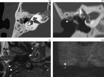
One patient (a 17-year-old man) had a partial breakdown of the canal wall, which resulted in a recurrent cholesteatoma. This was visualised on both high resolution CT and MRI (Figure 1). The recurrent cholesteatoma was clearly seen at the posterosuperior margin of the external auditory canal, with extension into the bony obliterated mastoid. The canal wall had broken down, with subsequent bone pâté resorption. This recurrent, external auditory canal cholesteatoma was treated 56.8 months after the primary bony obliteration technique surgery. During the revision surgery, the cholesteatoma was completely removed and the external auditory canal defect was closed using solid sculpted cortical bone and bone pâté. During subsequent follow up, no new recurrence was detected.
A 17-year-old man with a prior history of canal wall up bony obliteration technique surgery for cholesteatoma on the left side, and with suspected recurrent external auditory canal cholesteatoma on micro-otoscopy. Surgery revealed recurrent cholesteatoma with canal wall breakdown and partial bone pâté resorption. (a) Axial, high resolution computed tomography (CT) image at the level of the basal turn of the left cochlea. The external auditory canal wall is interrupted by a presumed recurrent cholesteatoma with marked bone pâté resorption (arrowheads). The remainder of the post-operative mastoid cavity is filled with bone pâté. (b) Coronal, reformatted CT image at the level of the lateral semicircular canal, showing canal wall breakdown and soft tissue enhancement in the bone-obliterated cavity (arrows). The remainder of the post-operative mastoid cavity is filled with bone pâté (asterisk). (c) Axial, late (45 minutes) post-gadolinium, T1-weighted magnetic resonance imaging (MRI) scan. A recurrent cholesteatoma is seen as a large defect in the external auditory canal surrounded by enhancing inflammatory and scar tissue (arrowheads). (d) Coronal, non-echo-planar, diffusion-weighted MRI scan at the level of the left temporal bone. The cholesteatoma is clearly seen (arrow) as a very hyperintense, horseshoe shaped lesion under the left temporal lobe. The keratin content has partially evacuated downward into the external auditory canal. Such a hyperintensity seen on non-echo-planar, diffusion-weighted scanning is pathognomonic for cholesteatoma.

Patient two
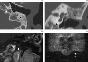
In a second patient (a 30-year-old man), micro-otoscopy showed a small mass positioned on top of the intact tympanic graft, suspected to be a cholesteatoma pearl (Figure 2). On high resolution CT, a small tissue opacification was seen just lateral to the superior margin of the tympanic graft, partially protruding into the external auditory canal and extending into the obliterated mastoid, supporting a diagnosis of residual cholesteatoma pearl (Figures 2a and 2b). This diagnosis was confirmed on non-echo-planar, diffusion-weighted MRI sequences (Figure 2d). However, on the late gadolinium-enhanced, T1-weighted MRI sequences the lesion could not be differentiated from the surrounding bony obliterated mastoids content. At reintervention, the cholesteatoma pearl measured 5–6 mm and was located at the superior margin of the external auditory canal, lateral to the tympanic membrane, with extension into the bony obliterated mastoids. It had presumably arisen from a fragment of the original cholesteatoma which had been covered by a piece of sculpted bone graft during canal wall reconstruction. During revision surgery, the residual cholesteatoma was completely removed, and the external auditory canal defect was closed using solid sculpted cortical bone and bone pâté.
A 30-year-old man with a prior history of canal wall up bony obliteration technique surgery for cholesteatoma on the right side, with micro-otoscopic suspicion of a residual cholesteatoma lateral and superior to the tympanic graft in the external auditory canal. Surgery revealed a residual cholesteatoma with canal wall breakdown and partial bone pâté resorption. (a) Axial, high resolution computed tomography (CT) image at the level of the right vestibule. A nodular lesion (black arrow) is seen eroding the surrounding bone-obliterated mastoid cavity (black asterisk). The tympanic cavity is well aerated. (b) Coronal, reformatted CT image at the level of the lateral semicircular canal. A clear, nodular lesion (white arrow) is seen lateral and superior to the tympanic graft. The remainder of the post-operative mastoid cavity is filled bone pâté (black asterisk). (c) Axial, late (45 minutes) post-gadolinium, T1-weighted magnetic resonance imaging (MRI) sequence. Note the confusing and often mixed signal intensities in the obliterated cavity (white arrows). Based on this sequence, a residual cholesteatoma cannot be confirmed or excluded. (d) Coronal, non-echo-planar, diffusion-weighted MRI sequence at the level of the left temporal bone. The cholesteatoma is clearly seen (white arrow) as a very hyperintense lesion under the left temporal lobe. The presence of such a hyperintensity on non-echo-planar, diffusion-weighted scanning is pathognomonic for cholesteatoma.

Patient three
In a third patient (a 69-year-old woman), non-echo-planar, diffusion-weighted MRI showed a characteristic hyperintense lesion set against a low intensity background, located in the apex of the temporal bone at the posterior margin of the internal carotid artery (see Figure 3). On standard MRI sequences, the lesion had the typical features of a cholesteatoma, namely a characteristic high signal intensity on T2-weighted scans and a hypointense signal with peripheral enhancement on late gadolinium-enhanced, T1-weighted scans. Retrospective evaluation of a high resolution CT scan taken prior to the primary bony obliteration in January 2001 revealed a (hitherto overlooked) small lesion in the petrosal apex. There was no clear connection between the original, acquired cholesteatoma, which had been successfully removed in 2001, and this more recently visualised, congenital cholesteatoma. Because the latter lesion had not grown since January 2001, no additional surgical treatment was planned. The patient has since been further monitored by non-echo-planar, diffusion-weighted MRI.
A 69-year-old woman with a prior history of canal wall up bony obliteration technique surgery for a left-sided cholesteatoma, with a normal micro-otoscopic investigation. (a) Axial, high resolution computed tomography (CT) image at the level of the left internal auditory canal. A non-specific, nodular lesion (white arrow) is seen in the petrosal apex. Note the homogeneously obliterated mastoid cavity (black asterisk). Based upon this image, a petrosal cholesteatoma cannot be confirmed or excluded. (b) Coronal, reformatted CT image at the level of the lateral semicircular canal. Again, a non-specific, nodular lesion (black arrow) is seen in the petrosal apex. Note the homogeneously obliterated mastoid cavity (black asterisk). (c) Axial, late (45 minutes) post-gadolinium, T1-weighted magnetic resonance imaging (MRI) sequence. Note the large, hypointense, non-enhancing, nodular lesion (white arrows), enabling diagnosis of a large petrosal cholesteatoma. (d) Coronal, non-echo-planar, diffusion-weighted image at the level of the left temporal bone. The cholesteatoma is clearly seen (white arrow) as a very hyperintense lesion. The presence of a hyperintensity on non-echo-planar, diffusion-weighted images is pathognomonic for cholesteatoma.

Imaging interpretation
High-resolution computed tomography
An aerated middle ear was found in 64.7 per cent of patients (33/51). A soft tissue obliterated middle ear was seen in 35.3 per cent of patients (18/51). Middle-ear cavity opacification was complete in 17.6 per cent (9/51) and partial in 17.6 per cent (9/51). In the bony obliterated mastoids, non-specific soft tissue opacification was visualised in 25.5 per cent (13/51), while 74.5 per cent (38/51) were homogeneously filled with bone. The soft tissue opacification in the middle-ear cavity and within the bony obliterated mastoids could not be further characterised using high resolution CT.
Magnetic resonance imaging
Non-echo-planar, diffusion-weighted MRI was performed on all patients, and detected three cholesteatomas, one of which was a clinically unsuspected petrosal (congenital) cholesteatoma. In all the other patients, no marked hyperintensity was visualised on the non-echo-planar, diffusion-weighted MRI sequences. Magnetic resonance imaging evaluation of bony obliterated mastoids relied mainly on the non-echo-planar, diffusion-weighted sequences. Indeed, on standard MRI sequences (including late gadolinium-enhanced, T1-weighted images) we found mixed and confusing signal intensities in the obliterated mastoids, probably due to the mixed presence of bone pâté and scar tissue; this made cholesteatoma diagnosis in obliterated mastoids impossible on standard MRI sequences. No false positive findings were seen on the non-echo-planar, diffusion-weighted MRI images in this series.
Discussion
The main goals of cholesteatoma surgery include total eradication of pathology and prevention of recurrent cholesteatoma.Reference Shelton and Sheehy1 The canal wall up bony obliteration technique, when correctly executed, is effective in lowering the recurrence rate of cholesteatoma.Reference Offeciers, Vercruysse, De Foer, Casselman, Somers and Ars2–Reference Roberson, Mason and Stidham6
However, the problem of residual cholesteatoma is inherent in all cholesteatoma surgery, regardless of the surgical approach and reconstructive technique applied. Therefore, the long-term safety of the ear is of primary concern in cholesteatoma surgery. This is especially the case when bony obliteration techniques are used, because they carry the potential risk of burying residual cholesteatoma beneath the obliteration material. It may then take years before the residual pathology becomes clinically evident, and major destruction and complications may ensue.
To avoid these problems, most authors advocate surgical staging after canal wall up surgery, in order to detect residual cholesteatoma.Reference Shelton and Sheehy1 The major drawback of this approach is the need for a second operation.
Over the last decade, attempts have been made to replace second stage surgery with imaging follow up. Until recently, high resolution CT was the imaging technique of choice for post-operative follow-up evaluation of mastoids undergoing bony obliteration.Reference De Foer, Vercruysse, Pouillon, Somers, Casselman and Offeciers7, Reference Yung and Karia8 High resolution CT using bone window settings is considered the method of choice for examination of the middle-ear structures. It provides excellent contrast between osseous structures, air and soft tissues, and it has a high spatial resolution. This enables visualisation of subtle osseous details, and allows good identification of associated bony erosions and good delineation of the pathology with respect to bony surroundings and air. Although it may take years, small cholesteatoma pearls may eventually become detectable in bone-obliterated mastoids by high resolution CT, appearing as punched-out lesions in the bone density.Reference Yung and Karia8 However, high resolution CT cannot differentiate cholesteatoma from other soft tissues such as scar tissue, cholesterol granuloma, granulation tissue or fluid. Therefore, high resolution CT has a low sensitivity and specificity for cholesteatoma.
In our study, high resolution CT revealed soft tissue lesions within obliterated mastoids in 25.5 per cent (13/51) of patients. Theoretically, such lesions could represent either residual cholesteatoma or scar tissue replacement after partial bone resorption, or incomplete bony obliteration of the mastoid cavity. In addition, high resolution CT revealed that 35 per cent of all our patients' middle-ear cavities were partially or completely opacified, due to lack of aeration and/or the presence of fibrous and/or inflammatory tissue. Further CT characterisation of these soft tissues in the middle-ear cavity and obliterated mastoid was impossible. Therefore, we conclude that high resolution CT is virtually useless for the non-invasive follow up of cholesteatoma.
In contrast, two newly available MRI protocols – late gadolinium-enhanced, T1-weighted sequences and non-echo-planar, diffusion-weighted sequences – enable specific characterisation of residual and recurrent cholesteatomas.Reference De Foer, Vercruysse, Pilet, Michiels, Vertriest and Pouillon9–Reference Ayache, Williams, Lejeune and Corré12 Using these MRI protocols, cholesteatoma can be unambiguously distinguished from other soft tissue such as scar tissue, cholesterol granuloma, granulation and fluid. The non-echo-planar, diffusion-weighted MRI sequence is an important improvement over the echo-planar, diffusion-weighted sequence, as the latter has a much lower contrast resolution and is made less reliable by susceptibility artefacts, thicker slices and a lower imaging matrix.Reference De Foer, Vercruysse, Pouillon, Somers, Casselman and Offeciers7, Reference De Foer, Vercruysse, Bernaerts, Maes, Deckers and Michiels10, Reference De Foer, Vercruysse, Bernaerts, Deckers, Michiels and Pouillon11, Reference Vercruysse, De Foer, Pouillon, Somers, Casselman and Offeciers13,Reference Vercruysse, De Foer, Somers, Casselman and Offeciers14 Non-echo-planar, diffusion-weighted MRI has three advantages over late gadolinium-enhanced, T1-weighted sequences, namely: more reliable lesion characterisation (due to better contrast resolution) resulting in a high specificity and selectivity; no need for contrast; and a shorter examination time. However, a disadvantage of non-echo-planar, diffusion-weighted MRI, compared with late gadolinium-enhanced, T1-weighted sequences, is its lower spatial resolution, which makes cholesteatoma localisation more difficult.
The current study also confirmed our previously reported findings, namely the difficulty of detecting and evaluating residual cholesteatoma within obliterated mastoids using standard MRI techniques (including late gadolinium-enhanced, T1-weighted scans), due to the frequent presence of mixed and confusing signal intensities.Reference De Foer, Vercruysse, Pouillon, Somers, Casselman and Offeciers7 Following a canal wall up bony obliteration technique procedure, the presence of a hyperintense lesion on non-echo-planar, diffusion-weighted MRI indicates the need for additional imaging (namely high resolution CT and late post-gadolinium, T1-weighted images) in order to determine the exact location and extension of any residual or recurrent cholesteatoma. If the hyperintense lesion is situated in the tympanic cavity, standard MRI sequences (including T2-weighted and late post-gadolinium, T1-weighted sequences) can provide additional information on the localisation of the lesion and its differentiation from any associated soft tissue in the middle-ear cavity. If the hyperintense lesion is situated in the obliterated mastoid, we advise high resolution CT scanning to exactly localise the soft tissue lesion, in order to facilitate surgical planning.
In canal wall up bony obliteration technique cases, the confusing and often mixed signal intensities seen on standard T2-weighted and late post-gadolinium, T1-weighted MRI images have led us to the working hypothesis that it is better to use non-echo-planar, diffusion-weighted MRI sequences for initial residual cholesteatoma screening. We allow for the possibility that it takes more than one year for a residual cholesteatoma to develop into a detectable lesion; therefore, we routinely image our post-operative cholesteatoma ears after one and five years.
Remarkably, we did not detect residual cholesteatoma within bone-obliterated spaces, even over long-term follow up; this is in concordance with other authors' findings.Reference De Foer, Vercruysse, Pouillon, Somers, Casselman and Offeciers7–Reference De Foer, Vercruysse, Pilet, Michiels, Vertriest and Pouillon9 This is probably due to the unfavourable local trophic conditions for keratinocyte survival and growth within the bone-obliterated space. Indeed, before obliteration we carefully removed all soft tissues until only healthy bone remained. It is known from clinical experience that skin only reluctantly covers an area of bare bone, but easily covers the same area if it is first lined with a thin layer of fascia. An animal study by Hinohara et al. gives some support to the hypothesis that bony obliteration interferes with the trophic conditions needed to allow residual keratinocytes to develop into a growing cholesteatoma pearl.Reference Hinohira, Gyo, Yanagihara, Bredberg and Alsterborg15 However, given that the rate of residual cholesteatoma following primary surgery is not negligible, strict follow up of cases remains necessary.
• The canal wall up bony obliteration technique lowers the incidence of recurrent cholesteatoma, but carries the potential risk of masking residual cholesteatoma
• The aim of this study was to report long-term follow-up imaging findings after canal wall up bony obliteration technique surgery, in order to detect residual and/or recurrent cholesteatoma
• Long-term follow up indicated that the canal wall up bony obliteration technique was a safe method of treating primary and recurrent cholesteatoma and reconstructing unstable cavities
• Magnetic resonance imaging, including delayed contrast and non-echo-planar, diffusion-weighted sequences, enabled detection of recurrent and residual cholesteatoma
Since operated ears quite often have residual soft tissue at the level of the middle ear and bony obliterated mastoids, which high resolution CT fails to characterise, we evaluate obliterated mastoids using non-echo-planar, diffusion-weighted MRI sequences. Such imaging has totally replaced exploratory second stage surgery in our department.
Conclusion
Long-term clinical and radiological follow-up after canal wall up bony obliteration technique procedures for cholesteatoma shows that this is a safe surgical treatment for primary and recurrent cholesteatoma, and for reconstruction of unstable cavities. Its meticulous application in our department greatly reduced the incidence of recurrent pathology.
After canal wall up bony obliteration technique procedures, long-term imaging follow up frequently shows associated soft tissue in the obliterated mastoid and middle-ear cavity. This soft tissue can be easily differentiated using non-echo-planar, diffusion-weighted MRI. Exploratory second stage surgery can now be safely replaced by MRI scanning, provided the non-echo-planar, diffusion-weighted MRI sequence is used.