




No CrossRef data available.
Article contents
The immunogenicity of Echinococcus granulosus antigen 5 is determined by its post-translational modifications
Published online by Cambridge University Press: 11 July 2005
Abstract
Since its early introduction as a marker for the immunodiagnosis of hydatid disease, antigen 5 (Ag5) has been regarded as one of the more relevant antigens of Echinococcus granulosus, and it is still widely used in different confirmation techniques. In this work we prepared 2 recombinant forms of the antigen, namely, rAg5 (corresponding to the unprocessed polypeptide chain of the antigen) and rAg5-38s (corresponding to its 38 kDa subunit). Their antigenicities were compared to that of the native antigen using a human serum collection. There was a major drop in the reactivity of the sera, particularly against rAg5-38s, which was confirmed by analysis of the cross-reactivity of 2 panels of monoclonal antibodies specific for rAg5-38s and the native antigen. Using the chemically deglycosylated native antigen, we demonstrated that the reduced antigenicity of the recombinants is due to the loss of the sugar determinants, and not to their misfolding. Inhibition experiments using phosphorylcholine confirmed that this moiety also contributes to the reactivity of the antigen, but to a much lesser extent. The presence of immunodominant highly cross-reactive glycan moieties in the Ag5 molecule may involve a parasite evasion mechanism.
Information
- Type
- Research Article
- Information
- Copyright
- © 2005 Cambridge University Press
INTRODUCTION
Antigen 5 is one of the main components of hydatid fertile cyst fluid. It is a 67 kDa glycoprotein that under reducing conditions separates into 2 components, of 38 kDa and 22–24 kDa (Lightowlers et al. 1989). Since its introduction as a marker of hydatid disease in the late 1960s, Ag5 has been regarded as one of the most valuable Echinococcus granulosus antigens (Capron, Vernes and Biguet, 1967). It is still widely used, particularly as a confirmatory assay, in techniques such as the Ouchterlony double-diffusion test or immunoelectrophoresis. In these techniques, Ag5 is visualized after the formation of a characteristic line of precipitation (arc 5), which in fact was the origin of the antigen's name. The utility of the antigen in serodiagnosis has been extensively evaluated using different sources of antigen, from crude extracts to purified antigen, and a wide variety of immunological techniques (ELISA, Western blot, immunoelectrophoresis, latex agglutination, etc). In that regard, the detection by immunoblot of the 38 kDa subunit, by far the most antigenic component of Ag5, has become one of the most popular ways of assaying patient sera against Ag5. In general, there is a good agreement about the good sensitivity attained with this antigen (Lightowlers et al. 1989; Chamekh et al. 1992; Barbieri et al. 1993), but its lack of specificity has been remarked upon as a matter of concern. Cross-reactions with other diseases were first demonstrated by Yarzabal et al. (1977) using sera from E. multilocularis-infected patients, and further revealed with sera from other helminthic infections, and even with normal sera (Di Felice et al. 1986).
Further progress in the use of recombinant forms of the antigen or synthetic peptides has been hindered by difficulties related to its cloning. Different attempts to clone Ag5 using antibodies failed, since they led to a molecule immunologically related to, but distinct from, Ag5 (Gonzalez et al. 2000). Recently, using sequence information derived from internal fragments of the antigen, we cloned the full-length Ag5 gene (Lorenzo et al. 2003). Ag5 is synthesized as a single polypeptide chain of 484 residues, which by removal of the leader peptide and digestion of a connecting dipeptide is processed into single disulphide-bridged 22 and 38 kDa subunits (165 and 296 residues, respectively). The smaller subunit binds calcium and possesses a glycosaminoglycan-binding motif that appears to be responsible for the observed binding of Ag5 to heparin. While this domain may provide interaction with cell surface and the intracellular matrix, the role of the 38 kDa subunits is unclear. This subunit is undoubtedly related to serine proteases of the trypsin family, but the catalytic Ser192 is substituted by Thr and no catalytic activity could be demonstrated so far. Putative N-glycosylation sites are predicted for each of the subunits at residues Asn55 and Asn213, which may explain its binding to concanavalin A. Direct evidence of N-glycosylation was found by March et al. (1991), who observed changes in the electrophoretic mobility of the antigen after treatment with N-glycosidase F. These authors also showed that the complex oligosaccharides of Ag5 do not contain terminal N-acetyl-D-glucosamine residues, and have no high-mannose structures (not sensitive to endo-beta-N-acetylglucosaminidase H). No putative O-glycosylation sites are predicted.
The presence of carbohydrate moieties, as well as the finding that Ag5 has phosphorylcholine epitopes (Shepherd and McManus, 1987), may account for the low specificity of the antigen. In addition, it has been reported that Ag5 bears epitopes that are reactive with anti-P1 blood group antigen antibodies, which could result in false-positive reactions when sera from P2 patients with suspected hydatid disease are tested by immunoblot or immunoprecipitation analysis. Consequently, the identification of discrete epitopes of the antigen that may overcome this drawback became a relevant research aim. The description by Liu et al. (1993) of Ag5-specific antibodies defining proteinaceous epitopes that do not cross-react with T. solium, E. multilocularis or E. vogeli suggests that these epitopes exist.
In order to advance in the molecular dissection of Ag5 antigenicity, we produced 2 recombinant forms of Ag5, namely its 38 kDa subunit and the whole Ag5 molecule. The diagnostic performance of these antigens was compared to that of the parent protein, and we found a markedly decreased antigenicity of the recombinant proteins. Using monoclonal antibodies (mAbs), chemical deglycosylation, and phosphorylcholine inhibition we demonstrated that the post-translational modification of the antigen, particularly the sugar moieties of Ag5, constitute the immunodominant epitopes of the antigen.
MATERIALS AND METHODS
Antigen 5 purification
Ag5 was purified from fertile bovine hydatid cyst fluid (HCF) by immunochromatography using the mAb 1D1 as described by Lorenzo et al. (2003). Briefly, HCF was applied to a 1D1-Sepharose column and the column was extensively washed with phosphate-buffered saline (PBS) containing 0·05% Tween 20 (PBS-T) followed by 0·5 M NaCl in PBS and finally with PBS. The Ag5 was eluted with 0·1 M glycine buffer, pH 2·5. The eluate was neutralized and extensively dialysed against PBS. Protein concentration was estimated using the bis-cinchonic acid protein assay (Pierce, Illinois, USA).
Antibodies
Hyperimmune rabbit and mouse sera against native Ag5 were prepared by standard protocols. mAbs reactive with Ag5 and rAg5-38s were prepared as described by Gonzalez et al. (1994). Briefly, mice were primed intraperitonally with 20 μg of immunopurified Ag5 or recombinant protein rAg5-38s in FCA, and boosted after 3 and 6 weeks with the corresponding antigen using FIA. Three days after the last booster each mouse was killed and the splenocytes were fused with SP2/0 cells. Cultures producing mAbs reactive with the corresponding antigen were selected by ELISA.
Human serum specimens
The following serum samples were used: 34 sera from surgically confirmed cystic hydatid patients, pre-selected on the basis of their reactivity with native Ag5 in ELISA; 18 serum samples from healthy donors; 18 serum samples from patients with alveolar hydatid disease and 18 serum samples form patients with Taenia solium cysticercosis.
Recombinant 38 kDa subunit and Ag5
Total RNA was isolated from freshly isolated E. granulosus protoescoleces, using Trizol reagent (Gibco BRL) according to the manufacturer's instructions. The reverse-transcription and PCR-amplification were performed in a single step with the One-step RT-PCR kit (Qiagen GmbH, Germany) following the manufacturer's instructions. The sequence encoding the 38 kDa subunit was amplified using the following primers: sense (5′-GGATCCGAATTCATTCTTGGTGGAAAAAGCGCCA-3′) and antisense: (5′-CCAAGCTTCATTAGACTGCGTAGCGGTTGATCC-3′). The PCR product was purified and digested with Eco RI and HindIII and cloned between the corresponding sites in the pMAL-c2X vector (New England Biolabs, Beverly, MA, USA). XL-1 Blue Escherichia coli cells were electroporated with the construct, and grown in ampicillin plates. Individual clones were sequenced using Ampli Taq FA according to standard procedures on an ABI Prism 377 DNA sequencer. The 38 kDa subunit fused to the C-terminus of maltose-binding protein (MBP) was purified from cell cultures on amylose-agarose (New England Biolabs) following standard procedures. The full-length Ag5 gene was amplified using the following primers: sense (5′-GGATCCGAATTCTTGGAGCTCACTCTCGATCCAG-3′) and antisense (5′-CCAAGCTTCATTAGACTGCGTAGCGGTTGATCC-3′). The PCR product was isolated (Quiaquick, Qiagen GmbH, Germany), cloned into pGEM T Easy (Promega). The fragment was then excised from pGEM by digestion with BamHI and HindIII, and purified and cloned between the corresponding sites in the pET-28a(+) vector (Novagen). Electrocompetent BL21-DE3 E. coli cells were transformed with the construct, the cells were grown in ampicillin plates and individual clones were submitted for sequencing. His-tagged Ag5 inclusion bodies were solubilized using 8 M urea, and the protein was purified on Ni-NTA agarose (Qiagen GmbH, Germany) under denaturing conditions. Refolding was achieved by diluting the urea from 8 M to 1 M by slow addition (1 ml/min) of 50 mM phosphate, 300 mM NaCl, pH 7·2, then allowing protein refolding at room temperature for 16 h with smooth agitation, followed by overnight dialysis at 4 °C against PBS.
Chemical deglycosylation
Non-selective removal of carbohydrates from immunopurified Ag5 was performed by reaction with trifluoromethanesulfonic acid (TFMS) (Edge, 2003). Glycofree™ Chemical Deglycosylation Kit (PROzyme, USA) was used essentially following the manufacturer's instructions. Briefly, a sample of Ag5 was thoroughly dialysed against 0·1% trifluoroacetic acid, then freeze-dried and treated with a mixture of TFMS/Toluene. After 4 h at −20 °C, the reaction was ended by neutralization. The deglycosylated polypeptide that precipitated upon neutralization was isolated directly by centrifugation and the pellet was dissolved in an appropriate buffer.
ELISA
Polystyrene ELISA plates (NUNC, Maxisorp, Denmark) were coated overnight at 4 °C with 250 ng of antigen per well and blocked with 5% of non-fat milk powder in PBS for 1 h at 37 °C. Serum samples (diluted 1/400) were assayed in triplicate (90 min, 37 °C). After washing, peroxidase-conjugated goat anti-human IgG (Sigma, USA) was added and incubated for 1 h at 37 °C, and then the wells were washed. Substrate solution containing H2O2 and 2,2′-azino-bis (3-ethylbenz-thiazoline-6-sulfonic) acid was added and absorbance was measured at 415 nm. The cutoff of the assay was established by Receiver Operating Characteristic (ROC) analysis (Swets, 1988) using the SPSS 10.0 software package (SPSS Inc. Illinois, USA). Inhibition with phosphorylcholine was performed as described above, using 20 mM Tris-HCl, pH 8·0 buffer, and comparing the reactivity of the sera in the presence or absence of 50 mM phosphorylcholine. Since this system exacerbated the cross-reactivity of the sera, only the cystic echinococcosis and normal sera were included in the ROC analysis.
Western blot
Native Ag5 and chemically deglycosylated Ag5 were resolved by electrophoresis on 12% SDS-PAGE gels under reducing conditions, then transferred onto nitrocellulose sheets (Bio-Rad). The nitrocellulose was blocked with 3% non-fat milk powder in PBS 1 h at 37 °C and washed. Fifteen human serum samples from surgically confirmed hydatid patients (diluted 1/100) were incubated with the antigens on nitrocellulose for 1 h at 37 °C, followed by extensive washing. Alkaline phosphatase-conjugated goat anti-human IgG (Sigma) (diluted 1/2500) was then incubated with the antigens for 1 h at 37 °C. After washing, a substrate solution containing 5-bromo-4-chloro-3’-indolylphosphate p-toluidine salt (BCIP) and nitro-blue tetrazolium chloride (NBT) was added according to the manufacturer's instructions (Sigma, USA).
Immunoelectrophoresis
Electrophoresis of antigens was performed on 0·9% agarose in 50 mM Tris buffer, pH 8·0. Then undiluted serum samples were dispensed in the tracks and allowed to migrate at 4 °C for 48 h. The agarose gels were extensively washed, dried and stained with Coomassie brilliant blue R 250 (sigma).
MS analysis of native and deglycosylated Ag5
The antigen was dissolved in 6 M guanidine-HCl, in the presence of DTT and was treated with an excess of iodoacetamide. After extensive dialysis against 0·1% trifluoroacetic acid the samples were lyophilized. This material was then rehydrated, during which process modified trypsin (Promega) was added. After overnight incubation at 30 °C, the digest was analysed by matrix-assisted laser desorption ionization time-of-flight (MALDI-TOF) MS on a Bruker Autoflex (Bruker Daltonics, Bremen, Germany). The instrument was optimized for peptides, and the spectra were internally calibrated using proteolytic peptide masses from a known sample.
RESULTS AND DISCUSSION
Production of recombinant Ag5 and its 38 kDa subunit
Since the Ag5 38 kDa subunit is clearly the most antigenic component detected by Western blotting under reducing conditions (Chamekh et al. 1990), we initially worked with this subunit to evaluate the potential diagnostic use of the recombinant forms of Ag5. This subunit was amplified by PCR as described, expressed as a fusion protein with MBP, and purified on amylose-agarose. The electrophoretic mobility of rAg5-38s agrees with the expected migration of a fusion protein constituted by MBP plus the 296 amino acid of the 38 kDa subunit. After partial digestion of rAg5-38s with factor Xa 3 main bands can be observed in SDS-gels, a strong band of about 76 kDa corresponding to undigested rAg5-38s, a second one corresponding to the MBP component (43 kDa) of the fusion protein and a component of about 33 kDa which corresponds to the recombinant 38 kDa subunit (calculated molecular mass=33008 Da). This was confirmed by the reactivity of the mouse anti-Ag5 serum (data not shown). Owing to the problems related to its cleavage, rAg5-38s was used for all purposes, as the fusion protein.
Unfortunately, the diagnostic performance of rAg5-38s was very poor, and for this reason we also prepared the recombinant form of the full length Ag5 gene (rAg5). rAg5 did not contain the leader peptide, but included the dipeptide Leu-Lys that connects the 22 kDa and 38 kDa in the unprocessed pro-Ag5 (Lorenzo et al. 2003). This recombinant protein formed inclusion bodies under all of the culture conditions assayed. Thus, purification of rAg5 required solubilization with urea followed by a refolding process. In SDS-gels the recombinant antigen showed increased electrophoretic mobility when compared with the native antigen, due to the fact that its expression in E. coli prevents posttranslational modifications. The dipeptide Leu-Lys was not susceptible to cleavage in E. coli, and thus rAg5 did not generate the 22 and 38 kDa subunits by reduction (data not shown).
rAg5-38s and rAg5 are poorly antigenic
Sera from healthy individuals and from patients with different parasitic diseases were used to evaluate the antigenicity of the recombinant proteins by ELISA (Fig. 1). Since our main concern was the recovery of the antigenic epitopes of native Ag5, we pre-selected our panel of hydatid disease serum samples on the basis of their positive reaction with this antigen. The recombinant antigens performed with an overall decrease in reactivity, most notably in the case of rAg5-38s. According to the cut-off estimated by ROC curves analysis, only 65% of the native Ag5-positive hydatid disease serum samples scored as positive when tested with rAg5, and just 21% gave positive results with rAg5-38s (table inserted in Fig. 1). Interestingly, the cross-reactivity with sera from related diseases was also greatly reduced. Using rAg5, the percentage of alveolar echinococcosis specimens that scored as positives dropped from 94 to 11% and, similarly, there was a drop from 39 to 11% for the cysticercosis serum samples. The recovery of some major antigenic determinants in rAg5 as compared to those of rAg5-38s was striking. Since we know that the 22 kDa subunit is poorly antigenic (Fig. 4), the better performance of rAg5 appears to be related to the occurrence of major discontinuous epitopes in the whole molecule. Nevertheless, despite the improved specificity, the reactivity of rAg5 is still far from that of native antigen, which may preclude its diagnostic applications.

Fig. 1. Reactivity of the human serum samples with native Ag5, and recombinant forms of the antigen assessed by ELISA. The serum panel consisted of: 18 sera from healthy donors (Ns), 34 sera from hydatid patients (Eg), 18 sera from patients with alveolar hydatidosis (Em) and 18 sera from patients with cysticercosis (Ts). The horizontal line represents the cut off value for each assay estimated by ROC curves. The inserted table summarizes the number (%) of serum samples classified as positive by ROC curve analysis.
Two main factors could be the cause of the reduced antigenicity of the recombinants, their improper folding, particularly in the case of rAg5 as a consequence of its recovery from inclusion bodies, and/or the presence of highly immunogenic post-translational modifications in the native antigen. We did a series of experiments to study this; first we used a panel of mAbs as probes to analyse the relevance of proteinaceous epitopes in the recombinants and the native antigen; second, we assessed the antigenicity of non-proteinaceous moieties of Ag5 by chemical treatment of its sugar moieties and inhibition with phosphorylcholine.
Production of monoclonal antibodies against proteinaceous epitopes of Antigen 5
Based on the high reactivity of the 38 kDa subunit of native Ag5 in Western blots, it is evident that its main epitopes are conserved after reduction, SDS treatment and heating, therefore, if they exist, the determinants sitting in the polypeptide moiety of the 38 kDa subunit should be conserved in its recombinant form. To study this, we prepared mAbs against rAg5-38s and analysed their cross-reactivity with native Ag5, on the other hand, we also prepared mAbs to the native antigen and used them to probe the conservation of their determinants in the recombinants. Monoclonal antibodies against rAg5-38s and native Ag5 were prepared using standard procedures. Positive hybridoma supernatants were primary selected (IgG isotype) from each fusion by screening against the corresponding immunizing antigen in ELISA. Seventeen clones reacting with rAg5-38s but not with MBP were selected from mice immunized with rAg5-38s, and 38 positive clones resulted from the fusion carried out with the donor mice immunized with native Ag5. The cross-reactivity of the anti-rAg5-38s antibodies with Ag5 is shown in Fig. 2A. Most of the clones did cross-react with the native antigen, showing that rAg5-38s and native Ag5 share proteinaceous epitopes that are accessible for binding in the native antigen. However, these epitopes are not immunogenic in the native molecule as could be observed when the opposite situation was examined (Fig. 2B). None of the mAbs raised against native Ag5 reacted with rAg5-38s (not shown), and there was an important fall in the reactivity of the mAbs against rAg5, indicating that major immunodominant epitopes of native Ag5 are absent in rAg5. The picture delineated with the use of the mAbs as probes is very much in line with that found with patient sera. We are aware that this is just an approach to the study of Ag5 immunogenicity, and bear in mind that our immunization protocol may not reproduce the immune response generated by the antigen in infected individuals. Nevertheless, we do not believe that the observed antibody response is an artifact, and conversely, the use of adjuvants has the potential to promote a stronger response against the polypeptide moiety of Ag5, which was not observed.

Fig. 2. Reactivity of the mAbs against native Ag5 and the recombinant forms of the antigen analysed by ELISA. (A) Analysis of 17 hybridoma supernatants raised against rAg5-38s. (B) Analysis of 38 hybridoma supernatants produced against native Ag5.
Asn55 and Asn213 are glycosylated in native Ag5
Our work with mAbs confirmed that the proteinaceous epitopes of the 38 kDa subunit are not immunogenic when this component is assembled in the native antigen, which suggests that the observed reactivity of this subunit in Western blot may be related to its post-translational modifications. Similarly, the results obtained with rAg5 also point in that direction, although in this case we need to consider the possibility of misfolding. To clarify this point, we destroyed the sugar moieties of Ag5 by chemical deglycosylation with TFMS acid. This procedure, removes O-linked oligosaccharides bound to a serine or threonine residue, N-linked glycans with N-acetylglucosamine-asparagine linkage, glycosaminoglycans attached to the core protein of proteoglycans and collagen saccharides attached to hydroxyproline by an acid-catalysed dehydration mechanism (Edge, 2003). This treatment leaves the protein backbone intact, with no alteration of the protein core immunoreactivity. The change in the electrophoretic mobility of deglycosylated Ag5 is shown in Fig. 3. A shift is observed for each of the subunits after reduction. Note that in the case of the smaller subunit of the antigen, the 22–24 kDa bands shifted to a single band of about 20 kDa (calculated molecular mass of the 22 kDa subunit=19556 Da), indicating that 2 different glycoforms exist for this subunit. The 38 kDa band exhibited a drop of about 5 kDa, which is in agreement with the calculated molecular mass (33008 Da) of the 38 kDa subunit. The electrophoretic mobility of this band is identical to that of the recombinant 38 kDa subunit.

Fig. 3. Reactivity of mAbs and human serum samples against the deglycosylated antigen (dAg5). Ag5 and dAg5 were analysed by ELISA with the 38 hybridoma supernatants raised against native Ag5 (A) and with our serum collection (B). The serum panel consisted of: 18 sera from healthy donors (Ns), 34 sera from hydatid patients (Eg), 18 sera from patients with alveolar hydatidosis (Em) and 18 sera from patients with cysticercosis (Ts). The horizontal line represents the cut off value for each assay estimated by ROC curves. The inserted figure shows the electrophoretic mobility of Ag5 after chemical deglycosylation. SDS-PAGE analysis of the native Ag5 (1), chemically deglycosylated Ag5 (2) and rAg5-38s digested with Factor Xa (3) run under reducing conditions.
Based on the predicted absence of putative O-glycosylation sites and previous information obtained with N-glycosidases (March et al. 1991) it can be assumed that the putative N-glycosylation sites (Asn55 and Asn213) predicted for each of the subunits are in fact glycosylated. By sequencing of internal fragments (Lorenzo et al. 2003), we have previously found evidence of glycosylation in Asn213. In order to obtain evidence of glycosylation in the 22 kDa subunit we performed a comparison of the MS-fingerprints of trypsin digests of the native and deglycosylated antigen. In general, no major differences were observed between the two spectra, with no signs of deamidation after TFMS treatment. In the digest corresponding to the deglycosylated antigen we could identify a fragment of 2516.1317 Da, which was absent in the Ag5 digest. The mass of this fragment only differs in 0.0494 Da from the calculated mass of the fragment ESHGGFYFYDSNGATLMFNR (2313.0029 Da) plus the mass of the innermost asparagine-linked N-acetylglucosamine residue (203·0794) which remains after TFMS deglycosylation, indicating that Asn55 of the 22 kDa subunit is glycosylated. This finding was supported by the presence of an accompanying peak in the spectrum of dAg5, with a mass increase of 16 Da, which is in agreement with the presence of an oxidized methionine.
The destruction of the carbohydrate moieties severely affects the antigenicity of Ag5
The deglycosylated antigen (dAg5) was assayed with our serum and mAb panels (Fig. 3). The reactivity of the mAbs with Ag5 and deglycosylated Ag5 was highly similar to that obtained with rAg5; when individually examined, basically, all clones reacting with the deglycosylated antigen were also reactive with rAg5, which indicates that refolding of rAg5 was highly successful. The reactivity of dAg5 with human sera also paralleled that obtained with rAg5. ROC analysis showed that after deglycosylation 30% of the cystic echinococcosis sera score as negative, and in addition there was a substantial drop in the absorbance readings, even for those that still score as positive. The loss of reactivity was still more striking in the case of the E. multilocularis and T. solium sera. The antigenicity of dAg5 was also analysed by Western blot; Fig. 4 shows the complete loss of the antigenic determinants of the 38 kDa subunit when the sugar moieties were removed. The faint residual reactivity shown by some of the serum samples is actually due to the fact that a small amount of non-deglycosylated antigen (undetectable in Coomasie-stained SDS-gels) remained. This is evidenced by the reactivity of the rabbit Ag5 specific serum used as control, which in addition to the 22 and 38 kDa deglycosylated subunit, also reacted with the remains of the glycosylated 38 kDa subunit. Finally, since the identification of arc 5 by immunoelectrophoresis is still a widely used diagnostic technique, we analysed the involvement of Ag5 sugar epitopes in the formation of this arc, and found that in their absence, the formation of arc 5 by a pool of patient sera is completely abolished (Fig. 5). This result was confirmed by individual analysis of a number of hydatid disease serum samples (not shown). Note that the hyperimmune rabbit antiserum, which also reacts against the protein core of Ag5, still forms a precipitation arc with dAg5, but with a different shape, surely because the electrophoretic mobility is changed by the elimination of the antigen glycoforms.

Fig. 4. Western blot analysis of Ag5 (a) and deglycosylated antigen (b) run under reducing conditions. Fifteen serum samples from patients with hydatid disease and a hyperimmune rabbit Ag5 specific antiserum (Rb) were used for detection. Notice that in addition to the smaller component of the deglycosylated antigen, Rb reveals the presence of the deglycosylated 38 kDa subunit and that of small remains of the glycosylated subunit. Except for serum 14, none of the human sera reacted against the 22 kDa component of the antigen.

Fig. 5. Immunoelectrophoresis analysis. After electrophoresis of deglycosylated Ag5 (top) and native Ag5 (bottom), a pool of hydatid disease sera (Panel 1) and a rabbit Ag5 specific antiserum (Panel 2), were allowed to migrate on the agarose gel and the bands were Coomassie stained.
The role of phosphorylcholine moieties
Anti-phosphorylcholine antibodies are found in a wide variety of parasitic infections as well as in normal sera. The presence of phosphorylcholine is well known in bacteria (Tomasz, 1967) as a modification of the polysaccharides of the cell wall and cell membrane lipoteichoic acid. In eukaryotic organisms it has been detected in a wide variety of pathogens such as protozoa, fungi and helminths (Harnett and Harnett, 1999), where it is linked to sugar moieties of structural glycolipids or N-linked glycans of soluble glycoproteins (Haslam et al. 1997). The presence of phosphorylcholine moieties in the 38 kDA subunit of Ag5 has been documented before (Shepherd and McManus, 1987; Lightowlers et al. 1989) and has been indicated as an important source of cross-reactivity. Owing to the fact that phosphorylcholine occurs as a modification of the N-linked glycans, deglycosylation with TFMS also removes this modification. Therefore, to evaluate the contribution of the phosphorylcholine epitopes we performed a competitive ELISA on plates coated with native Ag5. As seen in Fig. 6 and, the phosphorylcholine determinants of Ag5 account for a small fraction of the antigen reactivity, but have a considerable role in the cross-reactivity with sera from other diseases, notably in the case of the cysticercosis sera where cross-reactivity decreased up to 50%. This is in line with the observations of Shepherd and McManus (1987), who also found that the phosphorylcholine epitopes were a main source of cross-reactivity with trematodes and nematodes.

Fig. 6. Inhibitory effect of free phosphorylcholine (PC) on the reactivity of human sera with Ag5 assessed by ELISA. Serum samples were diluted (1/400) in 20 mM Tris buffer with or without 50 mM PC. The serum panel consisted of: 18 sera from healthy donors (Ns), 34 sera from hydatid patients (Eg), 18 sera from patients with alveolar hydatidosis (Em) and 18 sera from patients with cysticercosis (Ts). The horizontal line represents the cut off value estimated by ROC curves. Inserted are shown the number (%) of human serum samples scored as positive by ROC analysis.
Conclusions
Since its introduction in hydatid serology Ag5 has been regarded as one of the most valuable E. granulosus antigens. Recent progress in its molecular characterization paved the way for a more standardized use of the antigen, by allowing its production by recombinant technology. This appeared as an attractive goal for several reasons (i) its use in routine serological assays requires its production in large and reproducible amounts, and its occurrence in hydatid cyst fluid is scant; (ii) during our studies on the characterization of Ag5, we found that all DNA clones or the amino acid sequences of internal fragments obtained from different parasite sources were identical, suggesting that no polymorphic forms of the antigen exist, as it has been the case for the other E. granulosus major antigen, namely antigen B; and (iii) its expression in E. coli could be of benefit considering that the non-proteinaceous epitopes of E. granulosus had been regarded as a common source of cross-reactivity in hydatid serology (Di Felice et al. 1986). However, due to the fact that these epitopes are crucial components of Ag5 immunoreactivity, this latter potential advantage turned out to be a major obstacle to the diagnostic use of the recombinant forms of Ag5. Though the use of eukaryotic expression systems could promote a glycosylation pattern that may resemble the dominant saccharidic epitopes of the native molecule, this is not an attractive goal because, as we have seen, these determinants are a major source of cross-reactivity.
In spite of the disappointing diagnostic performance of the recombinants, they contributed to a better understanding of Ag5 immunogenicity. It is now clear that the immunoreactivity of Ag5 is determined by a combination of post-translational and proteinaceous epitopes. The latter are mostly of discontinuous nature and require that both subunits are assembled in the native molecule. Although we could confirm a role for the phosphorylcholine moiety of Ag5, the striking loss of immunoreactivity caused by deglycosylation of the antigen clearly showed that the saccharidic epitopes are the major immunodominant determinants in the course of the hydatid infection. Phosphorylcholine-containing glycoprotein have been shown to possess a plethora of immunomodulatory activities (Harnett and Harnett, 1999) and it is well know that in different parasitic infections, carbohydrates play a major role in the host-parasite relationship (Hokke and Deelder, 2001; Khoo and Dell, 2001). Further progress on the knowledge of Ag5's role in parasite survival will provide the basis to ascertain whether the observed immunodominance of Ag5 post-translational modifications is biologically meaningful.
This work was supported by an NIH Fogarty International Center Grant TW05718. We would like to thank María Noel Miguez for technical assistance and Dr Ulf Hellman for the MALDI-TOF MS analysis.
References
REFERENCES
Barbieri, M.,
Sterla, S.,
Battistoni, J. and
Nieto, A. (
1993).
High performance latex reagent for hydatid serology using an Echinococcus granulosus lipoprotein antigen fraction purified from cyst fluid in one step.
International Journal for Parasitology
23,
565–572.CrossRefGoogle Scholar
Capron, A.,
Vernes, A. and
Biguet, J. (
1967).
Le Diagnostic Immuno-Electrophorétique de l'Hydatidose.
SIMEP,
Lyon.
Chamekh, M.,
Facon, B.,
Dissous, C.,
Haque, A. and
Capron, A. (
1990).
Use of a monoclonal antibody specific for a protein epitope of Echinococcus granulosus antigen 5 in a competitive antibody radioimmunoassay for diagnosis of hydatid disease.
Journal of Immunological Methods
134,
129–137.CrossRefGoogle Scholar
Chamekh, M.,
Gras-Masse, H.,
Bossus, M.,
Facon, B.,
Dissous, C.,
Tartar, A. and
Capron, A. (
1992).
Diagnostic value of a synthetic peptide derived from Echinococcus granulosus recombinant protein.
Journal of Clinical Investigation
89,
458–464.CrossRefGoogle Scholar
Di Felice, G.,
Pini, C.,
Afferni, C. and
Vicari, G. (
1986).
Purification and partial characterization of the major antigen of Echinococcus granulosus (antigen 5) with monoclonal antibodies.
Molecular and Biochemical Parasitology
20,
133–142.CrossRefGoogle Scholar
Edge, A. S. (
2003).
Deglycosylation of glycoproteins with trifluoromethanesulphonic acid: elucidation of molecular structure and function.
The Biochemical Journal
376,
339–350.CrossRefGoogle Scholar
Gonzalez, G.,
Orn, A.,
Cazzulo, J. J. and
Gronvik, K. O. (
1994).
Production and characterization of monoclonal antibodies against the major cysteine proteinase of Trypanosoma cruzi.
Scandinavian Journal of Immunology
40,
389–394.CrossRefGoogle Scholar
Gonzalez, G.,
Spinelli, P.,
Lorenzo, C.,
Hellman, U.,
Nieto, A.,
Willis, A. and
Salinas, G. (
2000).
Molecular characterization of P-29, a metacestode-specific component of.
Molecular and Biochemical Parasitology
105,
177–184.CrossRefGoogle Scholar
Harnett, W. and
Harnett, M. M. (
1999).
Phosphorylcholine: friend or foe of the immune system?
Immunology Today
20,
125–129.Google Scholar
Haslam, S. M.,
Khoo, K. H.,
Houston, K. M.,
Harnett, W.,
Morris, H. R. and
Dell, A. (
1997).
Characterisation of the phosphorylcholine-containing N-linked oligosaccharides in the excretory-secretory 62 kDa glycoprotein of Acanthocheilonema viteae.
Molecular and Biochemical Parasitology
85,
53–66.CrossRefGoogle Scholar
Hokke, C. H. and
Deelder, A. M. (
2001).
Schistosome glycoconjugates in host-parasite interplay.
Glycoconjugate Journal
18,
573–587.CrossRefGoogle Scholar
Khoo, K. H. and
Dell, A. (
2001).
Glycoconjugates from parasitic helminths: structure diversity and immunobiological implications.
Advances in Experimental Medicine and Biology
491,
185–205.CrossRefGoogle Scholar
Lightowlers, M. W.,
Liu, D. Y.,
Haralambous, A. and
Rickard, M. D. (
1989).
Subunit composition and specificity of the major cyst fluid antigens of Echinococcus granulosus.
Molecular and Biochemical Parasitology
37,
171–182.CrossRefGoogle Scholar
Liu, D.,
Rickard, M. D. and
Lightowlers, M. W. (
1993).
Analysis of taeniid antigens using monoclonal antibodies to Echinococcus granulosus antigen 5 and antigen B.
Parasitology Research
79,
82–85.CrossRefGoogle Scholar
Lorenzo, C.,
Salinas, G.,
Brugnini, A.,
Wernstedt, C.,
Hellman, U. and
Gonzalez-Sapienza, G. (
2003).
Echinococcus granulosus antigen 5 is closely related to proteases of the trypsin family.
The Biochemical Journal
369,
191–198.CrossRefGoogle Scholar
March, F.,
Enrich, C.,
Mercader, M.,
Sanchez, F.,
Munoz, C.,
Coll, P. and
Prats, G. (
1991).
Echinococcus granulosus: antigen characterization by chemical treatment and enzymatic deglycosylation.
Experimental Parasitology
73,
433–439.CrossRefGoogle Scholar
Shepherd, J. and
McManus, D. (
1987).
Specific and cross-reactive antigens of Echinococcus granulosus hydatid cyst fluid.
Molecular and Biochemical Parasitology
25,
143–154.CrossRefGoogle Scholar
Swets, J. A. (
1988).
Measuring the accuracy of diagnostic systems.
Science
240,
1285–1293.CrossRefGoogle Scholar
Tomasz, A. (
1967).
Choline in the cell wall of a bacterium: novel type of polymer-linked choline in Pneumococcus.
Science
157,
694–697.CrossRefGoogle Scholar
Yarzabal, L. A.,
Bout, D. T.,
Naquira, F. R. and
Capron, A. R. (
1977).
Further observations on the specificity of antigen 5 of Echinococcus granulosus.
Journal of Parasitology
63,
495–499.CrossRefGoogle Scholar

Fig. 1. Reactivity of the human serum samples with native Ag5, and recombinant forms of the antigen assessed by ELISA. The serum panel consisted of: 18 sera from healthy donors (Ns), 34 sera from hydatid patients (Eg), 18 sera from patients with alveolar hydatidosis (Em) and 18 sera from patients with cysticercosis (Ts). The horizontal line represents the cut off value for each assay estimated by ROC curves. The inserted table summarizes the number (%) of serum samples classified as positive by ROC curve analysis.

Fig. 2. Reactivity of the mAbs against native Ag5 and the recombinant forms of the antigen analysed by ELISA. (A) Analysis of 17 hybridoma supernatants raised against rAg5-38s. (B) Analysis of 38 hybridoma supernatants produced against native Ag5.

Fig. 3. Reactivity of mAbs and human serum samples against the deglycosylated antigen (dAg5). Ag5 and dAg5 were analysed by ELISA with the 38 hybridoma supernatants raised against native Ag5 (A) and with our serum collection (B). The serum panel consisted of: 18 sera from healthy donors (Ns), 34 sera from hydatid patients (Eg), 18 sera from patients with alveolar hydatidosis (Em) and 18 sera from patients with cysticercosis (Ts). The horizontal line represents the cut off value for each assay estimated by ROC curves. The inserted figure shows the electrophoretic mobility of Ag5 after chemical deglycosylation. SDS-PAGE analysis of the native Ag5 (1), chemically deglycosylated Ag5 (2) and rAg5-38s digested with Factor Xa (3) run under reducing conditions.

Fig. 4. Western blot analysis of Ag5 (a) and deglycosylated antigen (b) run under reducing conditions. Fifteen serum samples from patients with hydatid disease and a hyperimmune rabbit Ag5 specific antiserum (Rb) were used for detection. Notice that in addition to the smaller component of the deglycosylated antigen, Rb reveals the presence of the deglycosylated 38 kDa subunit and that of small remains of the glycosylated subunit. Except for serum 14, none of the human sera reacted against the 22 kDa component of the antigen.
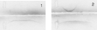
Fig. 5. Immunoelectrophoresis analysis. After electrophoresis of deglycosylated Ag5 (top) and native Ag5 (bottom), a pool of hydatid disease sera (Panel 1) and a rabbit Ag5 specific antiserum (Panel 2), were allowed to migrate on the agarose gel and the bands were Coomassie stained.
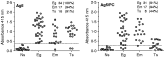
Fig. 6. Inhibitory effect of free phosphorylcholine (PC) on the reactivity of human sera with Ag5 assessed by ELISA. Serum samples were diluted (1/400) in 20 mM Tris buffer with or without 50 mM PC. The serum panel consisted of: 18 sera from healthy donors (Ns), 34 sera from hydatid patients (Eg), 18 sera from patients with alveolar hydatidosis (Em) and 18 sera from patients with cysticercosis (Ts). The horizontal line represents the cut off value estimated by ROC curves. Inserted are shown the number (%) of human serum samples scored as positive by ROC analysis.
 You have
Access
- 34
- Cited by