


Introduction
Helicobacter pylori is a microaerophilic, Gram-negative bacterium, composed of spiral-shaped rods. It is associated with gastric ulcer, cancer and gastritis.Reference Ozdek, Cirak, Samim, Bayiz, Safak and Turet1 It is one of the most common human bacterial infections in the world, affecting more than half of the entire population.Reference Kariya, Okano and Nishizaki2 Although the main localisation of this bacterium is the stomach, recent studies have analysed an association between H pylori and upper airway diseases.Reference Kariya, Okano and Nishizaki2–Reference Siupsinskiene, Jurgutaviciute, Katutiene, Janciauskas, Vaitkus and Adamonis6
It has been determined that the bacterium releases two key virulence factors: CagA (cytotoxin-associated gene A), which results in the accumulation of genetic changes in the cell, and VacA (vacuolating cytotoxin A), which causes immunosuppression and blocks T-cell proliferation.Reference Kariya, Okano and Nishizaki2, Reference Malfertheiner, Megraud, O'Morain, Gilbert, Kuipers and Axon7 H pylori affects the epithelium, causes an immune response, increases cytokines, triggers chronic inflammation and eventually increases the risk of gastric cancer.Reference Malfertheiner, Megraud, O'Morain, Gilbert, Kuipers and Axon7 Some authors argue that similar changes may develop in the upper airway as in the gastrointestinal tract.Reference Hagymási and Tulassay8
The role of H pylori infection in chronic rhinosinusitis is not yet completely understood, but it is known that this infection can be detected in nasal and sinus mucosa.Reference Ozdek, Cirak, Samim, Bayiz, Safak and Turet1–Reference Kurtaran, Uyar, Kasapoglu, Turkay, Yilmaz and Akcay4 There are three theories explaining the bacterium's appearance in the nasal mucosa: (1) the nasal and sinus mucosa act as a reservoir for H pylori bacterium; (2) the oral cavity is a reservoir for this bacterium, and H pylori enters the nasal cavity through oropharyngeal reflux; and (3) H pylori enters the nasal cavity through gastroesophageal reflux.Reference Ozdek, Cirak, Samim, Bayiz, Safak and Turet1–Reference Kurtaran, Uyar, Kasapoglu, Turkay, Yilmaz and Akcay4, Reference Katra, Kabelka, Jurovcik, Hradsky, Kraus and Pavlik9 Because nasal polyposis is a local mucosal reaction to inflammation, it usually forms at the ostiomeatal complexes. Under the effect of inflammation-causing agents, the mucous membrane of the sinuses becomes hyperplastic and intersected, which causes mucociliary clearance dysfunction, increases oedema of the nasal mucosa and promotes hyperplasia.Reference Morinaka, Ichimiya and Nakamura10, Reference Bansal, Sharma, Agarwal and Saha11
Recently, H pylori bacterium itself has been proposed as a possible cause of chronic rhinosinusitis.Reference Ozdek, Cirak, Samim, Bayiz, Safak and Turet1–Reference Yemisen, Mete, Kanbay, Balkan and Ozaras3 There are speculations that H pylori can damage the nasal mucosa, which becomes more sensitive to other chronic rhinosinusitis causing agents. However, chronic inflammation of the sinus and nasal mucosa may be a beneficial medium for this bacterium, and in this case H pylori can be a result rather than a cause of chronic rhinosinusitis.Reference Ozdek, Cirak, Samim, Bayiz, Safak and Turet1, Reference Morinaka, Ichimiya and Nakamura10
Although recent research has investigated the possibility of H pylori as an aetiological factor in different nasal diseases, the numbers of studies and investigated patients in this area are not sufficient to support this claim. There is also no clear link between H pylori in the upper airway and gastroesophageal reflux or laryngopharyngeal reflux. Therefore, the primary aim of this study was to determine the prevalence of H pylori infection in nasal biopsy specimens from patients with chronic rhinosinusitis with polyposis versus that in control patients. The secondary aim was to assess the correlations between H pylori infection identified in nasal tissue and patients’ sociodemographic data and reflux-related symptoms and signs.
Materials and methods
The study was performed at the Hospital of Lithuanian University of Health Sciences, Kaunas, from January 2011 to July 2012. A total of 45 adult patients (16 females and 29 males), aged 18–85 years (with an average age (± standard deviation (SD)) of 51.8±14.9 years), who underwent endoscopic sinus surgery for chronic rhinosinusitis with nasal polyposis, and agreed to participate in the study, were selected for inclusion in the clinical group. Patients were excluded from the study if: they were on antibiotics or proton pump inhibitors (PPIs) four weeks prior to surgery; or they had antrochoanal polyps, Samter's triad, an immunological disorder, or unilateral, allergic or fungal sinusitis.
All patients had received local steroid therapy for three months, with no regression of the polyps. A diagnosis of chronic rhinosinusitis with nasal polyposis was made on the basis of clinical examination (nasal symptoms and nasal endoscopy) and sinus computed tomography findings. Nasal biopsy samples were obtained during endoscopic sinus surgery under general anaesthesia.
The control group consisted of 30 patients (6 females and 24 males), aged 19–78 years (average age (± SD) 41.6 ± 17.6 years), who were admitted to the hospital because of nasal septum deviation. The absence of nasal polyps was confirmed by nasal endoscopy. Nasal biopsy samples were collected from the lateral nasal wall below the middle turbinate during septoplasty. Only patients whose histological examinations of the removed nasal mucosa revealed normal tissue were selected for inclusion in the control group.
The study was conducted after receiving permission from the Kaunas Regional Biomedical Research Ethics Committee (number P1-86/2011). Informed consent was obtained from all the patients investigated.
Patient evaluation questionnaire
Prior to surgery, all patients were required to complete a specially designed questionnaire. This assessed demographics, unhealthy habits, and reflux-associated information including their history of gastroesophageal reflux disease (regular use of PPIs reported in the anamnesis and/or reflux-related findings on upper gastrointestinal endoscopy).
Reflux-related symptoms
These were assessed with a standardised, validated questionnaire and the reflux symptom index. The reflux symptom index examines nine symptoms common in laryngopharyngeal reflux: hoarseness; throat clearing; excess throat mucus; difficulty in food swallowing; coughing after eating or after lying down; choking episodes; troublesome cough; lump in the throat; and heartburn, chest pain, indigestion or regurgitation.Reference Belafsky, Postma, Amin and Koufman12 The symptoms were self-rated on a six-point scale from 0 (not a problem) to 5 (a severe problem). A reflux symptom index of 13 points or more was considered abnormal.Reference Belafsky, Postma, Amin and Koufman12
Video rhinolaryngoscopy
Clinical examination of the condition of the patient's ear, nose, throat and larynx was carried out using a specially designed form. For all patients, diagnostic rhinolaryngoscopy was performed in the out-patient department with a flexible 3.2 mm endoscope (RLS; Kay Elemetrics, Lincoln Park, New Jersey, USA), according to standard protocol. The same examiner used the same sterilised equipment for each patient.
A validated reflux finding score scale was used to determine the presence of subglottic oedema, ventricular obliteration, erythema, vocal fold oedema, diffuse laryngeal oedema, posterior commissure hypertrophy, granuloma and thick endolaryngeal mucus.Reference Belafsky, Postma and Koufman13 A reflux finding score of more than 7 was considered abnormal.Reference Belafsky, Postma and Koufman13
The video recordings were rated by an observer, who was unaware of the patient's identity. The intra-rater correlation for the video recordings (calculated over a 1-month time interval, using 10 patient video recordings randomly selected from the list) was 0.86.
H pylori identification methods
H pylori infection was identified via a histochemical examination and a rapid urease test. The samples were collected from nasal polyps for the clinical group patients and from nasal lateral wall mucosa for the control group patients, using sterile instruments. A rapid urease test was performed at the beginning of nasal surgery according to the manufacturer's instructions. The biopsy samples were kept in paraffin blocks until needed for the histological and histochemical examinations.
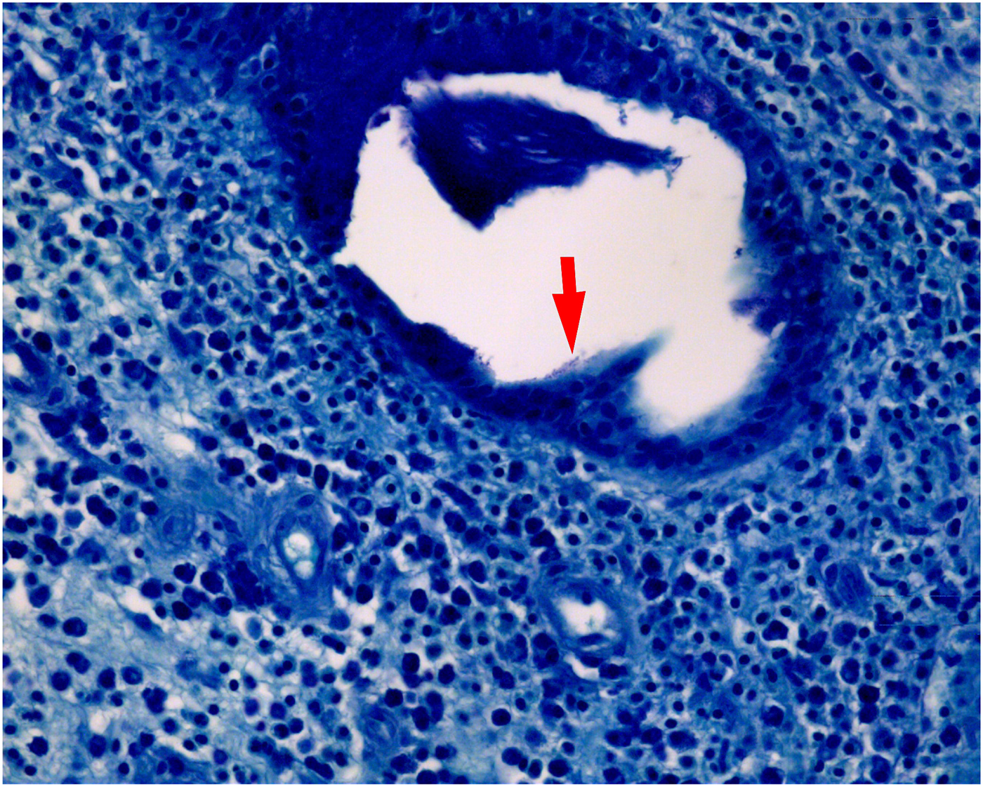
Regarding the histochemical examination, the biopsy material from the nasal tissues was subjected to Giemsa staining. Specifically, two 4-μm thick, paraffin-embedded sections were prepared and stained using the modified methodology (H pylori acquires a dark blue colour) (Figure 1).Reference Jančiauskas14
Histopathological image of a biopsy specimen taken from nasal polyps (arrow indicates H pylori on the submucosal gland epithelium). (Modified Giemsa stain; ×400)
Regarding the rapid urease test, at the beginning of surgery, the sterile bioptate (minimum 2 × 2 mm pieces) was placed into a medium with urea and a pH indicator. When the material contained H pylori, the urease enzyme changed the endogenous urea into ammonia and carbon dioxide. Moreover, the pH was changed by the released ammonia, and the indicator colour shifted from yellow to orange, red or purple. The campylobacter-like organism test (Kimberly-Clark, New Milford, Connecticut, USA) was used. The changes in colour were evaluated after 20 minutes, and after 1 and 3 hours, as recommended.Reference Tseng, Wang and Wu15 If the results of the two tests were positive, H pylori was considered present in the biopsy material of the nasal tissues.
Statistical analysis
Statistical analysis was performed using SPSS software, version 17 for Windows (SPSS, Chicago, Illinois, USA). The differences in qualitative parameters were calculated using the chi-square or Fisher's exact tests. The unpaired student's t-test (two-tailed) was used to compare the differences between the quantitative data of the two groups. Analysis examining the correlations between H pylori infection identified in the nasal tissue and different variables, such as demographics (age, gender, body mass index), unhealthy habits (alcohol consumption, smoking), a history of gastroesophageal reflux disease, reflux-related symptoms (assessed by the reflux symptom index) and reflux-related signs (assessed by the reflux finding score), was performed using the Pearson's or Spearman's correlation co-efficient (r). An α level of 0.05 was considered statistically significant. The Spearman's correlation co-efficient was used for an assessment of intra-rater reliability.
Results
Our study showed a significantly higher positive H pylori rate in the chronic rhinosinusitis with nasal polyposis group as compared to the control group. H pylori infection in the biopsy material from the nasal polyps was found in 28.9 per cent (13 of 45) of the patients from the clinical group. These results differ significantly from those of the control group, where H pylori infection in the biopsy material from the non-inflammatory nasal mucosa was found in only 3.3 per cent (1 of 30) of patients (p = 0.005) (Figure 2).
Distribution of H pylori positive results in the biopsy material of removed nasal polyps or normal nasal mucosa (n = 75). There was a statistically significant difference between the groups (p < 0.05).

A data comparison of patients in whom H pylori was detected in the biopsy specimens from the nasal tissues (13 patients with chronic rhinosinusitis with polyposis and 1 control patient) and the patients in whom H pylori was not detected (32 patients with chronic rhinosinusitis with polyposis and 29 control patients) showed no statistically significant differences with regard to patients’ gender, age, body mass index, smoking habits, alcohol consumption, history of gastroesophageal reflux disease, and summaries of reflux symptom index and reflux finding score (p > 0.05) (Table 1). Interestingly, we found that a reasonably high number of both H pylori positive and H pylori negative patients had a pathological reflux symptom index score (57.1 per cent and 41.0 per cent, p = 0.27) and pathological reflux finding score (64.3 per cent and 54.1 per cent, p = 0.48), respectively.
Characteristics of H pylori positive and negative patients

*Patients with identified nasal H pylori (n = 14); †patients with unidentified nasal H pylori (n = 61). BMI = body mass index; GERD = gastroesophageal reflux disease
The analysis of correlations between H pylori infection identified in the nasal tissue and different variables, which included separate reflux symptom index symptoms and reflux finding score signs, similarly showed no statistically significant relationships between intranasal H pylori infection and: patients’ demographic data (r = 0.05–0.21, p > 0.05), unhealthy habits (r = 0.006–0.02, p > 0.05) or laryngopharyngeal reflux related symptoms (r = −0.07–0.15, p > 0.05). However, there was a significant yet weak positive relationship between H pylori positivity and posterior commissure hypertrophy of the larynx (r = 0.24, p = 0.04). Thus, the presence of intranasal H pylori was weakly related to the manifestation of a common laryngopharyngeal reflux related sign. The data for the analysis of correlations between intranasal H pylori infection and patients’ laryngopharyngeal reflux related symptoms and signs are presented in Table 2.
Correlations between H pylori infection in nasal tissue and patients’ laryngopharyngeal reflux related data*
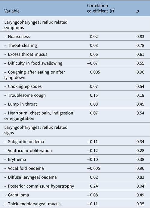
*n = 75. †Pearson's correlation co-efficient was used for parametric data and Spearman's for non-parametric data. ‡Statistically significant (p < 0.05)
Discussion
Our findings show that H pylori is present in the sinonasal tissue in nearly one-third of patients with chronic rhinosinusitis with nasal polyposis, and is significantly more frequent than in samples of control patients with healthy nasal mucosa. These results are in agreement with previously published data.Reference Bansal, Sharma, Agarwal and Saha11, Reference Cvorovic, Brajovic, Strbac, Milutinovic and Cvorovic16 In 2008, Cvorovic et al. used the same diagnostic kit as in our study, and found H pylori in 26.1 per cent of 23 patients with chronic rhinosinusitis with nasal polyposis, and in 0 per cent of 15 control group patients.Reference Cvorovic, Brajovic, Strbac, Milutinovic and Cvorovic16 The most recent study, published in 2016, by Bansal et al., presented similar results: H pylori was found in 22.8 per cent of 35 patients with chronic rhinosinusitis with nasal polyposis, and in only 2.9 per cent of the 35 control group patients.Reference Bansal, Sharma, Agarwal and Saha11
Recent studies that used the most sensitive H pylori diagnostic method, polymerase chain reaction, did not demonstrate uniform results. However, the majority of these studies detected H pylori and the most virulent CagA gene in nasal polyps specimens.Reference Koc, Arikan, Atasoy and Aksoy17–Reference Burduk, Kaczmarek, Budzynska, Kazmierczak and Gospodarek20
Some authors have investigated patients with chronic rhinosinusitis without nasal polyposis, and these studies have revealed similar results: the prevalence of H pylori was higher in those patients than in the patients without chronic rhinosinusitis.Reference Ozdek, Cirak, Samim, Bayiz, Safak and Turet1, Reference Morinaka, Ichimiya and Nakamura10, Reference Nikakhlagh, Samarbafzadeh, Jahani, Poostchi, Kayedani and Naghashpoor21
A significantly higher rate of H pylori in inflammatory sinonasal mucosa than in normal mucosa may suggest a positive role of H pylori in chronic rhinosinusitis and nasal polyposis. Moreover, a recent study assessing functional endoscopic sinus surgery results showed greater improvement in patients with chronic rhinosinusitis and H pylori sinonasal colonisation.Reference Jelavic, Grgić, Cupić, Kordić, Vasilj and Baudoin22
There are several possible explanations for more frequent detection of intranasal H pylori in patients with chronic rhinosinusitis. Chronic inflammation of the sinus and nasal mucosa may create a more suitable environment for this bacterium to survive than normal nasal mucosa. Nevertheless, the presence of H pylori alone in the nasal tissues cannot prove a causal relationship between chronic rhinosinusitis and polyposis. It is known that H pylori, and species with the CagA gene in particular, could modify the normal gastric mucosa surface;Reference Rook23 therefore, we could speculate that H pylori infection may modify the mucosa of the nose and maxillary sinus as well. Such modifications may not only facilitate colonisation by H pylori, but could also provide a target for an antibody response.Reference Morinaka, Ichimiya and Nakamura10, Reference Rook23
In 2003, Morinaka et al. established that H pylori may play a role as an antigen in patients who have chronic sinusitis with infiltration of eosinophils and neutrophils.Reference Morinaka, Ichimiya and Nakamura10 Similar data are also available in the recent Bansal et al. study.Reference Bansal, Sharma, Agarwal and Saha11 The authors examined samples from 35 nasal polyposis patients and 35 controls with normal nasal mucosa, and assessed the morphological changes. They found that inflammatory symptoms – lymphocyte infiltration forming lymphocyte aggregates and epithelial hyperplasia – were significantly more frequent in H pylori positive samples from nasal polyps than in samples from nasal polyps in which H pylori was not detected.Reference Bansal, Sharma, Agarwal and Saha11 Thus, we can hypothesise that H pylori itself may cause or contribute to the chronic inflammation. The prevalence of H pylori and the virulent CagA gene might also depend on demographics – a high incidence of infection with CagA-positive H pylori has been observed in Western Asia and Eastern Europe.Reference Burduk, Kaczmarek, Budzynska, Kazmierczak and Gospodarek20, Reference Ozyurt, Gungor, Ergunay, Cekin, Erkul and Haznedaroglu24
However, there are contradictory results regarding the role of H pylori in chronic rhinosinusitis and nasal polyps, based on the fact that H pylori and even the CagA gene can also be found in normal nasal mucosa, and similarly in nasal polyps.Reference Burduk, Kaczmarek, Budzynska, Kazmierczak and Gospodarek20, Reference Ozyurt, Gungor, Ergunay, Cekin, Erkul and Haznedaroglu24 Researchers have argued that the nose could serve as a reservoir for H pylori, which may be transmitted directly into the nasal cavity from saliva and dental plaque, or from any extragastric location, including the adenoids or tonsils.Reference Kariya, Okano and Nishizaki2, Reference Siupsinskiene, Katutiene, Jonikiene, Janciauskas and Vaitkus5, Reference Dinis and Subtil25 Any of the extragastric sites, including the nose, could serve as a reservoir for gastric re-infection rather than being a cause of chronic rhinosinusitis and polyps, as recurrence after antimicrobial therapy is not uncommon.Reference Dinis and Subtil25
The presence of H pylori in patients with chronic rhinosinusitis with polyposis could be linked to pathological reflux of the upper airway. It has been suggested that gastroesophageal reflux might play a role in the pathogenesis of chronic rhinosinusitis.Reference Dinis and Subtil25, Reference Sella, Tamashiro, Anselmo-Lima and Valera26 Our study shows a high prevalence of laryngopharyngeal reflux related symptoms and signs among H pylori positive patients (mainly patients with chronic rhinosinusitis with polyposis) and H pylori negative patients (equal numbers of patients with chronic rhinosinusitis with polyposis and controls). However, we found a significant yet weak positive relationship between intranasal H pylori infection and the most common laryngopharyngeal reflux related sign, posterior commissure hypertrophy of the larynx.
It is not surprising that we found a significant relationship between intranasal H pylori and the most common laryngopharyngeal reflux related sign, despite similar results for pathological reflux symptoms and signs in H pylori positive and H pylori negative patients. A possible explanation is that not all episodes of extra-oesophageal reflux could reach the nasopharynx and nasal cavity, which has been proven by diagnostic tests with 24-hour nasopharyngeal pH-metry using a four-channel pH probe.Reference Wong, Omari, Myers, Rees, Nair and Jamieson27 Additionally, perhaps not all patients had intragastric H pylori that could be transmitted to the upper airway.Reference Campbell, Kilty, Hutton and Bonaparte28 Furthermore, nasopharyngeal reflux is more commonly severely refractory to treatment in chronic rhinosinusitis patients.Reference Sella, Tamashiro, Anselmo-Lima and Valera26, Reference DelGaudio29 Unfortunately, in our study we did not use objective 24-hour pH-metry or multichannel impedancometry to prove the laryngopharyngeal reflux diagnosis or to estimate the character of reflux episodes in the upper airway. The reflux symptom index and reflux finding scores that were developed for quantification of the laryngopharyngeal reflux symptoms and signs are more concerned with the larynx than how the reflux enters the nasal cavity.Reference Belafsky, Postma, Amin and Koufman12, Reference Belafsky, Postma and Koufman13 However, our study shows that patients with hypertrophy of the posterior commissure of the larynx – one of the primary signs of laryngopharyngeal reflux – have a greater probability for positive H pylori in the nasal cavity. This fact could suggest the importance of reflux as the transmitter of H pylori to the upper airway.
The scientific literature linking H pylori infection identified in the nasal cavity to reflux is limited. Although pepsin and pepsinogen 1 has been detected in sinonasal samples,Reference Dinis and Subtil25 and some authors found that intranasal H pylori in patients with chronic rhinosinusitis was related to classical gastroesophageal reflux disease symptoms,Reference Ozdek, Cirak, Samim, Bayiz, Safak and Turet1, Reference Bansal, Sharma, Agarwal and Saha11 others did not find a significant relationship between H pylori in nasal polyps and chronic rhinosinusitis in patients without classical gastroesophageal reflux disease symptoms or signs.Reference Nemati, Mojtahedi, Naghavi, Banan and Zia30
These findings could suggest that gastric juice infected with H pylori, and not H pylori itself, is involved in the development of chronic rhinosinusitis with or without nasal polyposis.Reference Kariya, Okano and Nishizaki2 However, the scientific literature reveals several hypotheses concerning the possible influence of gastroesophageal reflux and H pylori on the pathogenesis of chronic rhinosinusitis and nasal polyposis. Numerous researchers have reported that gastroesophageal reflux and H pylori infection may be an interrelated phenomenon.Reference Ozdek, Cirak, Samim, Bayiz, Safak and Turet1, Reference Ozyurt, Gungor, Ergunay, Cekin, Erkul and Haznedaroglu24, Reference Campbell, Kilty, Hutton and Bonaparte28 It is possible that, as previously mentioned, chronic inflammation in the nasal cavity caused by reflux may constitute a hypoxic and acidic environment for H pylori colonisation.Reference Ozdek, Cirak, Samim, Bayiz, Safak and Turet1, Reference Koc, Arikan, Atasoy and Aksoy17 In addition, the H pylori bacterium itself could act as an inflammatory co-factor, similar to gastroesophageal reflux, by promoting inflammation, increasing production of interleukin-1 β, altering the sinonasal mucosa and contributing to the reactive hyperplasia of the epithelium.Reference Ozdek, Cirak, Samim, Bayiz, Safak and Turet1, Reference Katra, Kabelka, Jurovcik, Hradsky, Kraus and Pavlik9, Reference Koc, Arikan, Atasoy and Aksoy17, Reference Dinis and Subtil25 Recent analytical studies summarising the data on the role of H pylori in the upper airway also indicated that gastric juices infected with H pylori, and systemic immune responses to gastric H pylori infection, might play a causative role in upper respiratory diseases.Reference Kariya, Okano and Nishizaki2, Reference Yemisen, Mete, Kanbay, Balkan and Ozaras3, Reference Kurtaran, Uyar, Kasapoglu, Turkay, Yilmaz and Akcay4, Reference Campbell, Kilty, Hutton and Bonaparte28
No relationship between H pylori and other factors such as patients’ age, gender or unhealthy habits was established in this study or in previously reported data on patients with laryngeal and tonsillar diseases.Reference Siupsinskiene, Katutiene, Jonikiene, Janciauskas and Vaitkus5, Reference Siupsinskiene, Jurgutaviciute, Katutiene, Janciauskas, Vaitkus and Adamonis6 Dinis and Subtil also found that none of the variables examined, such as age, gender, nasal side, disease site, disease extension or the co-presence of allergy, were related to bacterial colonisation.Reference Dinis and Subtil25
Summarising the results of our study and the literature data, we argue that H pylori infection, regardless of the route of transmission, is potentially related to chronic rhinosinusitis with polyposis. However, the effect of this bacterium on the chronic inflammatory process in the nasal and sinus mucosa, and its relationship with laryngopharyngeal reflux, are not yet clear and need further investigation. The data presented in the literature are quite controversial. The findings are difficult to compare because of the different methods of H pylori identification used and other factors, including small sample sizes, various patient selection criteria and demographic conditions. Further well-designed, large-scale, multicentre studies are needed to determine the role of H pylori in the aetiology of different nasal and sinus diseases.
• This study investigated the incidence of intranasal Helicobacter pylori infection in patients with chronic rhinosinusitis with polyposis
• Associations with patients’ sociodemographic and laryngopharyngeal reflux related data were also examined
• H pylori was more prevalent in nasal tissue of chronic rhinosinusitis with polyposis patients than in control patients with a deviated septum
• Intranasal H pylori was associated with posterior commissure hypertrophy of the larynx
Conclusion
Our findings show that H pylori infection is potentially related to chronic rhinosinusitis with polyposis. H pylori was identified in the biopsy material of nasal polyps in nearly one-third of all patients who underwent endoscopic sinus surgery for chronic rhinosinusitis with polyposis, and it was significantly more frequent in these patients than in patients with normal nasal mucosa. The presence of H pylori in the nasal tissues was weakly associated with the manifestation of laryngopharyngeal reflux related posterior commissure hypertrophy of the larynx.
Competing interests
None declared



