




No CrossRef data available.
An integrative approach to the neurophysiological substrates of social withdrawal and aggression
Published online by Cambridge University Press: 13 April 2004
Abstract
An integrative model of the neurophysiology of aggression and social withdrawal is proposed. A detailed overview of the limbic–hypothalamic–pituitary–adrenal (LHPA) axis is presented first, because we consider it to be a critical system that interacts with a variety of physiological processes to modulate affect-related behaviors. This detailed analysis of the LHPA axis is then used to clarify the research literature that links aggression and social withdrawal to LHPA functioning. We then review the role of amygdala and prefrontal cortex functioning in modulating aggression and social withdrawal. Particular attention is paid to how the amygdala and the prefrontal cortex interact with the LHPA system and the environment to produce specific behavioral tendencies throughout development. A brief overview of the implied methodological and theoretical model is provided. We explain how a detailed understanding of specific physiological processes is essential in order to develop appropriate research protocols. In addition, we suggest that future research should focus on the mapping of distinct integrative biosocial profiles that are related to specific behaviors during different developmental stages.The authors would like to thank Sarah Dunphy–Lelii and Michael MacKenzie for their comments and suggestions during the preparation of this manuscript. This research was partially supported by Research Grants MH59396, HD/DK37431, and MH42251.
Information
- Type
- Research Article
- Information
- Copyright
- © 2004 Cambridge University Press
A growing body of research has linked individual differences in early behavioral maladjustment, such as aggression and social withdrawal, to the function of at least three key biobehavioral systems: the limbic–hypothalamic–pituitary–adrenal axis (LHPA), the amygdala, and the prefrontal cortex (PFC). Although our understanding of the affective function of these structures has greatly increased during the past decade, less is known about how these physiological processes interact and may be related to emotional regulation mechanisms and related behavioral manifestations. Specifically, little is known on how interactive patterns among these physiological processes and the environment may result in different types of behavioral maladjustment.
In this article we focus on the effects of specific physiological systems on two types of problem behaviors: aggression and social withdrawal. We do this for several reasons. Researchers studying the neurobiological substrates of these two forms of problem behaviors have focused on the same biological structures and events, suggesting a conceptual link between aggression and social withdrawal. In addition, both of these constructs have been associated with variations in emotional regulation patterns (Bradley, 2000). Consequently, we believe that these two concepts are in fact by-products of very similar neurophysiology–environment interactive profiles, which are related to a shared critical concept: the dysregulation of emotional experience and expression.
This review does not attempt to provide a comprehensive summary of all physiological processes that have been linked to aggression and social withdrawal. Instead, we focus on three physiological processes: the LHPA axis, amygdala, and prefrontal functioning. We do so because we believe them to have an important role in emotional regulation and related behavioral manifestations, specifically aggression and social withdrawal. In addition, the conceptual link between these physiological processes and childhood patterns of aggression and social withdrawal allows us to present a specific model of biobehavioral relationships. This model suggests that behavioral manifestations are mediated by specific neurophysiological profiles. These profiles are composed of finite patterns of interactions between biological systems, which, when interacting with key environmental variables, result in distinct patterns of behavioral maladjustment.
Emotional Regulation and Aggression and Social Withdrawal
We view emotional regulation as a series of neurocognitive processes that modulate the perception, processing, and reaction to emotional stimuli. In its most basic form, emotional regulation involves the regulation of arousal (Bradley, 2000). However, other dimensions of emotional regulation have also been identified such as attention, emotional memory, cognitive interpretations of events, and so forth (Thompson, 1994). In this paper, we agree with Bradley in viewing the regulation of physiological arousal as a central component of emotional regulation, which is directly related to other neurocognitive processes such as the perception of and reaction to fear stimuli and the cognitive processing and interpretation of negative and positive events.
Emotional regulation can occur at several levels. At a structural biological level our organisms have systems that result in physiological activation during specific stressful conditions. This activation is mediated by a number of systems such as the reticular activating system, the LHPA, the amygdala, and so forth. Regulation of this activation can occur internally through specific mechanisms such as the mineralocorticoid and glucocorticoid receptor negative feedback mechanisms of the LHPA axis or externally through neurocognitive processes. Furthermore, active neurocognitive processes, such as cognitive strategies, can alter our internal working models, which in turn alter our self-perception, sense of efficacy, and insight, all of which are important in affecting our perception and interpretation of stimuli (Bradley, 2000).
Deficits in emotional regulation have been associated with the development of both externalizing and internalizing behaviors problems (Cole, Michel, & Teti, 1994; Eisenberg, Cumberland, Spinrad, Fabes, Shepard, Reiser, Murphy, Losoya, & Guthrie, 2001; Shields & Cicchetti, 2001). In this work we focus on two specific types of externalizing and internalizing problems: social withdrawal and aggression. We narrow the focus of this paper to these two specific behaviors to help us more appropriately connect the animal neurophysiological literature to human findings. However, we warn the readers that some of the literature that we review here deals with the global concepts of externalizing and internalizing problems. We do this only when we believe that such literature helps us understand the relationship between biobehavioral systems and aggression and social withdrawal.
We define social withdrawal using an extension of Jerome Kagan's concept of inhibition. Kagan and collaborators (Kagan, 1989; Kagan, Reznick, & Snidman, 1988; Kagan, Reznick, Snidman, & Gibbons, 1988) have studied behavioral inhibition as a stable trait and have defined it in relation to children's responses to unfamiliar stimuli. Inhibited children, when exposed to novel situations, “show a pattern of restraint and avoidance, together with signs of wariness or fear of unfamiliar people, objects, and events” (Robinson, Kagan, Reznick, & Corley, 1992, p. 1030). Conversely, uninhibited children “display a relatively rapid and fearless approach to unfamiliar people, places, and objects” during similar situations. We define social withdrawal as a pattern of behavioral inhibition during social situations.
Dysregulation of emotional experience has also been associated with the phenomenology of aggressive behavior (Shields & Cicchetti, 2001). Although the theoretical positions that we take in this paper regarding the association between several physiological systems and aggression can be applied to a variety of externalizing problems (conduct disorder, oppositional defiant disorder, etc.), we focus our explanations on actual physical aggressive behavior. That is, we are interested in understanding the inner workings of the biosocial profiles that are related to tendencies to engage in physically aggressive behaviors (fighting, bullying, etc.).
In summary, this review describes three physiological systems that serve specific functions during emotional regulation processes and that affect the development and phenomenology of aggression and social withdrawal. We review the LHPA axis as a central component of the human stress response system, which affects the cognitive and subjective experience of emotionally charged situations and which has motivational properties by directly influencing the behavioral responses to stress. We then describe the neurophysiology of the amygdala and its relation to the LHPA, particularly as they relate to two components of emotional regulation: the processing and behavioral reaction to fearful stimuli and the consolidation of emotional, dependent memories. Finally, we present a summary of the functioning of the prefrontal lobes in the processing of positive and negative emotions as it relates to the subjective experience of negative emotionality. It is our hope that a detailed understanding of the neurophysiology of these constructs can result in an integrative model of the neurophysiology of aggression and social withdrawal
LHPA Axis
The LHPA axis is the major regulatory system for stress in humans and most mammals. It is activated during stress to produce a series of hormones, which leads to the release of corticoids. Cortisol, the end product of this activation, is the major stress steroid in humans that modulates several neurophysiological processes, such as termination of digestive activity, increase in heart rate, and mobilization of glucose as an energy source for utilization under a “flight or fight” type of events (de Kloet, 1991; Johnson, Kamilaris, Chrousos, & Gold, 1992). These processes have a survival function, priming the animal for action. Before we can discuss how the LPHA axis may be related to emotional regulation and some of its behavioral manifestations, such as aggression and social withdrawal, we must understand specific details of the LPHA axis activation and regulation.
The LHPA process begins when the paraventricular nucleus (PVN) of the hypothalamus produces and secretes corticotropin-releasing hormone (CRH), which in turns activates the anterior pituitary gland's corticotroph cells to secrete andrenocorticotropic hormone (ACTH; see Vazquez, 1998, for an extensive review of the functioning and development of this system). ACTH in turn activates the release of cortisol by the adrenal glands (adrenocortical stress response). Cortisol is also its own regulator, activating feedback mechanisms whereby the LHPA shuts down the release of activating hormones and consequently cortisol's own release. This cascade of hormonal activating events is triggered by stress signals received by multiple brain circuits specific for each stress modality that converge in the PVN hypothalamic area (see Figure 1). However, in addition to acting as an alarm system, the LHPA axis also has intrinsic activity, which maintains an underlying 24-hr cortisol rhythm that is independent of stress responses and linked to activity level. As such, this circadian rhythm consists of high cortisol levels that are observed during the start of the wake cycle (morning) in humans, which then decrease through the afternoon and are lowest at nighttime, at the start of the sleep cycle.

The basic model of the activation and regulation of the LHPA axis.
Although unbound levels of cortisol are viewed as an indication of LHPA axis activation, one has to consider whether the unbound cortisol levels represent basal LHPA circadian activity or stress-induced adrenocortical activation. These two processes have unique and independent functions; hence, one can expect that cortisol determinations may reflect, depending on cortisol's activating source, a variety of physiological events that may or may not have specific behavioral correlates. Thus, the context and time of day become critical factors in the interpretation of samples. For example, cortisol levels taken during nonstressful situations reflect a significantly different pattern of neural activation than that reflected by cortisol levels taken after stressful situations. Stress-induced cortisol is produced after an acute activation of the PVN of the hypothalamus. The basal levels of cortisol are governed by neuronal projections originating in brain nuclei associated with biological clocks, which also converge at the PVN; but these pathways appear to be exquisitely sensitive to the inhibitory control of unbound levels of corticoids.
The LHPA inhibitory control system is controlled by two types of corticoid receptors, mineralocorticoid receptors (MRs) and glucocorticoid receptors (GRs). MRs are mostly located in limbic structures, particularly the hippocampus. The hippocampal effects on LHPA functioning are mostly inhibitory and mediated by MR activation. In the brain, MRs have a high affinity for corticoids and are therefore activated to inhibit hormonal secretion by very low levels of corticoids (de Kloet, 1991). Because of MRs' high affinity to corticosteroids, these receptors are believed to control the tonic basal activity of cortisol by inhibiting CRH release. Conversely, GRs are localized throughout the brain and are activated by high levels of stress-induced corticoids (Heuser, Deuschle, Weber, Stalla, & Holsboer, 2000; Oitzl, van Haarst, & de Kloet, 1997). During stress, such as that experienced during some emotionally charged situations, high levels of cortisol saturate brain MR receptors first and then begin to bind to GR receptors (Oitzl et al., 1997). Although it is usually believed that GRs have an inhibitory function, recent data suggest that GRs may first help maintain the elevated levels of cortisol that may be necessary during dangerous situations (Oitzl et al., 1997). Thus, GR antagonists (blocking GRs) can result in blunted cortisol secretion, perhaps by increasing MR activation (implications of such findings are discussed later).
Therefore, individual variations in cortisol levels do not simply reflect a clear LHPA activation process. Instead, variations in cortisol levels can reflect a variety of events, including individual variations in the patterns of tonic LHPA functioning, the degree of LHPA activation during stress due to varying activations of brain cortical and/or subcortical signaling, individual differences in sensitivity to CRH and ACTH, and MR–GR dependent regulatory functions controlling the sensitivity to cortisol feedback inhibition. Figure 2 presents an expanded model of the LHPA axis functioning.

The expanded model of the activation and regulation of the LHPA axis.
LHPA, emotional dysregulation, and psychopathology
Because cortisol secretion is the end product of the activation of the LHPA system, the study of cortisol in a variety of contexts provides important information about various aspects of a person's stress response system. First, extremely high or low levels of cortisol at rest (basal) or induced by stress can be viewed as reflecting a dysregulation of the LHPA system (Heim, Ehlert, & Hellhammer, 2000) and they have been associated with distinct emotional regulation problems (Checkley, 1996; Gunnar, 2001; Tobin, 2001; ver Ellen & Van Kammen, 1990). Second, individual differences in normative cortisol levels have been linked to specific affect-related behaviors. This suggests that both normative and dysregulated LHPA function may have direct links with behavior and personality traits that are directly related to emotional regulation (Guerra, Nucci, & Huesmann, 1994; Gunnar, 1994).
For example, LHPA dysregulation, both in terms of resting (basal) hormonal activity and abnormalities of the activation or inhibition of hormonal levels after a stressor (reactive), has been linked to a variety of affective psychiatric disorders. Major depressive disorder and posttraumatic stress disorder are among the psychopathological disorders associated with LHPA abnormalities (Checkley, 1996; Gunnar, 2001; Tobin, 2001; ver Ellen & Van Kammen, 1990). A common interpretation of these findings is that a dysfunctional LHPA system may result in extreme physiological activation during possible stressful situations or elevated activation during day to day activities. When combined with dysfunctional emotional regulation processes (such as distorted cognitive appraisals of situations), this elevated stress response may create problems with the emotional reaction to both daily events and actual stressful situations, ultimately producing atypical levels of negative emotionality.
The literature linking dysregulation of the LHPA axis to affective disorders is extensive and continues to grow in a rapid manner. This literature underscores the importance of LHPA axis functioning on the regulation of emotional experience and expression. However, here we are interested in examining the evidence linking normal variations of LHPA functioning to individual differences in patterns of emotional regulation, social withdrawal, and aggression. Thus, we present evidence suggesting that normal LHPA circadian activity, as well as activation and regulation of the system during stress, are important components of the phenomenology of social withdrawal and aggression via affecting a number of emotional regulation processes.
LHPA and social withdrawal
The development of social withdrawal, or the tendency to avoid and withdraw from social interactions, has been linked to a number of neurophysiological processes, including the functioning of the LHPA system. Schmidt, Fox, Rubin, and Sternberg (1997) found that 4-year-old children with high morning salivary cortisol engaged in more wary behavior during peer play and were reported as shy by their mothers. In another study of LHPA functioning throughout the day, Dettling, Gunnar, and Donzella (1999) found that shyness was a predictor of high basal cortisol levels during the day among 6- to 8-year-old children. Furthermore, conduct disordered children with high basal levels of cortisol also reported high levels of anxiety (McBurnett, Lahey, Frick, Risch, Loeber, Hart, Christ, & Hanson, 1991). The association between cortisol and anxious behaviors appears to be present during infancy, suggesting a relatively stable developmental trajectory. For example, studies by Kagan and colleagues (Kagan et al., 1988; Kagan, Reznick, & Snidman, 1988) showed that infants who displayed high levels of negative affect to sensory stimulation were later identified as withdrawn in a variety of contexts when they were 4 years old. These children also presented higher basal cortisol levels than more highly sociable children. Consistent with these results, high levels of cortisol were also found among withdrawn anxious infants after the acute stressful challenge of a mother–infant separation (Tennes, Downey, & Vernadakis, 1997). The implications of these findings are considered in a later section, but it is possible that these children's overactive LHPA functioning (high basal cortisol levels) serves as a behavioral motivator by creating subtle but consistent levels of social stress. Children may attempt to regulate this stress by avoiding social situations.
The studies described above suggest that high cortisol levels may be a biological marker that differentiates shy from uninhibited children throughout many stages of development from infancy to adolescence and beyond. However, other studies have provided the opposite results, suggesting that specific biosocial contexts may significantly alter the relationship between cortisol and stress, at least during the school-age period. For example, Gunnar and colleagues presented a series of studies showing that behavioral inhibition may actually blunt LHPA activation during specific social situations, which results in a relative elevation of cortisol secretion among extroverted children (Gunnar, 1994; Gunnar, Tout, de Haan, & Pierce, 1997). They repeatedly found that extroverted children display higher levels of cortisol during the early stages of a school year when compared to shy and inhibited children (Gunnar, 1994; Gunnar, Tout, de Haan, & Pierce, 1997). Davis, Donzella, Krueger, and Gunnar (1999) also examined the cortisol levels of 7- to 12-year-old children during the first week of a new school year. They found highly assertive and social children had higher levels of cortisol than shy children, but this trend was reversed after the formation of peer groups. Similarly, de Haan and colleagues (1998) found that cortisol measures taken during a similar school period were correlated with assertive behavior in children. Likewise, high levels of cortisol were found among extroverted children after engaging in a competitive task (Donzella, Gunnar, & Krueger, 2000).
How can understanding the complexity of the LHPA axis help us clarify the findings linking high levels of cortisol to both inhibited and uninhibited children? First, most studies linking high levels of cortisol with shy and withdrawn behaviors have used basal resting levels of cortisol (Gunnar, 1994; Gunnar et al., 1997; Kalin, Larson, Shelton, & Davidson, 1998; McBurnett et al., 1991; Schmidt et al., 1997). Second, conversely, studies linking high levels of cortisol to outgoing and assertive behavior have measured cortisol after or during prolonged stressful situations (Davis et al., 1999; de Haan et al., 1998; Donzella et al., 2000; Gunnar, 1994; Gunnar et al., 1997). It is possible that inhibited children may have an overactive basal LHPA system (low basal inhibitory tone), which may be linked to increased levels of anxiety, withdrawn, and avoidant behaviors. When placed in social situations that may result in increased arousal (such as during the initial stages of group formation), these children may regulate negative arousal by avoiding social interactions. This may contribute to reduced activation of their LHPA system. In addition, it is possible that any activation of the LPHA may be difficult to observe because these children already present high basal levels of cortisol. Thus, the activation of the LPHA by a stressor should be measured in the context of each child's prestress cortisol levels.
In contrast, uninhibited children may have a readily active tonic inhibition of their LHPA system that results in low basal cortisol levels. This may lead to a high stress threshold that allows the child to engage in more stress-inducing behaviors, which in turn maximally elevates his/her cortisol levels (because the child is already starting from a low cortisol level under a resting condition). Thus, it is possible that the individual differences in cortisol activation found in various contexts among children with different levels of social withdrawal are partly related to MR functioning, which leads to different patterns of basal cortisol secretion and behavioral correlates.
Several animal models provide more insight into the role of GRs and MRs in the manifestation of inhibited and uninhibited behaviors (Kabbaj, Devine, Savage, & Akil, 2000). When rats are exposed to novel environments, they can be categorized according to their explorative behavior. High responders engage in high levels of exploratory behavior in a novel environment, whereas low responders engage in limited exploration (Kabbaj et al., 2000). These behaviors resemble the sensation seeking styles that have been identified in humans (Gerra et al., 1997, 1999; Gerra, Zaimovic, Timpano, Zambelli, Delsignore, & Brambilla, 2000). MRs and GRs are thought to be involved in the generation of exploratory behavior among rats because high responders and low responders differ in the density of GR expression in the hippocampus and because artificial manipulation of GR and MR activity results in a reversal of these behavioral characteristics. In a very influential study of the physiological makeup of high and low responder rats, Kabbaj and colleagues (2000) found that in a basal nonstressed state, compared to low responder rats, high responders have increased levels of CRH in the PVN but lower CRH levels in the central nucleus of the amygdala. High responder animals also have lower basal GR expression in the hippocampus compared to low responder animals. In contrast, both high and low responder animals have equal levels of MR expression in the hippocampus. It is interesting that high and low responder animals were found to have equal basal corticosterone levels. However, they differed in the magnitude of the corticoid response to novel stress. Although we may intuitively think that high levels of exploration would be associated with a low corticosterone stress response in high responder rats, these animals have a high corticosterone response to novelty whereas low responder animals have a blunted response. When further scrutinizing the biological markers described above, this is not entirely unexpected. The profile described in high responder animals is consistent with increased responsiveness to stress (high CRH in PVN), coupled with decreased anxiety behavior (low CRH levels in the amygdala, a structure associated with fear and anxiety, where CRH is not directly related to LHPA activity). In addition, low GR hippocampal expression also suggests a diminution in the inhibitory capacity observed as a sustained corticosterone release after novelty, which has been reported by some investigators in high responder animals (Kabbaj, Piazza, Le Moal, & Maccari, 1996; Piazza, Deroche, Deminiere, Maccari, Le Moal, & Simon, 1993).
GRs appear to be key in modulating the corticosterone response induced by novelty stress. Direct antagonism of hippocampal GR (blocking GR action) changes the behavior of a low responder rat that has high GR expression in the hippocampus into behaviors consistent with those of a high responder rat. This is evidenced by changes in their exploratory behavior and their corticosterone response in a novel environment (Kabbaj et al., 2000; Maccari, Piazza, Deminiere, & Angelucci, 1991; Strohle, Poettig, Barden, Holsboer, & Montkowski, 1998). This suggests that high responder rats engage in exploratory behaviors, despite experiencing high levels of stress during exploration. It is possible that high responders engage in exploratory behaviors precisely because of such physiological reactions, which drives risk-taking behavior. Favoring this possibility is the vast evidence suggesting that corticoids are reinforcing in rats, which lead them to engage in self-administration of psychostimulants or to substitute stress for psychostimulant administration (Piazza et al., 1993). Finally, given the high level of distribution of GRs in the brain, including the limbic system, the activation of GRs may have an impact on multiple neuronal pathways, which may result in avoidant or escape behaviors. Thus, blocking of GR activation or a reduced number of GRs may prevent an organism from activating avoidant or escape behaviors, resulting in more exploratory and risk-taking activity.
These animal models may provide insight into the underlying neural mechanisms that modulate inhibited and uninhibited human behavior. Although we must be careful in translating the significance of novelty as a stressor in rodents to its equivalent in human children, we could extrapolate based on the high–low responder animal model that perhaps extroverted children should not have an underactive LHPA response to stress when exposed to unfamiliar surroundings or novel social interactions. As with the high responder rats, these children may experience a physiological stress reaction when engaging in assertive social behaviors. Their high levels of assertive social behaviors cannot be explained by an “underactive” LHPA response to stressful situations. Instead, their behavior could be explained by a sensitive LHPA system, perhaps high levels of CRH in the PVN that are just waiting to be released upon demand, decreased levels of CRH in fear-related centers (amygdala), and reduced GR expression in the hippocampus that limits the occurrence of avoidant behaviors. As in the high responder animals, this combination would result in increased or normal responsiveness to stress but a decrease in anxiety-like behaviors when exposed to or prior to novel social environments. Likewise, it is plausible that inhibited children have high levels of CRH in the amygdala and high GRs in the hippocampus, both of which could explain their nonexploratory freezing behavior that is displayed during stressful situations and their low levels of cortisol that are secreted during these situations because their actual exposure to the novel situation is self-regulated through avoidant behaviors.
The application of animal models to human data appears adequate because the interpretation of these results is consistent with the work by Gunnar and colleagues, who have shown that children with different temperamental characteristics have different LHPA activation when placed in specific environmental contexts. During situations that may lead to stressful situations, such as the start of a school year, inhibited children display very low levels of cortisol whereas uninhibited children display high levels of cortisol (Davis et al., 1999; Donzella et al., 2000; Gunnar, 1994; Gunnar et al., 1997). They postulate that inhibited children engage in nonsocial behaviors in order to avoid possible stressful situations, which results in decreased levels of cortisol. Conversely, uninhibited children actively seek social interactions despite their normal LHPA response to stress, which in novel contexts may explain their high LHPA activation during these encounters. Considering the multiple functions that cortisol subserves, it is possible that elevations of cortisol prior to these encounters lead inhibited children to engage in socially avoidant behaviors. Instead, uninhibited children seek these social interactions that activate their LHPA axis. Their active LHPA axis mobilizes fuel sources (glucose) for heightened activity during these encounters. In addition, facilitation of cognitive function and association, consolidation, and memory processes are linked to elevated (but optimal) levels of corticoids in plasma during learning tasks (de Kloet, 1991). It is possible that during social encounters the uninhibited children are consolidating and processing information about social situations that would allow them to be much more adept in future social interactions. Indeed, this may very well explain the lower cortisol levels detected in uninhibited children later in the school year once peer groups are established.
LHPA and aggression
The literature linking LHPA functioning and aggression is also seemingly contradictory. Although a large number of studies have found an inverse association between cortisol and aggression, other studies have failed to find this association or have found the opposite results. Thus, both blunted and overactive LHPA functioning has been found among aggressive children of different ages. McBurnett, Lahey, Rathouz, and Loeber (2000) studied the relationship between basal cortisol levels and aggression among elementary school boys (ages 7–12 years) participating in a longitudinal study. They found that low basal cortisol levels in the morning were associated with high levels of aggressive conduct disorder, peer ratings of aggression, and symptoms of oppositional defiant disorder. Other studies of young children have indicated that low levels of morning cortisol were associated with hostility toward teachers and aggression toward peers (Tennes & Kreye, 1985; Tennes, Kreye, Avitable, & Wells, 1986). Low morning cortisol levels were also found among school-aged children reporting clinically significant levels of externalizing behavior problems (Cicchetti & Rogosch, 2001b). In addition, studies of adolescents and young adults have provided similar results. For example, Vanyukov, Moss, Plail, Blackson, Mezzich, and Tarter (1993) found that low morning basal cortisol levels were associated with increased conduct disorder symptoms among 150 adolescents.
However, a small but notable number of studies have failed to replicate these findings (Gerra et al., 1997; Kruesi, Schmidt, Donnelly, Hibbs, & Hamburger, 1989; McBurnett et al., 1991; Schulz, Halperin, Newcorn, Sharma, & Gabriel, 1997). Kruesi et al. (1989) compared adolescents with aggression and conduct problems to nonaggressive adolescents. They reported no significant differences in urinary cortisol levels among these groups. It is important to note, however, that Kruesi and colleagues measured 24-hr urinary cortisol output, which integrates intraday variations that could eliminate differences among the groups that may be present in discrete times of the day. A similar study of aggressive and nonaggressive adolescents also failed to find a relationship between morning plasma basal cortisol levels and aggression (Schulz et al., 1997). Furthermore, some studies indicate that high levels of cortisol are associated with high levels of aggression in some contexts. Gerra et al. (1997) experimentally induced aggression in young adults (18–19 years old) and found that participants with high basal cortisol levels responded more aggressively than participants with low basal cortisol. McBurnett et al. (1991) also found that 9-year-old children who display both conduct problems and anxiety also have higher levels of cortisol than normal children. Nevertheless, we should note that cortisol collection in this study was conducted during a clinic visit, which varied in time during the day. Thus, it is not clear whether such findings represent variations in basal or stress-induced cortisol levels.
The same principles that help explain the relationship between LHPA functioning and social withdrawal may explain the relationship between LHPA and aggression. LHPA functioning does not have a direct causal relationship with aggression. Instead, it affects emotional regulation by determining chronic and event-related stress levels and by eliciting regulatory behaviors. When these LHPA activation patterns interact with other physiological systems and environmental contexts, the likelihood of occurrence of specific aggressive responses is maximized. For example, children with low basal cortisol levels may have a higher anxiety threshold (Kruesi et al., 1989), which would prevent them from experiencing anxiety and guilt when engaging in inappropriate behavior and/or in anticipation of discipline encounters. Their basal, MR mediated system may be overactive (high basal tone). This overactivation of MRs would result in reduced basal cortisol levels around their circadian clock. However, their LHPA response to stress may be intact. When a child with high basal tone of the LHPA system (low basal cortisol levels) is placed in a context where aggression is either expected or necessary, he/she may lack the inhibitory capacity to engage in escape or avoidant behaviors and may proceed to engage in aggressive behaviors. Cortisol levels would then become maximally elevated as the result of the aggressive encounter (greater delta and greater mobilization of glucose for energy use).
Therefore, the data on cortisol and aggression may be highly sensitive to the contextual sampling procedure and the coping mechanisms evoked. At least during early childhood, if cortisol levels are measured in a familiar, nonstressful setting, low levels of cortisol may be more likely to predict high levels of aggression. However, if taken in a context where aggressive interactions are prevalent, high levels of cortisol may better indicate high levels of aggression. Because the LHPA axis is a dynamic system, these relationships may change over time as the LHPA axis is exposed to these patterns of secretion. Thus, in the adolescent and young adult the neuroendocrine system may have a new “setpoint” at which basal cortisol levels are now elevated as the result of “downregulation” or decreased expression of corticoid receptors in key brain regions as a direct consequence of high cortisol response for many years. Although higher basal cortisol levels may prevail in this age group in comparisons to nonaggressive controls, the peak response to stressful challenge between these groups may not differ significantly. Rather, the change between basal to peak levels would be different (i.e., a small change in aggressive adolescents and a large change in nonaggressive adolescents; Dallman, Akana, & Scriber, 1991).
Another possible explanation of the conflicting findings concerning the relationship between cortisol and aggression may be found in non-human primate models of aggression. Similar to Dodge's concept of proactive and reactive human aggression (Dodge & Coie, 1987; Dodge, Lochman, Harnish, Bates, & Petit, 1997), Kalin (1999b) and colleagues (1998) suggest that there are two specific types of primate aggression: offensive and defensive. Defensive aggression is motivated by excessive fear, whereas offensive aggression has its roots in difficulty with impulse control and underarousal. Monkeys display these two types of aggression and the neuroendocrinological correlates for each aggression type are unique (Kalin, 1999a). Monkeys displaying defensive aggression have increased asymmetry of their PFC, an area of the brain that filters and associates multiple neural pathways to allow the formal execution of a task or behavior (executive function). Monkeys displaying defensive aggression also have high morning basal levels of cortisol, exhibit a stable state of hypervigilance, and display aggressive behaviors in response to minor threats (Kalin, 1999a, 1999b). Conversely, monkeys displaying offensive aggression have low serotonin levels in their cerebrospinal fluid, high plasma testosterone levels, and low basal cortisol. Monkeys displaying offensive aggression are uninhibited and active and display aggressive behaviors in an impulsive and unpredictable manner.
Low brain serotogenic activity and high plasma testosterone levels have been consistently associated with decreased impulse control and increased levels of aggression in humans (Cleare & Bond, 1997, 2000; Gerra et al., 1997; Kalin, 1999b; Van Goozen, Frijda, & Van de Poll, 1994; Van Goozen, Matthys, Cohen–Kettenis, Thijssen, & van Engeland, 1998). The low levels of cortisol found among monkeys displaying offensive aggression may also create a state of underarousal, which could result in predatory aggression when combined with low brain serotonin levels and high circulating testosterone. However, monkeys with low levels of basal cortisol also displayed high levels of defensive aggression in particular contexts. This suggests that, although LHPA overactivation (high basal cortisol) is associated with increased levels of defensive aggression only, LHPA underactivation (low basal cortisol) is associated with increased levels of both defensive and offensive aggressive responses.
Therefore, it appears that the relationship between LHPA and aggression is not only highly sensitive to the context in which the cortisol samples are taken but also may be closely related to the subtype of aggressive behavior displayed by the individual. We can again extrapolate these findings to understand the literature on humans. It is possible that the fairly consistent findings associating low basal levels of cortisol to aggression among children and adolescents may be a by-product of the methodology used on these studies. The method use to measure aggressive behavior may oversample events of “offensive aggression.” Given the non-human primate findings, even if the methodology used equal samples of offensive and defensive aggression, it would not be surprising to find an association between low levels of basal cortisol and aggression because such an LHPA profile is associated with high levels of both offensive and defensive aggression. Instead, high levels of cortisol and aggression would be expected among children and adolescents engaging in mostly defensive aggressive acts and during aggressive interactions or highly aggressive contexts.
Amygdala
Several structures within the LHPA system have direct and indirect connections to the amygdala (Afifi & Bergman, 1998). For example, the amygdala sends signals to the hippocampus through direct, temporal lobe white matter pathways. The amygdala also sends signals to the hypothalamus and PFC through the ventral amygdalofugal pathways. Input signals are received from the PFC, all sensory systems, and the thalamus. Because of the strong links between the amygdala and limbic structures, it is not surprising that the amygdala has been associated with emotional regulation, and consequently, with both aggression and social withdrawal, mostly through its role in modulating fear-induced behaviors. The study of the amygdala and its relation to emotion, memory, social behaviors such as reproduction and aggression, and the modulation of the autonomic and neuroendocrine systems follows three different lines of research. These include case studies of patients with anterior temporal lobe damage, lesion studies using rodents and non-human primates, and more recent imaging studies using clinical and nonclinical human populations. Based on these findings, the amygdala has been viewed as a key component in the circuitry of emotional regulation and experience (see Davidson, Jackson, & Kalin, 2000, for a review). We now present a brief review of this literature, paying special attention to research linking the amygdala to the activation and regulation of the LHPA axis.
The amygdala appears to play a key role in the processing of emotional sensory information. For example, clinical neuropsychological studies examining the cognitive effects of bilateral damage to the amygdala indicate impaired processing of facial emotions. Specifically, bilateral lesions to the amygdala can result in an inability to recognize negative emotions and difficulty in learning procedures that involve emotional components (Adolphs, Tranel, Hamann, Young, Calder, Phelps, Anderson, Lee, & Damasio, 1999; Boucsein, Weniger, Mursch, Steinhoff, & Irle, 2001; Calder, Young, Rowland, Perrett, Hodges, & Etcoff, 1996; Hamann & Adolphs, 1999).
In the largest study of humans with amygdala damage, Adolphs and colleagues (1999) found that subjects with bilateral amygdala damage were significantly impaired in recognizing negative facial emotions, particularly fear. However, the subjects were not impaired in recognizing neutral or positive emotions such as happiness. Adolphs et al. suggested that the amygdala is a component of a complex neural system that recognizes facial cues signaling the presence of danger. They argued that this is consistent with animal studies indicating that damage to the amygdala results in an inability to recognize potentially dangerous stimuli. However, they also noted that individual performance on the facial recognition task varied from complete impairment to normal performance. It is not clearly understood why some patients with complete bilateral amygdala lesions did not have problems recognizing all facial expressions including fear. However, it is possible that, given the length of time between the time of the injury and the time of assessment, some patients had already developed compensatory strategies or new neural pathways to help them identify negative facial emotions. Hamann and Adolphs (1999) examined the ability of two subjects with complete bilateral amygdala damage to recognize similarities among similar emotions. These patients were able to correctly identify the similarities among all emotions. This suggests that the inability to recognize negative emotional expressions among people with bilateral damage to the amygdala may be related to an inability to provide meaning to specific emotions rather than an inability to recognize the features of an emotional expression. Furthermore, there is also evidence that the amygdala's role in the recognition of negative emotions is not limited to facial expressions, because bilateral damage to the amygdala also appears to affect recognition of auditory stimuli that represent danger or anger (Scott, Young, Calder, Hellawell, Agglenton, & Johnsons, 1997). More recent functional magnetic resonance imaging (fMRI) studies provide similar results indicating that, when clinical and nonclinical subjects are exposed to stimuli that are associated with fear or anxiety, there is significant activation of the amygdala (Morris, Frith, Perrett, & Rowland, 1996; Phillips et al., 1997; Rauch, van der Kolk, Fisler, & Alpert, 1996). In particular, the exposure to faces showing expressions of fear has been found to consistently activate the amygdala (Morris et al., 1996; Phillips et al., 1997).
Studies of non-human primates suggest that the amygdala can also modulate the behavioral response to fearful stimuli. Although early studies have reported that lesions to the amygdala result in drastic behavioral and personality changes usually known as the Kuver–Busy syndrome (reduced fear responses, hypersexuality, and hyperorality; Aggleton & Passingham, 1981), more recent studies indicate that lesions to the amygdala reduce behavioral responses to some unconditioned fear responses (such as the exposure to a snake), yet fear responses to some social human interactions remain intact (Kalin, Shelton, Davidson, & Kelley, 2001). For example, Kalin and colleagues (2001) were able to lesion the amygdala of several sexually mature rhesus monkeys while sparing adjacent areas as well as fibers passing through the amygdala. In adult male monkeys, these lesions reduced fear responses after exposure to a snake. However, lesioned monkeys did not reduce their freezing behavior during a human intruder paradigm and had levels of frontal lobe asymmetry similar to those of nonlesioned monkeys. Kalin suggested that the amygdala mediates behavioral responses to acute fear, but it does not affect behaviors that reflect more stable behavioral traits such as social anxiety. These findings have important implications about the role of the amygdala in influencing the behavioral responses to fear and its relationship to social behaviors such as withdrawal and aggression.
The point in development at which neuronal pathways are interrupted also appears to be important. Amaral (2002) is currently following a group of macaque monkeys that endured bilateral amygdalectomy shortly after birth. In contrast to monkeys that had amygdala lesions as adults, these monkeys do not show a blunted behavioral response to a social stressor. Instead, they appear extremely fearful when exposed to an unfamiliar animal. This indicates that there are discrete temporal windows in which the amygdala is developing its connections to other brain structures and cortical associative areas that are important for the development and modulation of fear-related behaviors. Thus, this implies that the time that an insult is created during development, be it anatomical, toxic, or “experiential,” may lead to very different behavioral and perhaps neuroendocrine manifestations later in life.
The amygdala appears to be related to social withdrawal and aggression in at least two specific ways. Because the amygdala plays a role in the activation of the LHPA, dysregulation of the amygdala may affect LHPA functioning. Although there are few direct amygdala connections to the PVN in the hypothalamus, a number of researchers have argued that social inhibition may be the result of an overstimulated amygdala (much like the “freezing” response to fear in monkeys). Freezing behavior or social inhibition may underlie the observed elevated basal levels of cortisol found among inhibited–shy children in particular contexts (Kagan, 1994; Kagan et al., 1988; Kagan, Reznick, & Snidman, 1988; Schmidt et al., 1997). Thus, the amygdala may indirectly affect LHPA functioning and thus influence both social withdrawal and aggression.
There are some conflicting reports about the role of the amygdala in affecting LHPA functioning, which may again be related to the extent of the lesions performed by the investigator and/or the age of the animal at the time of the intervention. Nevertheless, stimulation of the amygdala results in increased corticoid production (Feldman, Conforti, & Siegel, 1982), whereas damage to this structure (specifically, the central nucleus of the amygdala) results in blunted LHPA activity (ACTH release) in response to physical stress (Van de Kar, Piechowski, Rittenhouse, & Gray, 1991). Despite the blunted ACTH response, the adrenocortical response to stress (cortisol response) is unaffected by bilateral amygdala lesions in adult rhesus monkeys (Kalin et al., 2001), suggesting that the adrenal response is sensitized to low ACTH levels following amygdala damage. Furthermore, basal tonic cortisol levels are likewise unaffected (Kalin et al., 2001). In conclusion, these studies suggest that, although the amygdala may be related to LHPA functioning, it is neither necessary nor sufficient for the regulation of tonic LHPA function or the adrenocortical response to stress.
The amygdala has also been implicated in the facilitation of fear-induced learning and the expression of fear-related behavior. Particularly, animal and human studies suggest that the amygdala facilitates the learning of emotional information as well as stimulus–response (SR) contingencies related to fear (Goldstein, Rasmusson, Bunney, & Roth, 1996; Rosen, Hamerman, Sitcoske, Glowa, & Schulkin, 1996). For example, Goldstein and colleagues (1996) found that amygdala lesions in rats prevented the learning of simple SR contingencies and blocked previously learned responses. Damage to the amygdala may therefore prevent the animal's anticipation of the possible consequences of behavior.
It is possible that this property of the amygdala directly affects the development of aggression in some contexts. For example, some researchers have explained the evidence linking low cortisol levels to aggression to be a function of reduced stress response during discipline encounters (Kruesi et al., 1989). Kochanska (1993, 1994, 1995, 1997) has suggested that children with different temperamental predispositions may benefit from different types of parenting styles. It is possible that some children's failure to learn SR contingencies and the inability to consider the possible consequences of behavioral responses may be partly related to the level of amygdala function and may be impacted by specific parenting styles. Thus, the low cortisol levels found among some of these children may be related to the differential development of the functional amygdaloid–hippocampal circuitry that indirectly influences key regulatory elements of the LHPA axis. Recent advances in fMRI technology could help answer this question. It would be revealing to observe the pattern of amygdala activation during SR learning paradigms among normal children and those with externalizing behavior problems. Unfortunately, most of the research on amygdala functioning has been based on animals or humans with severely damaged amygdalas. Moreover, studies focusing on normal variations of amygdala functioning and the developmental trajectory of this structure are noticeably absent.
Prefrontal lobe asymmetry
The amygdala and other limbic structures have unilateral and reciprocal connections with the forebrain, particularly with the prefrontal cortical regions (Afifi & Bergman, 1998). It is therefore relevant that a number of studies of the neurophysiology of emotional expression have implicated several areas of the PFC in the processing and expression of both negative and positive emotions. Specifically, activation of the left prefrontal areas is associated with the experience and processing of positive emotions whereas activation of the right prefrontal areas is associated with negative emotions (Gainotti, 1972; Gladue, 1991; Morris, Robinson, Raphael, & Hopwood, 1996). Extensive reviews of this relationship have already been presented (Davidson et al., 2000). Thus, we only provide a brief summary of the literature and focus on discussing how understanding the neural projections between the PFC and several limbic structures can help us clarify the relationship between cerebral asymmetry and emotional regulation as it pertains to aggression.
The role of cerebral asymmetry in the regulation of emotional expression has been reported in the clinical neuropsychology literature since the early 1970s. These studies showed that patients with damage to the left side of their brain tended to report more depressive symptoms than did patients with right hemisphere injuries (Gainotti, 1972). It has been argued that the disruption of specific circuitry within the left cortex may preclude the expression of positive affect, which in turn may result in depressive symptomatology. In addition, damage to the left hemisphere results in a marked cerebral asymmetry favoring activation of the right hemisphere, which controls the regulation of negative affect (Davidson, 1993; Gainotti, 1972; Morris et al., 1996).
A number of studies using EEG recordings of cerebral activation have implicated the right frontal lobe in the activation of negative affect and related behavioral responses. Nathan Fox and colleagues have found evidence linking frontal asymmetry in the regulation of emotions, for example, 7- to 22-month-old infants showed marked right frontal activation during a mother–infant separation paradigm (Fox, 1994; Fox, Bell, & Jones, 1992; Fox, Calkins, & Bell, 1994; Fox, Rubin, Calkins, Marshall, Coplan, Porges, & Long, 1995; Fox, Schmidt, Calkins, & Rubin, 1996). Conversely, socially outgoing toddlers showed increased left frontal activation during social situations (Fox et al., 1992). This group of investigators also found that relative right frontal asymmetry is associated with later behavioral problems among both socially inhibited and uninhibited toddlers, suggesting that right frontal asymmetry is related to negative emotionality (Fox et al., 1996; Rubin, Coplan, Fox, & Calkins, 1995).
Other experimental studies with adults have shown similar results (Davidson, Ekman, Saron, Senulis, & Friesen, 1990; Wittling & Genzel, 1995). Davidson et al. (1990) found a marked activation of the right PFC and right temporal lobe after the presentation of an aversive film and a contralateral activation of the same regions after the presentation of a film designed to elicit positive affect. By presenting aversive films to subjects targeting specific hemispheres, Wittling and Genzel (1995) expanded on Davidson et al.'s findings. They were able to present an aversive or neutral film to either the left or the right visual field of a subject and found that a physiological reaction to the aversive film occurred only after it was presented to the left visual field. This suggested that the right hemisphere is the predominant regulatory site for negative emotions. There is also evidence linking basal patterns of cerebral asymmetry to specific temperaments and social behaviors. Particularly, relative right prefrontal asymmetry has been found among infants with more difficult temperaments and in socially inhibited children (Davidson & Fox, 1989; Schmidt et al., 1997; Schmidt, Fox, Schulkin, & Gold, 1999; Schmidt, Fox, Sternberg, Gold, Smith, & Schulkin, 1999).
In order to understand how PFC asymmetry relates to emotional regulation and aggression, we must explore the relationship between the PFC and other subcortical structures previously identified as major regulators of emotional experience and expression. In addition, given the role of the amygdala and LHPA axis in the regulation of emotional experience, we can expect the PFC to be functionally and anatomically related to these two systems. In fact, a number of investigators have recently proposed ways to explain the possible circuitry between the PFC and these subcortical structures (Davidson & Irwin, 1999; Feldman, Conforti, & Weidenfeld, 1995). Specifically, reciprocal neural projections between the PFC and the amygdala can be used to explain the role of the PFC in regulating amygdala function and the role of these two systems in the processing and integration of aversive SR contingencies. Furthermore, specific neural projections from the PFC to several subcortical structures related to the LHPA axis (e.g., hippocampus, subiculum) may explain how the PFC may influence the regulation of the LHPA system (Feldman et al., 1995).
The PFC has direct neural projections from a variety of limbic structures that are directly linked to the amygdala (Afifi & Bergman, 1998). There are input projections from the thalamus to the PFC, and these connections contain information arising from the temporal cortex and the amygdala. Direct reciprocal connections from the PFC to the amygdala have also been identified (Afifi & Bergman, 1998). Output projections to the amygdala are both excitatory and inhibitory in nature. However, damage to the PFC results in an overactivation of the amygdala, suggesting that the effect of the PFC on the amygdala is predominately inhibitory (Gewirtz, Falls, & Davis, 1997; Morgan, Romanski, & LeDoux, 1993). How does this translate to the functional domain? Lesions to the PFC in rats reduce the PFC inhibitory action on the amygdala, resulting in increased difficulty in the facilitation of extinction of aversive responses (Morgan et al., 1993). In humans, damage to the PFC impairs the ability to anticipate future negative consequences (Bechara, Tranel, Damasio, & Damasio, 1996). Therefore, it appears that the PFC plays an important role in regulating the acquisition of aversive contingencies, the acquisition of new ones, and the extinction of aversive conditioned responses.
The role of the PFC in the acquisition and extinction of aversive conditioned behaviors may help us understand the relationship between cerebral asymmetry and emotional regulation. Right frontal lobe asymmetry has been associated with withdrawal-related behaviors, such as those activated to escape or avoid aversive stimuli (see Davidson & Irwin, 1999, for a recent review). These behaviors resemble aversive SR responses, whose extinction may be modulated by the PFC. Thus, it is possible that right prefrontal asymmetry results in reduced inhibitory signals toward the amygdala, which would then maintain aversive-related responses. This hypothesis can be applied to explain the relatively high rates of avoidant behaviors displayed by socially inhibited children. New social interactions are intrinsically stressful, thus explaining the findings showing that socially uninhibited children experience high levels of stress (as measured by cortisol secretion) during the early weeks of the school year when social groups are being formed (Davis et al., 1999; de Haan et al., 1998). The stress perceived by these children eventually subsides after repeated exposures to the same social situations. This can be viewed as a form of extinction or habituation. That is, with repeated social interactions, the events (or expectations of these events) that may have previously elicited stress (such as rejection and ridicule by unknown peers) are no longer present or expected, resulting in the extinction of the stress response. Children with right cerebral asymmetry may have difficulty habituating and reducing such stress response, which may result in increased rates of avoidant behaviors.
Paradoxically, damage to the PFC is also associated with the inability to regulate behavior based on possible aversive consequences. This can result in a pattern of impulsive behavior with little anticipation or recognition of the consequences, a pattern common among children with externalizing behavior problems. Because of their inability to anticipate negative consequences from their behavior, these children may be resistant to traditional methods of socialization, which require a basic understanding of SR relations. Therefore, severe asymmetry of PFC activation may be related to deficits in the extinction of aversive conditioned responses, which may explain the consistent association found between the PFC and negative affective style and withdrawal-related behaviors. Conversely, normal PFC functioning may be necessary to regulate behavior based on anticipated consequences, the lack of which could explain some externalizing behavior problems.
As mentioned previously, the PFC also appears to be involved in the regulation of LHPA activation. In a series of studies with non-human primates, Kalin and colleagues (1998) found an association between extreme right frontal asymmetry and elevated basal levels of cortisol. Although these findings do not provide concrete evidence explaining how these two processes may be related, the data from Wittling and Genzel's study (1995) suggest that right hemisphere activation is involved in the activation of the LHPA axis in humans. In this study, presentation of an aversive film to the right hemisphere (left visual field) resulted in increased cortisol levels. Presentation of the same film to the left hemisphere resulted in reduced cortisol levels, which were comparable to those produced by the presentation of a neutral film to each hemisphere. It appears that the processing of the aversive component of the film and/or the physiological reaction to the aversive film is partly under the control of the right hemisphere. Although there are direct connections between the PFC and the hypothalamus through the medial forebrain bundle (Afifi & Bergman, 1998), it is not clear if the excitatory effect of the right hemisphere on the LHPA axis is directly through PFC activation.
In conclusion, right prefrontal asymmetry has been linked to the processing and expression of negative emotions. Right prefrontal activation has also been associated with LHPA axis activation during the regulation of negative emotion. Most importantly, however, Fox and colleagues (1996) findings indicate that, although right frontal asymmetry is more commonly seen in socially inhibited children, such a pattern of activation predicts future behavioral problems for both socially inhibited and uninhibited children. These findings underscore the role of right frontal activation in the regulation, processing, and expression of negative emotions.
Conclusions and Integration
An integrative review of the neurophysiology of childhood aggression and social withdrawal was presented. We considered how environmental factors influence and interact with biological processes, as well as how these biological processes interact among themselves, resulting in numerous biobehavioral profiles. This integrative review contains at least three important implications for our field, which are briefly discussed below. These implications include (a) the need to create conceptual models that truly reflect the integrative nature of biosocial systems, (b) the need to review current methodological processes, and (c) the need to further and more systematically explore the role of development in our conceptualization and study of brain–behavior relationships.
Theoretical model
The theoretical model presented here follows work by previous biobehavioral theorists such as Norm Anderson (Anderson, 1997; Anderson & Scott, 1999) and Allan Booth (Booth, Carver, & Granger, 2000). It builds on a detailed understanding of neurophysiology to explain how more global social systems influence the phenomenology of aggression and social withdrawal. This approach to conceptualizing aggression and social withdrawal is not based on biological reductionism and does not represent a simple additive approach to biobehavioral relationships, where more biological and behavioral–environmental variables are simply added to models or are given equal attention when explaining the development of behavioral tendencies. Instead, this approach postulates that a number of distinct neurobiological factors, when interacting among themselves and with specific environmental variables, create unique biosocial systems (profiles) that increase the frequency of occurrence of specific behaviors, such as aggressive behaviors or social withdrawal. None of these neurobiological factors are by themselves necessary or sufficient to “cause,” or reliably affect, the phenomenology of specific behaviors. Thus, we suggest that the study of brain–behavior relationships must move away from the study of linear relationships between single biological systems and behaviors. Rather, researchers should move toward the mapping of individual integrative profiles within multiple biosocial systems and their associated behavioral manifestations within particular contexts.
Researchers may find that patterns of LHPA activation, amygdala functioning, or PFC asymmetry may be associated with specific behaviors such as aggression and social withdrawal. However, a linear analysis of the association between these variables may result in significant but weak and unreliable associations between the variables. In order to create more stable explanatory conceptual models we should systematically map the specific biosocial profiles associated with these behaviors. We now present a small picture of the neurophysiology of aggression and social withdrawal as an example of the type of integration model we propose.
Aggression
We propose that different patterns of LHPA functioning are related to aggressive responses only or primarily in the presence of specific biological and environmental factors. As described in Figure 3, we propose that the tendency to engage in aggressive behaviors partly depends on three interrelated biobehavioral phenomena, all of which can be considered components of emotional regulation: reduced inhibitory capacity, increased or decreased stress/arousal response, and fear. These three biobehavioral systems are directly related to most neurophysiological processes reviewed in this article.

The expanded model of the neurophysiological profile of aggression.
Damage and limited PFC functioning affect the ability to regulate behavior based on the possible aversive consequences, thus reducing inhibitory capacity (self-regulation; Bechara et al., 1996). In addition, reduced CRH in the amygdala appears to be associated with decreased inhibitory capacity in the rat. High responder rats, which have reduced CRH expression in the amygdala, display a marked disregard for fear cues and reduced avoidant behaviors (Kabbaj et al., 2000). Finally, serotonin has been extensively related to reduced inhibitory capacity, indicating that serotogenic functioning regulates aggressive responses through its effects on self-regulation (see Bernhardt, 1997, for a review).
Differential patterns of basal LHPA functioning may be related to unique physiological processes. An increased basal and stress-induced LHPA activation (high basal cortisol) may be mediated by multiple systems, such as a marked right frontal lobe asymmetry (Wittling & Genzel, 1995), overactive amygdala functioning (Feldman et al., 1982), and reduced MR mediated inhibitory signals coming from the hippocampus (de Kloet, 1991). Conversely, reduced basal and stress-induced LHPA activation may be mediated by increased tonic inhibitory signals from the hippocampus, which can reduce fear- and anxiety-related behaviors.
Therefore, we propose that the neurophysiological profile of aggression of the offensive type is characterized by (a) reduced inhibitory capacity, (b) reduced basal LHPA functioning, (c) increased testosterone, and (d) reduced fear-induced avoidant behaviors. In contrast, defensive aggression is characterized by (a) reduced inhibitory capacity, (b) increased basal LHPA functioning, (c) increased testosterone, and (d) fear. These patterns are in turn associated with a number of physiological events such as amygdala, serotogenic, and prefrontal functioning, as well as variations in CRH, MR, and GR expression throughout the brain. The profile that is built from the proposed model is not new and is consistent with that obtained by Kalin and colleagues (2001) in their study of the physiological characteristics of defensive and offensive aggressive rhesus monkeys. However, here we attempted to provide an integrative explanation of how these systems relate to each other and how their interactive profiles are related to specific aggressive responses.
Finally, these physiological profiles may not be sufficient to significantly increase the frequency of aggression if they occur within specific environmental contexts. For example, high basal levels of cortisol, even when occurring within a specific biological profile, may not be enough to facilitate the occurrence of defensive aggressive behaviors without interacting with environmental variables such as parenting styles. Accordingly, children with this type of biological profile may exhibit increased levels of aggression or externalizing problems when they also receive aggressive punishment from their parents. A harsh discipline style may interact with the sensitive LHPA and amygdala systems, resulting in an overstimulation of the child, precluding normal internalization. However, children with this same biological profile may not present high levels of aggressive behaviors if they are provided with socialization techniques that “fit” their pattern of physiological arousal.
Social withdrawal
Figure 4 provides a summary of the integrative model of the phenomenology of social withdrawal. We propose that the phenomenology of social withdrawal is strongly mediated by prefrontal lobe functioning, specifically by right frontal lobe asymmetry and reduced frontal lobe functioning. Prefrontal lobe functioning has been associated with amygdala activation, in that damage to the PFC affects inhibitory signals to the amygdala (Gewirtz et al., 1997; Morgan et al., 1993), which in turn may increase the frequency of avoidant behaviors. In addition, frontal lobe damage hampers extinction of responses to aversive stimuli and therefore fear-induced behaviors (Morgan et al., 1993), which can have a reciprocal influence on socially avoidant behaviors. That is, failure to extinguish fear-induced behaviors could lead to increased avoidant behaviors, which subsequently prevents the necessary exposure to fear stimuli (social situation) that would facilitate extinction. Moreover, right frontal lobe asymmetry has been related to increased LHPA activation (Wittling & Genzel, 1995), which could also lead to increased avoidant behaviors. In conclusion, reduced frontal lobe functioning and right frontal lobe asymmetry may result in increased amygdala activation, increased stress-induced LHPA functioning, and difficulty of extinction of fear-related behaviors. All of these events have been associated with the phenomenology of social withdrawal.

The expanded model of the neurophysiological profile of social withdrawal.
The type of research program that we suggest postulates that biobehavioral models must be addressed, at least conceptually, at multiple levels of analyses (Anderson & Scott, 1999). This means that the study of the phenomenology of a behavior must be approached from a multidisciplinary perspective that allows researchers to consider the nature of systems interacting at multiple levels. For example, the relationship between high basal cortisol and defensive aggression should be simultaneously understood at a structural environmental level by examining family dynamics; at a neurofunctional level by examining frontal lobe functioning; or at a cellular/endocrinological level by examining serotonin, MR and GR functions, and testosterone levels. This approach has been previously proposed as an important step in breaching the gap between purely biological and psychological views of behavior, psychopathology, and medical conditions (Anderson & Scott, 1999). However, we do not suggest that researchers must be experts in multiple fields. We suggest instead that research be conducted by multidisciplinary teams facilitating the development of hypotheses that are based on multidisciplinary and multilevel theoretical models. For example, our multidisciplinary research team is currently developing studies that address a number of questions proposed in this review such as whether offensive aggression is unique to a specific biosocial profile characterized by the presence of low basal cortisol levels, high levels of testosterone, right prefrontal lobe asymmetry, and permissive parenting styles.
Methodological considerations
The theoretical position presented here suggests that extreme care should be taken when designing studies, so that the instrumentation and procedures that are used reflect the highly complex interactive nature of these variables. The goal of this approach is to create multivariable profiles that best predict the phenomenology of a behavior. However, this approach does not simply involve entering more variables into a regression model. Instead, it involves the creation of complex but traditional cluster analytical and multivariate models that allow researchers to compare multiple groups representing specific biobehavioral profiles.
Because of space limitations it is not our intent to provide a detailed analysis of the most appropriate statistical tools that should be used when testing the type of models we describe. However, it is important to note that the testing of these models does not necessarily require the use of advanced statistical measures, such as multiple equation models or growth curve modeling, except when developing and testing longitudinal models. It does require, however, the use of multivariate statistics with significantly large sample sizes to accommodate the increased number of interactive terms required to test these types of hypotheses. For example, to test previously presented hypotheses researchers must create models that examine the profiles created by the interactions among basal cortisol, testosterone, and parenting styles. This 2 × 2 × 2 interaction results in a model with eight cells/profiles that would be compared. Although there are a number of ways to assign or include subjects into each cell/group (e.g., cluster analysis, multivariate interactive terms, a priori group formation, etc.), each of these profiles must be represented only by participants that meet specific criteria. These groups should be fairly equally represented and should contain a large enough sample size to produce significant power for appropriate statistical analysis. This is a methodological hurdle that must be overcome.
Biobehavioral researchers must also be aware of the underlying processes that may cause various biological events. A superficial understanding of these processes may result in inappropriate interpretation of the meaning of biological samples that are intended to represent specific neurophysiological processes. This underscores the need to approach research not only from a multidisciplinary but also a multilevel perspective. Appropriate interpretations of results is facilitated after an in-depth understanding of how events occurring at multiple levels of analysis affect each other.
As discussed earlier, similar results for neurophysiological measures, such as cortisol levels, do not necessarily represent a similar pattern of neural activation and subsequent behavioral correlates. One example is the common procedure used to measure LHPA functioning: saliva cortisol. Individual differences in saliva cortisol levels taken at specific times during the day are commonly interpreted as representing LHPA functioning and are interpreted as representing individual differences in global LHPA activation. However, extremely high or low levels of cortisol may result from a variety of physiological processes; and these measurements can be affected by events at many levels of the LHPA activation process, which may not have the same behavioral implications. When acting as a neurotransmitter, CRH has been found to be primarily involved in behavioral responses to stress. However, equal levels of CRH, which may result in equal pattern of behaviors, may terminate in significantly different levels of corticoids based on individual differences in adrenal gland sensitivity. In addition, acute levels of ACTH will activate all adrenal cortical zones, including reticulata where dehydroepiandosterone (DHEA) is produced (Engel & Grant, 2001). Although most DHEA produced by the adrenal is inactive (transformed to a sulfated form traveling as DHEAS), a fraction of DHEA is a precursor to and is reduced to testosterone in the adrenals and gonads. Both DHEA and testosterone have been related to aggression and other behaviors (Engel & Grant, 2001). Thus, researchers may fail to find a relationship between the LHPA axis and specific behaviors if they do not consider other levels of analysis within the LHPA axis and other possible by-products of LHPA activation.
Furthermore, these apparent “basal” measurements may not be reflective of basal tonic LHPA functioning but may instead be of stress-inducted LHPA activation resulting from undetected stressful variables at the time of sampling. The simple standardization of sample collecting procedures does not ensure that the biological sample collected reflects similar emotional states, because the meaning and the stressfulness of the collection procedure itself may be significantly different between research participants. Thus, it is necessary to critically analyze the collection procedures in regard to the time of the day, person collecting the sample, and events before and after the procedure, especially the extent to which the child anticipated and was affected by the anticipation of the procedure or other events.
Developmental perspective
Finally, research on the biological underpinnings of social withdrawal and aggression has generally neglected to look at these factors from a developmental perspective. We know little about how these biological variables interrelate and affect behavior, but we know even less about how development affects these relationships. Evidence from at least three research areas provide the impetus for the need to approach biosocial research from a developmental perspective.
First, animal and human research suggests that specific early aversive experiences, particularly acute stress, can lead to significant neuroanatomical changes, which can permanently affect future behavior. For example, Meaney and colleagues have extensively documented that early experiences affect the functioning of the LHPA system as well as other neurocircuitry such as the GABA and benzodiazepine receptor system (Anisman, Zaharia, Meaney, & Merali, 1998; Caldji, Francis, Sharma, Plotsky, & Meaney, 2000; Huot, Thrivikraman, Meaney, & Plotsky, 2001; Meaney, 1985a, 1985b; Meaney & Stewart, 1981). Human studies provide similar results. Children who have been maltreated during infancy and early childhood have been found to have altered LHPA functioning marked by increased morning and afternoon cortisol levels and reduced daily variations of cortisol (Cicchetti & Rogosch, 2001a, 2001b). Evidence is also accumulating suggesting that the LHPA changes produced by early aversive experiences remain stable throughout development and can be observed during adulthood (Heim, Newport, Bonsall, Miller, & Nemeroff, 2001; Heim, Newport, Heit, Graham, Wilcox, Bonsall, Miller, & Nemeroff, 2000). Little research is available that indicates how the functioning of these biobehavioral systems is affected when exposed to both severe and normative levels of stress during different developmental stages throughout the life span.
Second, animal lesion studies and human neuropsychological evidence indicate that the behavioral consequences of brain insults may drastically differ as a function of the time of the insult. As described earlier, Amaral (2002) is currently following a group of macaque monkeys that endured bilateral amygdalectomy shortly after birth (2 weeks of age) and were subsequently returned to and raised by their mothers. Amygdala lesions normally result in reduced fear responses to novel inanimate objects as well as during social interactions. Surprisingly, however, Amaral's findings indicate that when the lesion occurs in early infancy it does not preclude the fear response to social interactions. Although no data are currently available about the developmental progression of these monkeys, Amaral indicates that other brain areas may be able to elicit social fear responses in early infancy (Amaral, 2002). Human neuropsychology studies also indicate that the time of insult has differential behavioral and cognitive consequences (see Kolb, Gibb, & Gorny, 2001, for a recent review).
Third, developmental research has consistently shown that biobehavioral relationships are not stable throughout development. That is, both basic neourophysiological functioning and the behavior associated with a specific biological profile change throughout development. For example, Jerome Kagan and colleagues have presented extensive descriptive research showing how specific biological variables associated in infancy and early childhood with specific behavioral profiles did not remain stable throughout childhood (Kagan et al., 1988; Kagan, Reznick, & Snidman, 1988; Schmidt et al., 1997). Some children who displayed anxious and fearful temperament in early childhood were no longer anxious and fearful in middle childhood but kept the biological profile of anxious and fearful children. Although an extensive analysis of these findings is beyond the scope of this article, it is clear that the relationship between biological variables and behavior can be drastically altered throughout development, emphasizing the plasticity of biobehavioral systems.
In conclusion, the biosocial processes that explain the stability and changes of the relationship between biological systems and behavior are not understood and should be an important factor of future longitudinal studies. In particular, with regard to social withdrawal and aggression, there is little information about the natural developmental changes in LHPA functioning and how these changes interact with other variables to affect normal and abnormal development. Furthermore, future research needs to examine how individual developmental differences in the pattern of functioning of various physiological systems (LHPA, amygdala, PFC, etc.) throughout development are associated with specific behaviors. Finally, additional research is needed to determine how the timing of exposure to both aversive and positive environmental experiences affects the development and functioning of physiological systems and how changes in these systems affect behavioral expression throughout the life span.
References
REFERENCES
Adolphs, R.,
Tranel, D.,
Hamann, S.,
Young, A. W.,
Calder, A. J.,
Phelps, E. A.,
Anderson, A.,
Lee, G. P., &
Damasio, A. R.
(1999).
Recognition of facial emotion in nine individuals with bilateral
amygdala damage.
Neuropsychologia
37,
1111–1117.Google Scholar
Afifi, A., &
Bergman, R.
(1998).
Functional neuroanatomy.
New York:
McGraw–Hill.
Aggleton, J. P., &
Passingham, R. E.
(1981).
Syndrome produced by lesions of the amygdala in monkeys (Macaca
mulatta).
Journal of Comparative and Physiological Psychology
95,
961–977.Google Scholar
Amaral, D. G.
(2002).
The primate amygdala and the neurobiology of social behavior:
Implications for understanding social anxiety.
Biological Psychiatry
51,
11–17.Google Scholar
Anderson, N. B.
(1997).
Integrating behavioral and social sciences research at the National
Institutes of Health, U.S.A.
Social Science and Medicine
44,
1067–1071.Google Scholar
Anderson, N. B., &
Scott, P. A.
(1999).
Making the case for psychophysiology in an era of molecular
biology.
Psychophysiology
36,
1–13.Google Scholar
Anisman, H.,
Zaharia, M. D.,
Meaney, M. J., &
Merali, Z.
(1998).
Do early-life events permanently alter behavioral and hormonal
responses to stressors?
International Journal of Developmental Neuroscience
16,
149–164.Google Scholar
Bechara, A.,
Tranel, D.,
Damasio, H., &
Damasio, A. R.
(1996).
Failure to respond autonomically to anticipated future outcomes
following damage to prefrontal cortex.
Cerebral Cortex
6,
215–225.Google Scholar
Bernhardt, P. C.
(1997).
Influences of serotonin and testosterone in agression and
dominance: Convergence with social psychology.
Current Directions in Psychological Science
6,
44–48.Google Scholar
Booth, A.,
Carver, K., &
Granger, D. A.
(2000).
Biosocial perspectives on the family.
Journal of Marriage and the Family
62,
1018–1034.Google Scholar
Boucsein, K.,
Weniger, G.,
Mursch, K.,
Steinhoff, B. J., &
Irle, E.
(2001).
Amygdala lesion in temporal lobe epilepsy subjects impairs
associative learning of emotional facial expressions.
Neuropsychologia
39,
231–236.Google Scholar
Bradley, S. J.
(2000).
Affect regulation and the development of psychopathology.
New York:
Guilford Press.
Calder, A. J.,
Young, A. W.,
Rowland, D.,
Perrett, D. I.,
Hodges, J. R., &
Etcoff, N. L.
(1996).
Facial emotion recognition after bilateral amygdala damage:
Differentially severe impairment of fear.
Cognitive Neuropsychology
13,
699–745.Google Scholar
Caldji, C.,
Francis, D.,
Sharma, S.,
Plotsky, P. M., &
Meaney, M. J.
(2000).
The effects of early rearing environment on the development of
GABA-sub(A) and central benzodiazepine receptor levels and
novelty-induced fearfulness in the rat.
Neuropsychopharmacology
22,
219–229.Google Scholar
Checkley, S.
(1996).
The neuroendocrinology of depression.
International Review of Psychiatry
8,
373–378.Google Scholar
Cicchetti, D., &
Rogosch, F. A.
(2001a).
Diverse patterns of neuroendocrine activity in maltreated children.
Development and Psychopathology
13,
677–693.Google Scholar
Cicchetti, D., &
Rogosch, F. A.
(2001b).
The impact of child maltreatment and psychopathology on
neuroendocrine functioning.
Development and Psychopathology
13,
783–804.Google Scholar
Cleare, A. J., &
Bond, A. J.
(1997).
Does central serotonergic function correlate inversely with
aggression? A study using d-fenfluramine in healthy
subjects.
Psychiatry Research
69,
89–95.Google Scholar
Cleare, A. J., &
Bond, A. J.
(2000).
Ipsapirone challenge in aggressive men shows and inverse
correlation between 5-HT-sub(1A) receptor function and aggression.
Psychopharmacology
148,
344–349.Google Scholar
Cole, P. M.,
Michel, M. K., &
Teti, L. O.
(1994).
The development of emotion regulation and dysregulation: A clinical
perspective.
Monographs of the Society for Research in Child Development
59(2–3),
73–100.Google Scholar
Dallman, M. F.,
Akana, S. A., &
Scriber, K. A.
(1991).
Mortyn Jones Memorial Lecture: Stress, feedback and facilitation in
the hypothalamo–pituitary–adrenal axis.
Journal of Neuroendocrinology
4,
517–526.Google Scholar
Davidson, R. J.
(1993).
Cerebral asymmetry and emotion: Conceptual and methodological
conundrums.
Cognition and Emotion
7,
115–138.Google Scholar
Davidson, R. J., &
Fox, N. A.
(1989).
Frontal brain asymmetry predicts infant's response to maternal
separation.
Journal of Abnormal Psychology
98,
127–131.Google Scholar
Davidson, R. J.,
Ekman, P.,
Saron, C.,
Senulis, J., &
Friesen, W. V.
(1990).
Emotional expression and brain physiology I:
Approach/withdrawal and cerebral asymmetry.
Journal of Personality and Social Psychology
58,
330–341.Google Scholar
Davidson, R. J., &
Irwin, W.
(1999).
The functional neuroanatomy of emotion and affective style.
Trends in Cognitive Neurosciences
3,
11–21.Google Scholar
Davidson, R. J.,
Jackson, D. C., &
Kalin, N. H.
(2000).
Emotion, plasticity, context, and regulation: Perspectives from
affective neuroscience.
Psychological Bulletin
126,
890–909.Google Scholar
Davis, E. P.,
Donzella, B.,
Krueger, W. K., &
Gunnar, M. R.
(1999).
The start of a new school year: Individual differences in salivary
cortisol response in relation to child temperament.
Developmental Psychobiology
35,
188–196.Google Scholar
de Haan, M.,
Gunnar, M. R.,
Tout, K.,
Hart, J., &
Stansbury, K.
(1998).
Familiar and novel contexts yield different associations between
cortisol and behavior among 2-year-old children.
Developmental Psychobiology
33,
93–101.Google Scholar
de Kloet, E. R.
(1991).
Brain corticosteroid receptor balance and homeostatic control.
Frontiers in Neuroendocrinology
12,
95–164.Google Scholar
Dettling, A. C.,
Gunnar, M. R., &
Donzella, B.
(1999).
Cortisol levles of young children in full-day childcare centers:
Relations with age and temperament.
Psychoneuroendocrinology
24,
519–536.Google Scholar
Dodge, K. A., &
Coie, J. D.
(1987).
Social-information-processing factors in reactive and proactive
aggression in children's peer groups.
Journal of Personality and Social Psychology
53,
1146–1158.Google Scholar
Dodge, K.,
Lochman, J.,
Harnish, J.,
Bates, J., &
Petit, G.
(1997).
Reactive and proactive aggression in school children and
psychiatrically impaired chronically assaultive youth.
Journal of Abnormal Psychology
106,
37–51.Google Scholar
Donzella, B.,
Gunnar, M. R., &
Krueger, W. K.
(2000).
Cortisol and vagal tone responses to competitive challenge in
preschoolers: Associations with temperament.
Developmental Psychobiology
37(4).Google Scholar
Eisenberg, N.,
Cumberland, A.,
Spinrad, T. L.,
Fabes, R. A.,
Shepard, S. A.,
Reiser, M.,
Murphy, B. C.,
Losoya, S. H., &
Guthrie, I. K.
(2001).
The relations of regulation and emotionality to children's
externalizing and internalizing problem behavior.
Child Development
72,
112–134.Google Scholar
Engel, S. R., &
Grant, K. A.
(2001).
Neurosteroids and behavior.
International Review of Neurobiology
46,
321–348.Google Scholar
Feldman, S.,
Conforti, N., &
Siegel, R. A.
(1982).
Adrenocortical responses following limbic stimulation in rats with
hypothalamic deafferentations.
Neuroendocrinology
35,
205–211.Google Scholar
Feldman, S.,
Conforti, N., &
Weidenfeld, J.
(1995).
Limbic pathways and hypothalamic neurotransmitters mediating
adrenocortical responses to neural stimuli.
Neuroscience and Biobehavioral Reviews
19,
235–240.Google Scholar
Fox, N. A.
(1994).
Dynamic cerebral processes underlying emotion regulation.
Monographs of the Society for Research in Child Development
59(2–3),
250–283.Google Scholar
Fox, N. A.,
Bell, M. A., &
Jones, N. A.
(1992).
Individual differences in response to stress and cerebral
asymmetry.
Developmental Neuropsychology
8,
161–184.Google Scholar
Fox, N. A.,
Calkins, S. D., &
Bell, M. A.
(1994).
Neural plasticity and development in the first two years of life:
Evidence from cognitive and socioemotional domains of research.
Development and Psychopathology
6,
677–696.Google Scholar
Fox, N. A.,
Rubin, K. H.,
Calkins, S. D.,
Marshall, T. R.,
Coplan, R. J.,
Porges, S. W., &
Long, J.
(1995).
Frontal activation asymmetry and social competence at four years of
age: Left frontal hyper and hypo activation as correlates of social
behavior in preschool children.
Child Development
66,
1770–1784.Google Scholar
Fox, N. A.,
Schmidt, L. A.,
Calkins, S. D., &
Rubin, K. H.
(1996).
The role of frontal activation in the regulation and dysregulation
of social behavior during the preschool years.
Development and Psychopathology
8,
89–102.Google Scholar
Gainotti, G.
(1972).
Emotional behavior and hemispheric side of the lesion.
Cortex
8,
41–55.Google Scholar
Gerra, G.,
Avanzini, P.,
Zaimovic, A.,
Sartori, R.,
Bocchi, C.,
Timpano, M.,
Zambelli, U.,
Delsignore, R.,
Gardini, F.,
Talarico, E., &
Brambilla, F.
(1999).
Neurotransmitters, neuroendocrine correlates of sensation-seeking
temperament in normal humans.
Neuropsychobiology
39,
207–213.Google Scholar
Gerra, G.,
Zaimovic, A.,
Avanzini, P.,
Chittolini, B.,
Giucastro, G.,
Caccavari, R.,
Palladino, M.,
Maestri, D.,
Monica, C.,
Delsignore, R., &
Brambilla, F.
(1997).
Neurotransmitter–neuroendocrine responses to experimentally
induced aggression in humans: Influence of personality variable.
Psychiatry Research
66,
33–43.Google Scholar
Gerra, G.,
Zaimovic, A.,
Timpano, M.,
Zambelli, U.,
Delsignore, R., &
Brambilla, F.
(2000).
Neuroendocrine correlates of temperamental traits in humans.
Psychoneuroendocrinology
25,
479–496.Google Scholar
Gewirtz, J. C.,
Falls, W. A., &
Davis, M.
(1997).
Normal conditioning inhibition and extinction of freezing and
fear-potentiated startle following electrolytic lesions of medial
prefrontal cortex in rats.
Behavioral Neuroscience
111,
712–726.Google Scholar
Gladue, B. A.
(1991).
Aggressive behavioral characteristics, hormones, and sexual
orientation in men and women.
Aggressive Behavior
17,
313–326.Google Scholar
Goldstein, L. E.,
Rasmusson, A. M.,
Bunney, B. S., &
Roth, R. H.
(1996).
Role of the amygdala in the coordination of behavioral,
neuroendocrine, and prefrontal cortical monoamine responses to
psychological stress in the rat.
Journal of Neuroscience
16,
4787–4797.Google Scholar
Guerra, N. G.,
Nucci, L., &
Huesmann, L. R.
(1994).
Moral cognition and childhood aggression. In
L. R. Huesmann (Ed.),
Aggressive behavior: Current perspectives (pp.
13–33).
New York:
Plenum Press.
Gunnar, M. R.
(1994).
Psychoendocrine studies of temperament and stress in early
childhood: Expanding current models. In
J. E. Bates &
T. D. Wachs (Eds.),
Temperament: Individual differences at the interface of biology and
behavior (pp.
175–198).
Washington, DC:
American Psychological Association.
Gunnar, M. R.
(2001).
The role of glucocorticoids in anxiety disorders: A critical
analysis. In
Anonymous (Ed.),
The developmental psychopathology of anxiety.
New York:
Oxford University Press.
Gunnar, M. R.,
Tout, K.,
de Haan, M., &
Pierce, S.
(1997).
Temperament, social competence, and adrenocortical activity in
preschoolers.
Developmental Psychobiology
31,
65–85.Google Scholar
Hamann, S. B., &
Adolphs, R.
(1999).
Normal recognition of emotional similarity between facial
expressions following bilateral amygdala damage.
Neuropsychologia
37,
1135–1141.Google Scholar
Heim, C.,
Newport, D. J.,
Bonsall, R.,
Miller, A. H., &
Nemeroff, C. B.
(2001).
Altered pituitary–adrenal axis responses to provocative
challenge tests in adult survivors of childhood abuse.
American Journal of Psychiatry
158,
575–581.Google Scholar
Heim, C.,
Newport, D. J.,
Heit, S.,
Graham, Y. P.,
Wilcox, M.,
Bonsall, R.,
Miller, A. H., &
Nemeroff, C. B.
(2000).
Pituitary–adrenal and autonomic responses to stress in women
after sexual and physical abuse in childhood.
Journal of the American Medical Association
284,
592–597.Google Scholar
Heim, C.,
Ehlert, U., &
Hellhammer, D. H.
(2000).
The potential role of hypocortisolism in the pathophysiology of
stress-related bodily disorders.
Psychoneuroendocrinology
25,
1–35.Google Scholar
Heuser, I.,
Deuschle, M.,
Weber, B.,
Stalla, G. K., &
Holsboer, F.
(2000).
Increased activity of the
hypothalamus–pituitary–adrenal system after treatment with
mineralocorticoid receptor antagonist spironolactone.
Psychoneuroendocrinology
25,
513–518.Google Scholar
Huot, R. L.,
Thrivikraman, K. V.,
Meaney, M. J., &
Plotsky, P. M.
(2001).
Development of adult ethanol preference and anxiety as a
consequence of neonatal maternal separation in Long Evans rats and
reversal with antidepressant treatment.
Psychopharmacology
158,
366–373.Google Scholar
Johnson, E. O.,
Kamilaris, T. C.,
Chrousos, G. P., &
Gold, P. W.
(1992).
Mechanisms of stress: A dynamic overview of hormonal and behavioral
homeostasis.
Neuroscience and Biobehavioral Reviews
16,
115–130.Google Scholar
Kabbaj, M.,
Devine, D. P.,
Savage, V. R., &
Akil, H.
(2000).
Neurobiological correlates of individual differences in
novelty-seeking behavior in the rat: Differential expression of
stress-related molecules.
Journal of Neuroscience
20,
6983–6988.Google Scholar
Kabbaj, M.,
Piazza, P.,
Le Moal, M., &
Maccari, S.
(1996).
Individual differences in the noradrenergic regulation of
hippocampal corticosteroid receptors.
Society for Neuroscience Abstracts,
22, 18.Google Scholar
Kagan, J.
(1989).
Temperamental contributions to social behavior.
American Psychologist
44,
668–674.Google Scholar
Kagan, J.
(1994).
On the nature of emotion.
Monographs of the Society for Research in Child Development
59(2–3),
250–283.Google Scholar
Kagan, J.,
Reznick, J. S., &
Snidman, N.
(1988).
The physiology and psychology of behavioral inhibition in children.
Annual Progress in Child Psychiatry and Child Development
viii(686),
102–127.Google Scholar
Kagan, J.,
Reznick, J. S.,
Snidman, N., &
Gibbons, J.
(1988).
Childhood derivatives of inhibition and lack of inhibition to the
unfamiliar.
Child Development
59,
1580–1589.Google Scholar
Kalin, N. H.
(1999a).
Primate models and aggression.
Journal of Clinical Psychiatry Monograph Series
17(2),
22–24.Google Scholar
Kalin, N. H.
(1999b).
Primate models to understand human aggression.
Journal of Clinical Psychiatry
60(15),
29–32.Google Scholar
Kalin, N. H.,
Larson, C.,
Shelton, S. E., &
Davidson, R. J.
(1998).
Asymmetric frontal brain activity, cortisol, and behavior
associated with fearful temperament in rhesus monkeys.
Behavioral Neuroscience
112,
286–292.Google Scholar
Kalin, N. H.,
Shelton, S. E.,
Davidson, R. J., &
Kelley, A. E.
(2001).
The primate amygdala mediates acute fear but not the behavioral and
physiological components of anxious temperament.
Journal of Neuroscience
21,
2067–2074.Google Scholar
Kochanska, G.
(1993).
Toward a synthesis of parental socialization and child temperament
in early development of conscience.
Child Development
64,
325–347.Google Scholar
Kochanska, G.
(1994).
Beyond cognition: Expanding the search for the early roots of
internalization and conscience.
Developmental Psychology
30,
20–22.Google Scholar
Kochanska, G.
(1995).
Children's temperament, mother's discipline, and security
of attachment: Multiple pathways to emerging internalization.
Child Development
66,
597–615.Google Scholar
Kochanska, G.
(1997).
Multiple pathways to conscience for children with different
temperaments: From toddlerhood to age 5.
Developmental Psychology
33,
228–240.Google Scholar
Kolb, B.,
Gibb, R., &
Gorny, G.
(2001).
Cortical plasticity and the development of behavior after early
frontal cortical injury.
Developmental Neuropsychology
18,
423–444.Google Scholar
Kruesi, M. J.,
Schmidt, M. E.,
Donnelly, M.,
Hibbs, E. D., &
Hamburger, H. D.
(1989).
Urinary free cortisol output and disruptive behavior in children.
Journal of the American Academy of Child and Adolescent Psychiatry
28,
441–443.Google Scholar
Maccari, S.,
Piazza, P. V.,
Deminiere, J. M., &
Angelucci, L.
(1991).
Hippocampal type I and type II corticosteroid receptor affinities
are reduced in rats predisposed to develop amphetamine
self-administration.
Brain Research
548,
305–309.Google Scholar
McBurnett, K.,
Lahey, B. B.,
Frick, P. J.,
Risch, C.,
Loeber, R.,
Hart, E. L.,
Christ, M. A. G., &
Hanson, K. S.
(1991).
Anxiety, inhibition, and conduct disorder in children: II. Relation
to salivary cortisol.
Journal of the American Academy of Child and Adolescent Psychiatry
30,
197–201.Google Scholar
McBurnett, K.,
Lahey, B. B.,
Rathouz, P. J., &
Loeber, R.
(2000).
Low salivary cortisol and persistent aggression in boys referred
for disruptive behavior.
Archives of General Psychiatry
57,
38–43.Google Scholar
Meaney, M. J.
(1985a).
Early postnatal handling alters glucocorticoid receptor
concentrations in selected brain regions.
Behavioral Neuroscience
99,
765–770.Google Scholar
Meaney, M. J.
(1985b).
The effects of postnatal handling on the development of the
glucocorticoid receptor systems and stress recovery in the rat.
Progress in Neuro-Psychopharmacology and Biological Psychiatry
9,
731–734.Google Scholar
Meaney, M. J., &
Stewart, J.
(1981).
A descriptive study of social development in the rat (Rattus
norvegicus).
Animal Behaviour
29,
34–45.Google Scholar
Morgan, M. A.,
Romanski, L. M., &
LeDoux, J. E.
(1993).
Extinction of emotional learning: Contribution of medial prefrontal
cortex.
Neuroscience Letters
163,
109–113.Google Scholar
Morris, J. S.,
Frith, C. D.,
Perrett, D. I., &
Rowland, D.
(1996).
A differential neural response in the human amygdala to fearful and
happy facial expressions.
Nature
383(6603),
812–815.Google Scholar
Morris, P. L. P.,
Robinson, R. G.,
Raphael, B., &
Hopwood, M. J.
(1996).
Lesion location and poststroke depression.
Journal of Neuropsychiatry and Clinical Neurosciences
8,
399–403.Google Scholar
Oitzl, M. S.,
van Haarst, A. D., &
de Kloet, E. R.
(1997).
Behavioral and neuroendocrine responses controlled by the concerted
action of central mineralocorticoid (MRs) and glucocorticoid receptors
(GRs).
Psychoneuroendocrinology
22,
s87–s93.Google Scholar
Phillips, M. L.,
Young, A. W.,
Senior, C.,
Brammer, M.,
Andrews, C.,
Calder, A. J.,
Bullmore, E. T.,
Perrett, D. I.,
Rowland, D.,
Williams, S. C. R.,
Gray, J. A., &
David, A. S.
(1997).
A specific neural substrate for perceiving facial expressions of
disgust.
Nature
389(6650),
495–498.Google Scholar
Piazza, P. V.,
Deroche, V.,
Deminiere, J. M.,
Maccari, S.,
Le Moal, M., &
Simon, H.
(1993).
Cortisosterone in the range of stress induced levels possess
reinforcing properties: Implications for sensation seeking
behaviors.
Proceedings of the National Academy of Sciences
90,
11738–11742.Google Scholar
Rauch, S. L.,
van der Kolk, B. A.,
Fisler, R. E., &
Alpert, N. M.
(1996).
A symptom provocation study of posttraumatic stress disorder using
positron emission tomography and script-driven imagery.
Archives of General Psychiatry
53,
380–387.Google Scholar
Robinson, J. L.,
Kagan, J.,
Reznick, J. S., &
Corley, R.
(1992).
The heritability of inhibited and uninhibited behavior: A
twin study.
Developmental Psychology
28,
1030–1037.Google Scholar
Rosen, J. B.,
Hamerman, E.,
Sitcoske, M.,
Glowa, J. R., &
Schulkin, J.
(1996).
Hyperexcitability: Exaggerated fear-potentiated startle produced by
partial amygdala kindling.
Behavioral Neuroscience
110,
43–50.Google Scholar
Rubin, K. H.,
Coplan, R. J.,
Fox, N. A., &
Calkins, S. D.
(1995).
Emotionality, emotion regulation, and preschoolers' social
adaptation.
Development and Psychopathology
7,
49–62.Google Scholar
Schmidt, L. A.,
Fox, N. A.,
Rubin, K. H., &
Sternberg, E. M.
(1997).
Behavioral and neuroendocrine responses in shy children.
Developmental Psychobiology
30,
127–140.Google Scholar
Schmidt, L. A.,
Fox, N. A.,
Schulkin, J., &
Gold, P. W.
(1999).
Behavioral and psychophysiological correlates of self-presentation
in temperamentally shy children.
Developmental Psychobiology
35,
119–135.Google Scholar
Schmidt, L. A.,
Fox, N. A.,
Sternberg, E. M.,
Gold, P. W.,
Smith, C. C., &
Schulkin, J.
(1999).
Adrenocortical reactivity and social competence in seven year-olds.
Personality and Individual Differences
26,
977–985.Google Scholar
Schulz, K.,
Halperin, J. M.,
Newcorn, J. H.,
Sharma, V., &
Gabriel, S.
(1997).
Plasma cortisol and aggression in boys with ADHD.
Journal of the American Academy of Child and Adolescent Psychiatry
36,
605–609.Google Scholar
Scott, S. K.,
Young, A. W.,
Calder, A. J.,
Hellawell, D. J.,
Agglenton, J. P., &
Johnsons, M.
(1997).
Impaired auditory recognition of fear and anger following bilateral
amygdala lesions.
Nature
385,
254–257.Google Scholar
Shields, A., &
Cicchetti, D.
(2001).
Parental maltreatment and emotion dysregulation as risk factors for
bullying and victimization in middle childhood.
Journal of Clinical Child Psychology
30,
349–363.Google Scholar
Strohle, A.,
Poettig, M.,
Barden, N.,
Holsboer, F., &
Montkowski, A.
(1998).
Age and stimulus-dependent changes in anxiety-related behavior of
transgenic mice with GR dysfunction.
NeuroReport
9,
2099–2102.Google Scholar
Tennes, K.,
Downey, K., &
Vernadakis, A.
(1997).
Urinary cortisol excretion rates and anxiety in normal 1-year-old
infants.
Psychosomatic Medicine
39,
178–187.Google Scholar
Tennes, K., &
Kreye, M.
(1985).
Children's adrenocortical responses to classroom activities
and tests in elementary school.
Psychosomatic Medicine
47,
451–460.Google Scholar
Tennes, K.,
Kreye, M.,
Avitable, N., &
Wells, R.
(1986).
Behavioral correlates of excreted catecholamines and cortisol in
second-grade children.
Journal of the American Academy of Child Psychiatry
25,
764–770.Google Scholar
Thompson, R. A.
(1994).
Emotion regulation: A theme in search of definition.
Monographs of the Society for Research in Child Development
59(2–3),
250–283.Google Scholar
Tobin, J. P.
(2001).
Post traumatic stress disorder and the adrenal gland.
Irish Journal of Psychological Medicine
18,
27–29.Google Scholar
Van de Kar, L.,
Piechowski, R. A.,
Rittenhouse, P. A., &
Gray, T. S.
(1991).
Amygdaloid lesions: Differential effect on conditioned stress and
immobilization-induced increases in corticosterine and renin
secretion.
Neuroendocrinology
54,
89–95.Google Scholar
Van Goozen, S.,
Frijda, N., &
Van de Poll, N.
(1994).
Anger and aggression in women: Influence of sports choice and
testosterone administration.
Aggressive Behavior
20,
213–222.Google Scholar
Van Goozen, S. H. M.,
Matthys, W.,
Cohen–Kettenis, P. T.,
Thijssen, J. H. H., &
van Engeland, H.
(1998).
Adrenal androgens and aggression in conduct disorder prepubertal
boys and normal controls.
Biological Psychiatry
43,
156–158.Google Scholar
Vanyukov, M. M.,
Moss, H. B.,
Plail, J. A.,
Blackson, T. B.,
Mezzich, A. C., &
Tarter, R. E.
(1993).
Antisocial symptoms in preadolescent boys and in their parents:
Associations with cortisol.
Psychiatry Research
46,
9–17.Google Scholar
Vazquez, D. M.
(1998).
Stress and the developing
limbic–hypothalamic–pituitary–adrenal axis.
Psychoneuroendocrinology
23,
663–700.Google Scholar
ver Ellen, P., &
Van Kammen, D. P.
(1990).
The biological findings in post-traumatic stress disorder: A
review.
Journal of Applied Social Psychology
20,
1789–1821.Google Scholar
Wittling, W., &
Genzel, S.
(1995).
Brain asymmetries in cerebral regulation of cortisol secretion.
Homeostasis in Health and Disease
36,
1–5.Google Scholar
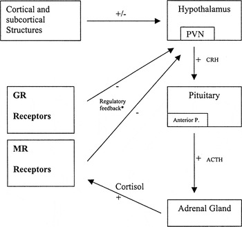
The basic model of the activation and regulation of the LHPA axis.
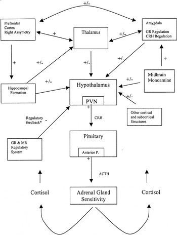
The expanded model of the activation and regulation of the LHPA axis.
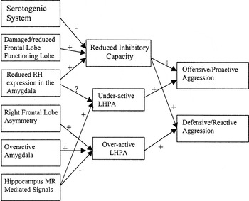
The expanded model of the neurophysiological profile of aggression.
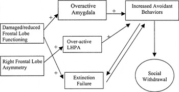
The expanded model of the neurophysiological profile of social withdrawal.
 You have
Access
- 38
- Cited by