Introduction
In François Jacob's (1977) famous metaphor, evolution is a tinkerer. It uses whatever genetic history has provided, and it exploits bits of genetic information that arrive by the chance occurrence of mutations. Natural selection, historically constrained and environmentally contingent, is the tinkerer’s workshop, but it is nevertheless a powerful and creative natural process. Its hallmark is the generation of novel complexity. But evolutionary outcomes are inevitably ephemeral. Environments change, and of the vast number of organisms that have existed during the last billion years of the earth’s 4.55 billion year history, only a tiny fraction are living today. This temporally modest sample nevertheless consists of millions of kinds of organisms, and it includes an enormous diversity of eyes. The presence of this diversity was recognized by Darwin in the Origin of Species and is recently discussed from evolutionary perspectives of a functional nature by Nilsson (Reference Nilsson2009).
Concerning homology
The morphological diversity of eyes obscures some seminal molecular homologies in whose discovery Drosophila has played a central role. Hox genes head the list, controlling the segmentation of animals during early stages of development, and more specifically, homologs of the transcription factor Pax-6 set in motion the development of eyes as different as those of mammals and fruit flies (Halder et al., Reference Halder, Callaerts and Gehring1995). Seen from the perspective of phenotypic function, however, opsins are the obvious examples of molecular homology, for opsins form the protein backbone of every visual pigment found in eyes. Over 1000 opsin genes have been sequenced, and some have roles in cells other than visual photoreceptors. To discuss the evolution of opsins would take us deeper into detail than can be accommodated in this paper, so here, we will focus on a few important generalizations about opsins before exploring more complex phenotypic examples of evolutionary tinkering
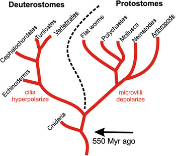
Fig. 1 is an evolutionary tree of the major extant groups of animals. To a first approximation, the opsins used in visual photoreceptors fall into two major subgroups that map onto these two branches: the deuterostomes and protostomes. The creatures on the left generally put their visual pigments into the membranes of modified cilia, and their photoreceptor cells hyperpolarize in the light. Those on the right put their visual pigments into microvilli of the plasma membrane with no relation to cilia. These rhabdomeric photoreceptors depolarize in the light. The two groups of opsins thus differ in the G proteins with which they combine and the transduction cascades they trigger. In ciliary photoreceptors, the Gt protein for the c-opsins interacts with phosphodiesterase, and the subsequent decrease of cyclic guanosine monophosphate (cGMP) causes cyclic nucleotide-gated ion channels to close and rods and cones to hyperpolarize. In rhabdomeric photoreceptors, the Gq protein for the r-opsins activates phospholipase C or an unknown messenger, which ultimately leads to the opening of Ca++ channels and depolarization of the photoreceptor. These pathways are reviewed in detail by Yau and Hardie (Reference Yau and Hardie2009).
Simplified evolutionary tree of the major living groups of animals. As described in the text, the ciliary photoreceptor cells of the eyes of Deuterostomes hyperpolarize in the light, whereas the rhabdomeric photoreceptors of the eyes of Protostomes depolarize in the light. The corresponding c-opsins and r-opsins thus stand at the head of different transduction cascades. Although this generalization is sufficiently accurate to be useful—all the instances of diverse evolutionary outcomes discussed in this paper, mostly from arthropods and vertebrates are consistent—there are numerous exceptions involving small eyes of polychaetes and molluscs or opsins expressed in neurons. For example, melanopsin in vertebrate ganglion cells is an r-opsin. Cnidaria have their own opsins. (After Yau & Hardie, Reference Yau and Hardie2009.)

But these two classes of photoreceptor do not represent monophyletic differences between chordates and the higher invertebrates. There are some important exceptions to the generalization of the preceding paragraph: some r-opsins occur in vertebrates and some c-opsins in invertebrates. As also noted above, opsins are present in other places than eyes. These exceptions are providing clues to the unresolved nature of ancestral photoreceptors existing 550 million years ago at the start of the Cambrian as well as functions and relationships of opsins yet poorly understood (Arendt, Reference Arendt2003; Nilsson, Reference Nilsson2009; Yau & Hardie, Reference Yau and Hardie2009; Porter et al., Reference Porter, Blasic, Bok, Cameron, Pringle, Cronin and Robinson2012).
Finally, multiple visual pigments absorbing in different regions of the spectrum occurred in the Cambrian, early in the evolution of multicellular organisms. Within the c-opsin lineage, vertebrates later derived four opsin lineages now associated with cones and color vision, and from one of the cone opsins, the rod pigment rhodopsin (reviewed by Collin et al., Reference Collin, Davies, Hart and Hunt2009). Subsets of arthropods, particularly the insects, have independently utilized ancient r-opsin pigment lineages for their own suites of color vision pigments (Briscoe & Chittka, Reference Briscoe and Chittka2001). Color vision in these major groups thus represents instances of convergent evolution, in which different pigment lineages have been independently employed for the same phenotypic function by vertebrates and insects.
In the evolution of visual systems, natural selection has utilized visual pigments with maximal absorption from 340 nm in the near UV to 620 nm in the red. Among terrestrial species, there are few cases of spectral tuning for ecological or other reasons; the matching of firefly photoreceptors to the lamps of their species, in this case enhanced with spectral filtering by accessory pigments, provides an example (Cronin et al., Reference Cronin and Goldsmith2000). In aquatic environments, tuning is more common because the spectral composition of the available light varies among habitats. The rhodopsins in the rods of deep-sea fish are blueshifted ≈20–30 nm, fitting better the spectral composition of residual sunlight at depth (Hope et al., Reference Hofstee1997). Similarly, cone pigments of fish are frequently matched to the space light in the water of their habitat (Loew & Lythgoe, Reference Loew and Lythgoe1978). As described in a later section, shifts in color vision pigments of teleost fish can occur by differential gene activation during development as well as through natural selection during speciation. Among invertebrates, mantis shrimp, equipped with an astonishing 16 different visual pigments, use colored filters to blueshift the long-wavelength receptors when they occupy deeper waters (Cronin et al., Reference Cronin, Jarvilehto, Weckstrom and Lall2001).
Color vision is nevertheless a general-purpose sensory system. When spectral composition is assessed by comparing the quantum catches of three or four visual pigments, the visual system can detect small color differences, greatly enriching visual perception. During the evolution of angiosperms, the evolution of colored flowers was likely driven by the presence of color vision in potential pollinators.
So how many times have eyes evolved independently? To paraphrase a famous politician, that depends on what you mean by “independently.” Sharing basic transcription factors that control developmental programs provides one perspective: one of ancient homology. Having structurally and functionally diverse phenotypes shaped by the tinkering of natural selection offers another. The question of independence, however, is less interesting than the problem to which it points: How does natural selection alter the shaping of diverse morphological phenotypes? Much is understood about the deep homologies triggering developmental subsystems—for example, transcription factors that direct local regions of the embryo to make an eye, even when the eye is experimentally induced in the wrong place or the molecular trigger has come from another species. Rather less is known about the molecular details of how natural selection impacts the fine-tuning of developmental outcomes. In evolutionary reasoning, this problem is traditionally framed in terms of selection for alternative alleles, but this perspective avoids the details of development, where patterns of temporal and spatial activations, inhibitions, and feedback loops engaging transcription factors, micro-RNAs, genes, and gene products interact to produce the phenotype. Put another way, how does natural selection tweak the regulatory processes of self-assembly that characterize development so as to produce small differences in heritably different phenotypes? Logically, there must be heritable variation in these regulatory processes that displays itself in the phenotype in order for natural selection to get traction. This is a central challenge for present and future experimental work. The following sections suggest some diverse phenotypic outcomes of increasing complexity whose existence poses interesting questions about the interplay of development and evolution.
Receptors and linearly polarized light
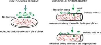
As shown in Fig. 2, a second functional difference between vertebrate and many rhabdomeric photoreceptors involves sensitivity to the plane of polarization of incident light, as it occurs in vivo. Rods and cones are insensitive because the absorption axes of the molecules of visual pigment are randomly oriented and free to diffuse in the planes of the disk membranes. If such membranes were rolled into cylinders, with absorption vectors randomly oriented in the tangent planes, absorption would be twice as great for the plane of polarization parallel to the cylinder axes as perpendicular (Moody & Parriss, Reference Moody and Parriss1961). This assumption is not realized in microvilli of crayfish, where dichroism and photo-induced dichroism indicate some orientation of the chromophores with the microvillar axes and some tilt into the plane of the membrane (Goldsmith, Reference Goldsmith, Snyder and Menzel1975; Goldsmith & Wehner, Reference Goldsmith and Warner1977). More dramatic evidence for axial orientation comes from intracellular measurements of polarization sensitivity of single photoreceptor cells of some insects and crustaceans (Shaw, Reference Shaw1969, Mote, Reference Mote1974, Labhart, Reference Labhart1980), with sensitivity ratios as high as 9 or 10.
The random order of rhodopsin chromophores close to planer with the disk membranes of the outer segment renders rods and cones insensitive to the plane of polarized light propagating along the axis of the outer segment. In organized rhabdomeric photoreceptors, where light is propagating at right angles to the microvillar axes, absorption by the visual pigment is dichroic, and the photoreceptor cell is sensitive to the plane of polarization. In the compound eyes of some arthropods, photoreceptor cells have polarization sensitivities as high as 10, indicating a degree of axial orientation of the chromophores with the microvillar axes.

Evolutionary adaptation and preadaptation
The outer segments of modified cilia and the microvillar structures of rhabdoms are both adaptations for packing membranes containing many molecules of visual pigment in the path of incoming light, increasing the probability of absorption and thus the sensitivity of the photoreceptor. But is sensitivity to the plane of polarization an adaptation? Conservatively, having photoreceptor membranes in the form of microvilli is an example of an exaptation, or preadaptation: a feature that exists for other reasons but is subsequently exploited for additional purposes. Photoreceptors with i) parallel microvilli, ii) chromophores of the visual pigment with a degree of axial orientation to the microvilli, and iii) a neural apparatus that exploits the sensory capacity to distinguish the plane of polarization (usefully demonstrated in behavior) collectively suggest morphological and neural refinements that deserve to be called adaptations.
The curious case of fish and polarized light
Despite the absence of dichroism for light propagating through the length of a rod or cone outer segment, fish present a conundrum: there is credible behavioral evidence that damselfish can be trained to associate food with either horizontally or vertically polarized light (Mussi et al., Reference Mussi, Haimberger and Hawryshyn2005) and trout trained to orient with respect to the plane of polarization of overhead light (Hawryshyn & Bolger, Reference Horváth, Clarkson and Pix1990). In addition, both the electroretinogram and trains of spikes in optic nerve fibers can be modulated by rotation of the plane of polarization of incident light (reviewed by Kamermans & Hawryshyn, Reference Kamermans and Hawryshyn2011). The latter might be dismissed as artifacts arising from unwanted reflections, but in fish, the only receptors sensitive to the plane of polarization are the UV cones, and fish that lose their UV cones as adults become insensitive to the plane of polarization. Flamarique et al. (Reference Flamarique, Hawryshyn and Hárosi1998) have offered the imaginative hypothesis that in the ordered mosaic of cones, UV-sensitive (UVS) single cones receive UV light laterally from adjacent double cones. (Double cones are discussed in more detail in a later section.) More specifically, the plane of fusion of the inner segments of double cones—which plane is at an angle to the long axis of the cells—functions as a mirror, reflecting light laterally and specifically onto the outer segments of UVS single cones. Critical experimental support for this model is lacking, and the idea remains in the realm of hypothesis.
Whatever the virtue of this hypothesis, there is one group of fish where a different mechanism exists, although it operates in the green and not the UV. Anchovies have unusual cones in which the disks are not transverse to the incident light but axial. They are thus sensitive to incident light polarized in the planes of the disks, just as the disk in Fig. 2 is dichroic when light comes from the side, with little absorption of light polarized perpendicular to the plane of the disk. These cells of the anchovy occur in pairs, with orthogonally oriented disks, thus possessing the features of an analyzer (Flamarique & Hárosi, Reference Flamarique and Hárosi2002).
Tinkering with the photochemistry
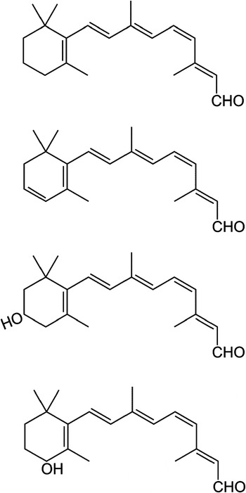
The photochemistry of eyes is the same everywhere, involving the isomerization of 11-cis retinoids. Four are known (Fig. 3). Retinal is the most common, but 3-dehydroretinal is found in a variety of animals, notably freshwater fish and turtles, premetamorphic amphibians, and some invertebrates. Because of the extra double bond in the ring, it absorbs at longer wavelengths than retinal, and so do the visual pigments that form when it combines with opsin. The presence of 3-dehydroretinal may be an adaptation for increased sensitivity in murky waters, where shorter wavelengths are absorbed by suspended organic material. Instances where 3-dehydroretinal is in some cells and retinal in others, or where the chromophores change at different stages in the life cycle (Collin et al., Reference Collin, Davies, Hart and Hunt2009), present interesting issues of molecular control.
The four retinals found in visual pigments. The 11-cis configuration associates with opsin in forming visual pigments. 3-dehydroretinal causes a bathochromic shift in spectrum relative to a retinal-based visual pigment with the same opsin. Evidence is growing that the hydroxyl groups can cause a hypsochromic shift. See the text for further details. As retinal is the aldehyde of vitamin A, the symbols A1–A4 are sometimes used as shorthand, but whether referring to the alcohol or the aldehyde may be clear only from the context.

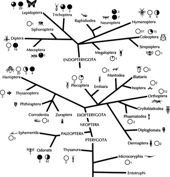
The use of 3-hydroxyretinal by insects and its absence in other arthropods indicates that it was a latecomer among the chromophores. Seki and Vogt (Reference Seki and Vogt1998) suggest its use dates from the late Paleozoic, during a rise in atmospheric oxygen, which presumably led to an increase in xanthophylls. It is not a reliable indicator of insect phylogeny, however, being prominent in Odonata, close to the base of the phylogenetic distribution of insect Orders, and again among Diptera and Lepidoptera, more recent groups. Even within an Order, some insects have retinal, some 3-hydroxyretinal, and some have both (Fig. 4; see also Seki & Vogt, Reference Seki and Vogt1998). Recently, the ninaB protein (named for a visual mutant in Drosophila) has been shown to have the double function in insects of cleaving and isomerizing zeaxanthin into all-trans- and 11-cis 3-hydroxyretinal (Oberhauser et al., Reference Oberhauser, Voolstra, Bangert, von Lintig and Vogt2008). This enzyme works as well with β-carotene as substrate, yielding all-trans and 11-cis retinal, and thus appears to be the entry point of retinoids into the insect visual system.
Phylogenetic tree of insect orders showing the distribution of retinal (A1, open circles) and 3-hydroxyretinal (A3, filled circles). Shading indicates average proportion found in individual eyes; numbers indicate number of species examined. A3 is the norm in Lepidoptera and Diptera, but it occurs together with A1 in many insects, particularly Odonata. (Modified from Smith & Goldsmith, Reference Smith, Freidman and Goldsmith1990.)

Why the hydroxyl group?
The least common chromophore, 4-hydroxyretinal, is known from a deep-sea bioluminescent squid, where it is accompanied by retinal and 3-dehydroretinal, forming three visual pigments with different absorbance spectra. The pigment with retinal as chromophore has λ max at 484 nm; the pigment with 4-hydroxyretinal, at 470 nm (Matsui et al., Reference Matsui, Seidou, Uchiyama, Sekiya, Hiraki, Yoshihara and Kito1988). Assuming the opsin is the same in both pigments, this 14-nm blueshift is caused by the difference in chromophore. Bleached salamander rods regenerated with 11-cis 4-hydroxyretinal contain a rhodopsin with λ max at 478 nm, blueshifted 31 nm from the native retinal-based rhodopsins (Frederiksen et al., Reference Frederiksen, Crouch, Nickle, Chakrabarti, Oprian, Koutalos, Boyer and Cornwall2012). Pigment regenerated in detergent from bovine opsin and 11-cis 3-hydroxyretinal is blueshifted 12 nm from rhodopsin regenerated with retinal (Gärtner et al., Reference Gärtner, Ullrich and Vogt1991). These observations suggest that retinoids with hydroxyl groups in the 3 or 4 positions produce a blueshift relative to retinal-based visual pigments.
One observation, indicating no spectral shift, is at variance with these results. Townson et al. (Reference Townson, Chang, Salcedo, Chadwell, Pierce and Britt1998) expressed the blue-sensitive visual pigment of honeybees heterologously in photoreceptors (R1–6) of flies, in which expression of all native visual pigments had been blocked genetically. The spectral sensitivity of these eyes matched well the blue receptors of honeybees. Although the chromophore was not identified, two indirect arguments suggest it was 3-hydroxyretinal. First, the spectral sensitivity function also showed the presence of the 350-nm sensitizing pigment, 3-hydroxyretinol, which is characteristic of flies, indicating the presence of 3-hydroxyretinoid. Second, in an analogous experiment, Abmad et al. (Reference Abmad, Natochin, Barren, Artemyev and O’Tousa2006) expressed bovine rhodopsin in the same photoreceptor cells of flies. They provided no spectral data on the pigment (which understandably did not activate the Gq protein of flies), but extracts of the eye contained 11-cis 3-hydroxyretinal.
If further work on insects shows that the substitution of 3-hydroxyretinal for retinal is commonly associated with a spectral shift, the use of the A1 and A3 chromophores in conjunction with three opsins can in principle lead to as many as six spectrally different visual pigments. The extent to which this diversity is realized is not known, but the wide use of both chromophores by Odonata is suggestive and begs for further study (Briscoe & Chittka, Reference Briscoe and Chittka2001).
Extending sensitivity at long wavelengths
As mentioned above, insects have three subsets of visual pigments sensitive in the UV, blue, and long-wavelength (L) regions of the spectrum. The L pigments are present as green-sensitive pigments in virtually all insect groups, but in Lepidoptera, red-sensitive (λ max > 565 nm) pigments have evolved four times (Briscoe & Chittka, Reference Briscoe and Chittka2001). The absorbance maxima of insect visual pigments therefore range from 340 to 600 nm. Gene duplication followed by selection seems to have been active in butterfly opsins, resulting in both red- and blueshifts in spectrum (Frentiu et al., Reference Frentiu, Bernard, Sison-Mangus, Brower and Briscoe2007).
Opsin evolution can be supplemented by other means to produce spectral shifts. For example, the butterfly Pieris brassicae has receptors maximally sensitive at 620 and 640 nm. The λ max of the visual pigment(s) lies at shorter wavelengths, but the sensitivity spectra are shaped by long-wavelength reflection from the tapetum and accessory red pigments adjacent to the rhabdom (Qiu & Arikawa, Reference Qiu and Arikawa2003). Other examples of shaping spectral sensitivity by accessory pigments are mentioned throughout this paper.
Tinkering with the structure of compound eyes
Well-developed compound eyes were present on trilobites 520 million years ago, before the appearance of vertebrates with well-developed eyes. Trilobites existed until the Ordovician, and they had multiple lineages and a variety of eyes (Horváth et al., Reference Hope, Partridge, Dulai and Hunt1997). Only the dioptric structures fossilize. Some trilobites lost their eyes as a result of living in deep water in the absence of light.
The earliest of these compound eyes (holochroal form, loosely meaning entire cornea) had thousands of hexagonal facets 100 μm in diameter, and images were likely created in the nervous system from a mosaic of light detectors corresponding to the mosaic of facets, much like the compound eyes of diurnal insects (see below). A later and derived form (schizochroal, divided cornea) had fewer much larger round facets separated by scleral rings. Each facet is the front surface of a calcite dioptric structure functioning as a doublet lens and focusing the light a short distance behind. The size and curvature of the facet and the quality of the lens suggest a useful image may have been projected onto a small retina. Functionally, the facets may thus represent an array of small eyes looking in slightly different directions but with overlapping visual fields.
A recent finding of fossilized compound eyes from the early Cambrian (515 million years ago) in Emu Bay shale of South Australia shows that compound eyes appeared earlier and in more sophisticated form than is revealed by trilobites (Lee et al., Reference Lee, Jago, Garcia-Bellido, Edgecombe, Gehling and Paterson2011). Unlike trilobite fossils, the organic material has been phosphatized and is exceptionally well preserved. The eyes are 7–9 mm on the longer axis and contain more than 3000 facets. The largest lenses are 150 μm in diameter and occur in a central flat part of the eye. These features indicate a region of high absolute sensitivity and small interommatidial angle, thus high spatial acuity. They are present in some contemporary species such as dragonflies and robber flies. The eyes (perhaps once borne on stalks?) are postulated to have belonged to a predatory species of arthropod.
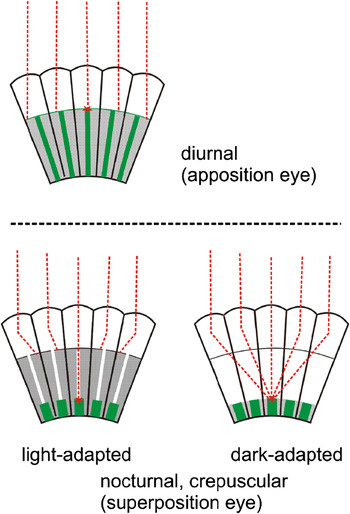
Like these fossils, contemporary compound eyes consist of a mosaic of ommatidia, each pointed in a slightly different direction. In diurnal, apposition eyes of insects (Fig. 5, top), each ommatidium has long photoreceptor cells (characteristically eight) arranged around the rhabdom like the wood surrounds the graphite of an ordinary pencil. The dioptric apparatus consists of a corneal lens on the apical surface and the “crystalline cone” just below. Light is focused on the distal end of the rhabdom, and the rhabdom has the properties of a light guide as light passes through its length. Sleeves of opaque screening pigment restrict sensitivity of each ommatidium to a narrow cone of near-axial light. This pigment is located variously in primary pigment cells around the crystalline cones, secondary pigment cells around the length of the photoreceptor (retinular) cells, or within the retinula cells. Movements of pigment in the primary pigment cells can function as an iris, controlling the amount of light that reaches the rhabdom.
Apposition and superposition eyes of insects and crustaceans. In apposition eyes, primary pigment cells around the crystalline cone and secondary pigment cells around the photoreceptors shield the rhabdom from light that does not enter the corneal facet at a small angle to the ommatidial axis. Superposition eyes have shorter rhabdoms, and pronounced movements of the screening pigments in the dark-adapted state allow light from a distant source to reach a rhabdom through a group of facets. As described in the text, superposition images can be achieved either through refraction or reflection.

In superposition eyes of nocturnal or crepuscular species (Fig. 5, below), the rhabdom is short and separated from the corneal surface and the underlying crystalline cones. Under low light conditions, the pigment sheaths retract, and a parallel beam of light is captured by an array of ommatidia and brought to a focus at a point on the rhabdom layer. This has the effect of enlarging the aperture and increasing sensitivity. But evolution has been inventive in the mechanisms for producing this result. In many insects and crustaceans, slightly off-axis light is refracted at the surface of the lens and internally by the crystalline cone, which functions as a lens cylinder in which refractive index is highest on the axis and decreases laterally (Exner, Reference Exner1891). In macruran decapods such as the crayfish, the facets are square, and light is reflected by multilayer mirrors in the distal part of the crystalline cone and again by total reflection at the walls of square mirror boxes formed by the crystalline cones (Vogt, Reference Vogt1980). In certain crabs, the walls of the crystalline cones are parabolic mirrors (Nilsson, Reference Nilsson1988).
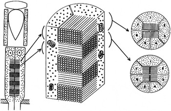
In crustaceans, the rhabdomeres form a box joint or mortise-and-tenon-like structure, shown in Fig. 6 for the superposition eyes of crayfish. These cells all have the same visual pigment: λ max is 533 nm in the crayfish Procambarus and Orconectes. There is also a small distal receptor cell (not illustrated) with a blue-sensitive visual pigment (Cummins & Goldsmith, Reference Cummins and Goldsmith1981).
Structure of the rhabdom in the compound eyes of the crayfish and other decapod crustaceans, from Eguchi (Reference Eguchi1965). Seven photoreceptor cells contribute microvilli to the rhabdom, three with microvilli oriented in one direction, and four with microvilli in the orthogonal direction. The interleaved tongues of microvilli prevent one set from screening the pigment in the orthogonal set, as would occur if one set were stacked in front of (distal to) the other. (An example of stacked rhabdomeres is illustrated in Fig. 7.) The interleaved arrangement causes the self-screening to be shared and makes each set of photoreceptors equally sensitive to light polarized in the axial plane of its microvilli as well as equally sensitive to unpolarized light.

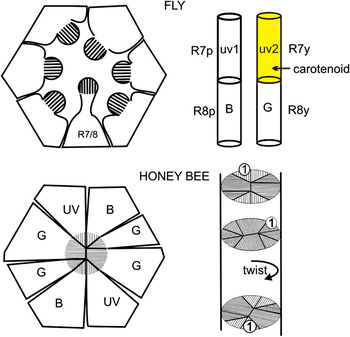
Fig. 7 shows examples of two ommatidia from apposition eyes of diurnal insects. The separate rhabdomeres of flies (Fig. 7, top left) run the length of the ommatidium; this “open” rhabdom is characteristic of Diptera, although it occurs in earwigs, aquatic Hemiptera, and some beetles as well. Six receptors (R1–6) have the same visual pigment, with λ max at 480 nm. The smaller seventh profile represents two shorter cells, one stacked over the other (Fig. 7, top right). R7 and R8 associate in two different tiers: pale (R7p/R8p) and yellow (R7y/R8y). Each tier has a different pair of visual pigments. R7 and R8 have long axons that bypass the first synaptic layer and equip flies for a modest capacity to distinguish between broad spectral regions (Fukushi, Reference Fukushi1994). In the dorsal rim of the eye, R7 and R8 have just the single UVS visual pigment that is expressed in R7p in the rest of the eye, and together, they can analyze polarized skylight (Hardie, Reference Hardie1984).
Above: open rhabdom of flies. Six of the cells (unlabeled) have the same visual pigment, are engaged in movement detection, and have a complex optical and synaptic interplay with surrounding ommatidia described in the text. Cell 7 is stacked over cell 8 (top right) and there are two R7/R8 pairs (Hardie, Reference Hardie1986). In the pale pair, R7p has a UV pigment (λ max 340 nm) and R8p a blue-sensitive pigment (λ max 437 nm). In the yellow pair, R7y has a different UV pigment (λ max 365 nm) together with a blue-absorbing light-stable mixture of carotenoids, whereas R8y has a green-sensitive rhodopsin (λ max 508 nm). The pigments in R7 screen those in cell R8, and the effect is dramatic in R7y/R8y: carotenoids in R7y depress sensitivity of R8y throughout the blue and move the wavelength of maximum sensitivity of R8y to about 550 nm. The compact rhabdom of the honeybee has eight retinular cells and three visual pigments (Fig. 7, lower left). The rhabdom functions as a single waveguide, and the spectral sensitivities of the several visual pigments are not distorted. The ommatidium is twisted 220 deg over its length, an adaption to eliminate sensitivity to the plane of polarization and the generation of “false colors.”

Optically, the retina of flies is more complex than it appears. Each of the rhabdomeres of R1–6 has a slightly different visual field. Moreover, each shares its visual field with five other rhabdomeres located in five different neighboring ommatidia (Kirschfeld, Reference Kirschfeld1967). All the cells with the same visual field converge on the same postsynaptic neuron (Braitenberg, Reference Braitenberg1967). Consequently, R1–6 of any single ommatidium project to six different synaptic cartridges, an arrangement referred to as neural superposition. R1–6 detect movement of external objects and the optical flow of the surround when the insect is in flight. They are likely involved in spatial vision as well. The short axons of these cells have different destinations from the long fibers of R7 and R8, and there is no evidence that R1–6 are involved in color vision. What passes for color vision in flies has separate circuitry from the system for motion detection.
The compact rhabdom of the honeybee (Fig. 7, bottom) is more typical of apposition eyes of insects and more like the analogy with a lead pencil. It has contributions from eight retinular (photoreceptor) cells, which share the same visual field. Four of the cells have a visual pigment maximally sensitive in the green (535 nm), two in the blue (440 nm), and two in the UV (370 nm), collectively supporting an excellent trichromatic color vision used in foraging from flowers (Daumer, Reference Daumer1956).
Some behaviors of insects are selectively driven by a spectral subset of receptors. Phototaxis, usually measured by forcing an insect to choose between two light sources and which is generally interpreted as an escape response towards open sky (or into darkness, depending on the species and the context), is strongly driven by the UV receptors, as is the dorsal light response, an orientation with respect to the horizon (Osorio, Reference Osorio1986). The optokinetic response to moving vertical stripes in the lateral visual field is driven primarily by R1–6 in flies and green-sensitive receptors in bees. In the bee, it is not clear whether the green-sensitive receptors involved in motion detection are also involved in color vision or are a distinct subset.
The ommatidia in the central part of the bee’s eye has an interesting 220 deg twist over its length (Fig. 7, bottom right), which renders its cells insensitive to the plane of polarization. This is interpreted as an adaptation to prevent light polarized by reflection, for example, from shiny surfaces of plants, from confounding color discriminations by creating false colors (Wehner & Bernard, Reference Wehner and Bernard1993). The famous polarization sensitivity of bees and ants, used for determining the position of the sun from a partial view of clear sky, is mediated by UV receptors in the dorsal rim of the eye, receptors that have no twist (Labhart, Reference Labhart1980).
Understanding the evolution of the twist in the ommatidia of the bee’s lateral eye should be enriched by experiments showing how the twist is generated during development. The superposition optics of Diptera pose a similar problem: how is the precise optical alignment of R1–6 achieved in the soft tissues of the pupa, and is it perfected by visual experience after escape from the pupil case? Knowing such details should help to deepen our understanding of precisely what natural selection selects in executing such precise structural tuning during development.
Tinkering with the visual cycle
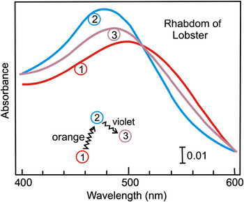
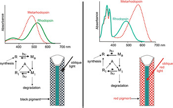
Unlike vertebrates, metarhodopsins of invertebrates characteristically do not bleach quickly. Light therefore creates a photosteady state populated by both rhodopsin and metarhodopsin (Fig. 8). Fig. 9 (left) shows how photoregeneration of rhodopsin typically functions in situ for green-sensitive visual pigments whose metarhodopsins absorb at shorter wavelengths. Light enters the ommatidia along their axes and initiates vision, but because metarhodopsin lingers in the eye, it can be photoconverted back to rhodopsin. The pigment that sheathes the rhabdoms and optically isolates the ommatidia is characteristically black and absorbs all wavelengths. In some cases, the eyes may not appear black because reflectance from the primary pigment cells around the crystalline cone reduces the contrast between the eyes and the body, producing cryptic coloration.
Interconversion of rhodopsin (R) and metarhodopsin (M) in an isolated rhabdom of a lobster (Homarus). Spectrum 1, mostly rhodopsin; spectrum 2, mostly metarhodopsin; spectrum 3, photosteady state of a mixture, whose composition depends of the relative spectral absorption of the actinic light by R and M. Note the presence of an isosbestic point, consistent with the presence of two molecular species and no loss of either during the irradiations. (After Bruno et al., Reference Bruno, Barnes and Goldsmith1977.)

Photochemical cycles in insects: Left, typical of many species; Right, Diptera. Left: the metarhodopsins of mid- and long–wavelength-sensitive pigments are blueshifted from the visual pigment (spectra), and the photosteady state induced by white light or long-wavelength light consists of more metarhodopsin than rhodopsin (diagram). In visual excitation, light (hυ) produces an active metarhodopsin (Ma) that initiates the transduction cascade, but activity is quickly inactivated (Mi) by the binding of arrestin (A). Lingering metarhodopsin (Mi) can be converted back to rhodopsin by the absorption of a second photon, and with the loss of arrestin, the rhodopsin can initiate another excitation. The rhabdom is sheathed by black accessory pigment that optically isolates the individual ommatidia. Leakage of the pigment sheaths would both convert rhodopsin to metarhodopsin and compromise image formation. Right: In flies, the metarhodopsin of R1–R6 is redshifted, and long-wavelength light drives the system from metarhodopsin to rhodopsin. The pigment in the pigment cells is red, and off-axis light at long wavelengths penetrates to the rhabdom and maintains a high level of rhodopsin. Vision is not impaired because long wavelengths do not initiate visual excitation. In the working eye, on-axis light arriving through the lens is necessary to convert rhodopsin to metarhodopsin and initiate visual excitation. The high absorption at short wavelengths in flies is caused by 3-hydroxyretinol, which functions as a sensitizing pigment. (Modified from Stavenga & Hardie, Reference Stavenga and Hardie2011.)

Flies are particularly interesting, not only for their superposition optics, but for how they exploit stray light. Fig. 9 (right) shows the absorbance spectrum of the visual pigment in R1–6. The metarhodopsin not only persists but also displays an unusually large spectral shift toward the red. In addition, 3-hydroxyretinol is associated with the visual pigment and sensitizes it to short (near UV) wavelengths. The interconversions of R and M takes place as indicated in the diagram, but photoregeneration of rhodopsin is greatly enhanced because the screening pigment in the sheath of secondary pigment cells is red, and stray, long-wavelength, off-axis light therefore penetrates to the rhabdom and shifts the photosteady state far toward the visual pigment. These leaks do not affect the rhodopsin, which is relatively insensitive to red light.
Dark processes for cycling
The photoregeneration of rhodopsin in the eyes of flies has suggested to some that there is no other process for regenerating 11-cis retinoids in invertebrates—that is, no visual cycle of retinoids as known in vertebrates. Accumulation of retinol in the eyes of bees during light adaptation has been known for nearly half a century (Goldsmith & Warner, Reference Goldsmith, Collins and Licht1964), and as we shall see shortly, there are several variations on the retinoid cycle among invertebrates. In this context, it is useful to keep in mind what makes an animal a useful “model”—in the case of Drosophila, the vast background of genetic information. But the insect Orders Diptera, Lepidoptera, and Hymenoptera have been on separate evolutionary paths for about 230 million years, long compared with the 65 million years of the adaptive radiation of modern mammals. Understanding evolution requires genomic studies that transcend the use of model animals (Porter et al., Reference Porter, Blasic, Bok, Cameron, Pringle, Cronin and Robinson2012).
In arthropods, metarhodopsin is lost from the rhabdom: with a half-time of about 2 h in the fly Calliphora (Schwemer, Reference Schwemer1984) and 13 h in Drosophila (Hofstee, Reference Hawryshyn and Bolger1996). Loss is much faster in the butterfly Vanessa, with a half-time 12.6 min (Bernard, Reference Bernard1983), is slower in other species of Lepidoptera, and is known to be temperature dependent. In the crayfish, metarhodopsin lingers for many hours or even days (Cronin & Goldsmith, Reference Cronin, Caldwell and Marshall1984). Following the bleaching or removal of metarhodopsin, how is the visual cycle completed?
Vertebrates have more than one dark process for generating 11-cis retinol from all-trans. In the pigment epithelium, the energy of hydrolysis of an all-trans retinyl ester is coupled to the process of isomerization generating 11-cis retinol (Bernstein et al., Reference Bernstein, Law and Rando1987). A second system associated with cones and Mueller cells uses a retinol isomerase to convert all-trans retinol to 11-cis. The reaction is pulled energetically uphill by esterifying the product, thus keeping the concentration of free 11-cis retinol very low (Mata et al., Reference Mata, Radu, Clemmons and Travis2002). The ninaB protein in insects has a homolog in vertebrates, but it is not yet clear whether it provides a source of 11-cis retinal in the vertebrate visual cycle (Oberhauser et al., Reference Oberhauser, Voolstra, Bangert, von Lintig and Vogt2008).
In an example of convergent evolution, decapod crustaceans also convert all-trans retinol to 11-cis by coupling the isomerization to the hydrolysis of retinyl esters, but the primary substrate is the retinyl ester of docosahexaenoate (Srivastava et al. Reference Srivastava, Lau and Goldsmith1996; Srivastava & Goldsmith, Reference Srivastava and Goldsmith1997). As mentioned earlier when discussing the acquisition of 3-hydroxyretinoids by insects, 11-cis retinal is produced when C40 carotenoids are cleaved by the ninaB protein. How much and under what conditions this reaction contributes 11-cis retinal to the visual cycle of insects is not yet clear.
Photoisomerases
A completely different mechanism present in invertebrates involves photoisomerases, of which there are at least two very different proteins, both working at the level of retinal rather than retinol. Retinochrome, the retinal photoisomerase of cephalopods (Hara & Hara, Reference Hara and Hara1973) and a marine gastropod (Ozaki et al., Reference Ozaki, Terakita, Hara and Hara1986), is a membrane protein, in fact an opsin. In the dark, it is mostly in the proximal region of the cephalopod photoreceptor cell, but in the light, about half of it moves into the distal region of the cell bearing microvilli and the visual pigment (Fig. 10).
Left: photoconversion of all-trans retinochrome to the 11-cis form (Hara & Hara, Reference Hara and Hara1973). Right: movement of a portion of the retinochrome from the inner region of the photoreceptor cell in the dark to the rhabdomeric region in the light (Hara & Hara, Reference Hara and Hara1976).

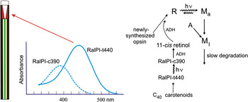
The fly Calliphora has a blue-sensitive retinal photoisomerase (Schwemer, 1984), but the protein has not been isolated. Evidence also exists for a retinal photoisomerase in Limulus (Smith et al., Reference Smith and Goldsmith1992). The retinal photoisomerase of honeybees is the best-known arthropod photoisomerase; it is a soluble protein, not an opsin (Schwemer et al., Reference Schwemer, Pepe, Paulsen and Cugnoli1984; Smith & Goldsmith, Reference Smith and Goldsmith1991a). When combined with all-trans retinal, it absorbs in the blue; on irradiation, the retinal isomerizes to 11-cis and the spectrum shifts to shorter wavelengths (Fig 11, left). The highest concentration of the photoisomerase is located in the primary pigment cells, where it can capture off-axis light that is not destined for the rhabdom (Smith & Goldsmith, Reference Smith and Goldsmith1991b).
Spectrum: The retinal photoisomerase of honeybees (RalPi) is a soluble protein, not an opsin. With all-trans retinal (t-440), it absorbs in the blue (λ max 440 nm); light isomerizes the retinal to 11-cis and shifts the spectrum to 390 nm. Half of the photoisomerase is concentrated in the primary pigment cells around the crystalline cone (arrow above the spectrum), but RalPi at much lower concentration occurs throughout the retinular cells. Diagram to the right: RalPi-t440 accumulates in the dark and accounts for 75–80% of the total retinal in the eye. The 11-cis retinal formed in the light is reduced to 11-cis retinol and bound to another protein that may function as a transport vehicle. Unlike decapod crustaceans, 11-cis retinol is not stored as esters of fatty acids. Conversion of 11-cis retinol back to retinal must occur, but its allocation between metarhodopsin stripped of retinal and newly synthesized opsin is not known. (After Smith & Goldsmith, Reference Smith and Goldsmith1991a,Reference Smith and Goldsmithb.)

In the dark-adapted state, 75–80% of the retinal in the eyes is associated with this protein, with accumulation of retinal likely from cleavage of C40 carotenoids (Fig. 11, right). In the light, vision is triggered by the isomerization of 11-cis to all-trans retinal associated with the visual pigment in the rhabdom, and 11-cis retinal is simultaneously formed from all-trans bound by the photoisomerase in the primary pigment cells. Newly formed 11-cis retinal is reduced to 11-cis retinol, which is then bound to another protein; only trivial amounts are esterified (Smith & Goldsmith, Reference Smith and Goldsmith1991a). The protein binding 11-cis retinol may be a transport vehicle, moving the retinol through the length of the photoreceptor cells where it can be oxidized to 11-cis retinal and bound to opsin, either newly synthesized, or recycled in the rhabdom, or both.
Evidence exists for a similar blue-sensitive retinal photoisomerase in the butterfly Papilio xuthus (Asian swallowtail). It is located in the distal part of the ommatidium, is active in the supernatant of an aqueous extract, precipitates in ammonium sulfate, and accepts either all-trans 3-hydroxyretinal (the native retinoid) or all-trans retinal as substrate (Shimazaki & Eguchi, Reference Shimazaki and Eguchi1995).
Evolutionary tinkering with vertebrate photoreceptors
Double cones and cone oil droplets
Double cones and cone oil droplets (Fig. 12) are common features of holostean fish, amphibians, reptiles, and birds, but neither is present in placental mammals (Walls, Reference Walls1942). The principal member of double cones characteristically has a cone oil droplet, which is absent in the accessory cone. The accessory member has an organelle called the paraboloid, which contains glycogen (e.g., Amemiya, Reference Amemiya1975), suggesting energy reserves. Both members have the same visual pigment. Relatively little is known about the roles of double cones; they are more common in diurnal retinas, where they can account for about half of the cones. In birds, they seem to be involved in processing optic flow and detecting movements (Campenhausen & Kirschfeld, Reference Campenhausen and Kirschfeld1998; Bhagavatula et al., Reference Bhagavatula, Claudianos, Ibbotson and Srinivasan2009). Significant separation of channels for color and movement is an evolutionary outcome found in insects (described above), fish (Schaerer & Neumeyer, Reference Schaerer and Neumeyer1996), birds (Campenhausen & Kirschfeld, Reference Campenhausen and Kirschfeld1998), and humans (Livingstone & Hubel, Reference Livingstone and Hubel1987; Cheadle & Zeki, Reference Cheadle and Zeki2011).
Double cones and cone oil droplets are present in the retinas of birds and reptiles but are absent in placental mammals. Relatively little is known about double cones, although in birds, they account for about half of the cone population. Single cones are involved in color vision, characteristically tetrachromatic.

Cone oil droplets are located in the inner segment of the cone, immediately adjacent to the base of the outer segment. Turtles (Leibman & Granda, Reference Liebman and Granda1975) and diurnal birds (Goldsmith et al., Reference Goldsmith and Wehner1984) have deeply colored oil droplets containing carotenoids in very high concentration. Oil droplets of birds are much paler or are absent in crepuscular or nocturnal species. Their role in color vision is discussed below.
Cones and color vision of fish
The teleosts comprise more than 95% of the species of present-day fish, but they are a derived group dating from the early Triassic, and their cones lack oil droplets. They do, however, have cones that occur in pairs. Twin cones have been recognized since the 19th century. They are fused along their inner segments (Fang et al., Reference Fang, Li, Wai, Kwong, Kung and Yew2005), and the two elements are morphologically identical. They are electrically coupled (Miller & Korenbrot, Reference Miller and Korenbrot1993), and the outer segments contain the same visual pigment (Britt et al., Reference Britt, Loew and McFarland2001, but see Loew & Lythgoe, Reference Loew and Lythgoe1978). Twin cones are more prevalent in species living in bright light, and their response functions are accordingly shifted to higher intensities (Miller & Korenbrot, Reference Miller and Korenbrot1993).
Walls (1942) distinguished between twin cones and pairs of nonidentical cones. His illustration of the later cites Butcher (Reference Butcher1938), who described this type of cone in Fundulus and referred to it as a double cone. Walls speculated that it was either transitional between double cones of other vertebrate classes and twin cones, or alternatively, was secondarily derived from twin cones. Fish double cones differ from twin cones in being fused only in the more proximal regions of the inner segment, being asymmetric (Fang et al., Reference Fang, Li, Wai, Kwong, Kung and Yew2005), and having a different visual pigment in each outer segment (Robinson et al., Reference Robinson, Schmitt, Hárosi, Reece and Dowling1993; Britt et al., Reference Britt, Loew and McFarland2001). Curiously, they are also reported as being electrically coupled, leading to the inference that they assess luminance rather than color differences (Marchiafava, Reference Marchiafava1985). Nevertheless, in trigger fish, the double cones (λ max at 530 and 480 nm) have been shown to participate in color discrimination along with a single cone (λ max at 413 nm) (Pignatelli et al., Reference Pignatelli, Champ, Marshall and Vorobyev2010). Fish double cones thus differ from the double cones of other vertebrates in lacking oil droplets, having two visual pigments, and participating in color vision. Their homology, if any, is not clear. To add confusion, one can read of twin cones with two visual pigments and double cones with only one visual pigment. The criteria for distinguishing fish double cones from twin cones may need to be reexamined (or alternatively, deserve more attention in application), but this is an issue for another place and another author.
Teleosts present much variation, and many species have UV receptors. Cyprinids (e.g., zebrafish, Robinson et al., Reference Robinson, Schmitt, Hárosi, Reece and Dowling1993) have double cones (λ max at 570 and 480 nm) and two classes of single cones (λ max at 415 and 362 nm). Behavioral experiments show that color vision in goldfish (a cyprinid) is tetrachromatic (Neumeyer, Reference Neumeyer1992). Reef fishes can be trichromatic (see above), and species with only single and twin cones should be dichromatic.
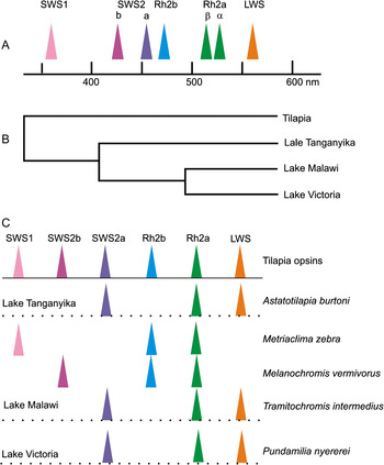
The riverine tilapia (Oreochromis niloticus) represents an ancestral outgroup to the recent adaptive radiations of lucustrine African cichlids (Fig. 13B). Spady et al. (Reference Spady, Parry, Robinson, Hunt, Bowmaker and Carleton2006) have found that tilapia has seven cone opsin genes, whose genetic lineages and spectral positions of cone pigments are shown in Fig. 13A. Some of the underlying gene duplications that led to this outcome date from the radiation of teleosts during the Mesozoic; the most recent was more than 10 million years ago. Usually, only three cone pigments occur in adult tilapia (SWS2a, Rh2, and LWS), but the other four are expressed at various times during ontogeny. Fig. 13C shows the representations of these pigment lineages in adult cichlids sampled from three lakes. Ontogenetic changes in cone pigments may be present in many species of fish; in the sample of cichlids studied, they occur on different schedules and in different patterns (Carleton et al., Reference Carleton, Spady, Streelman, Kidd, McFarland and Loew2008). This flexibility seems to provide a means for adjusting the visual system for differences in prey and/or the spectral nature of the aquatic environment at different stages in the life cycle as well as in different species or even in different populations of the same species.
Ontogenetic and species diversity of cone pigments in cichlid fish in the Great African Lakes. (A) Spectral positions and evolutionary lineages of the seven cone pigments expressed during the life cycle of tilapia. (B) Evolutionary relationships of the tilapia riverine outgroup to the lucustrine radiations of cichlids in the Great African Lakes. (C) Relation of cone pigments in several species of adult lake cichlids to the palette of cones in tilapia, based on absorption spectra. (After Spady et al., Reference Spady, Parry, Robinson, Hunt, Bowmaker and Carleton2006.)

Color vision of birds
Diurnal birds characteristically have four different cone pigments located in single cones: long-, middle-, and short-wavelength sensitive, plus a cone that can extend sensitivity into the UV (Bowmaker et al., Reference Bowmaker, Heath, Wilkie and Hunt1997; Cuthill et al., Reference Cuthill, Partridge, Bennett, Church, Hart and Hunt2000). This latter cone exists in two forms: violet sensitive (VS, λ max at 402–426 nm) and UVS (λmax at 363–373 nm). The VS form is the primitive state, but UVS forms have evolved at least four times by mutational changes among sites for three closely spaced amino acids. UVS cones (Fig. 14) are present in several Orders including Passeriformes (familiar song birds) and Psittcciformes (parrots) (Ödeen & Håstad, Reference Ödeen and Håstad2003). Fig. 14 shows the cone pigments of the budgerigar (Melopsittacus undulatus).
Spectra of the four cone pigments of the budgerigar (UVS and dashed curves) and their absorbance spectra after filtering by the cone oil droplets (solid curves). UVS, SWS, MWS, and LWS refer to UVS, short-, middle-, and long-wavelength sensitive, respectively. The UVS pigment is not filtered by a cone oil droplet. (Modified from Goldsmith & Butler, 2005.) Visual pigment data from Bowmaker et al., (1997); oil droplet data from Goldsmith et al., (Reference Goldsmith and Wehner1984).

The oil droplet of the UV cones contains no carotenoids, and the other oil droplets function as long-pass filters, narrowing the bandwidths of the visual pigments and shifting the λ max to longer wavelengths (Fig. 14). The principal member of double cones has red oil droplets, and both members contain the long–wavelength-sensitive visual pigment. As implied above, double cones appear not to be involved in avian color vision.
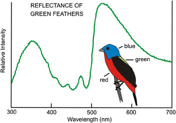
The budgerigar employs its four single cones in color vision (Goldsmith & Butler, Reference Goldsmith and Butler2005), and UV is perceived as a distinct color (Fig. 15). More generally among birds, UV is used in foraging and mate choice (Cuthill et al., Reference Cuthill, Partridge, Bennett, Church, Hart and Hunt2000). UV exists for birds to see in unexpected places; for example, Fig. 16 shows UV reflectance from the green back of a male painted bunting.
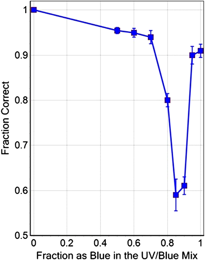
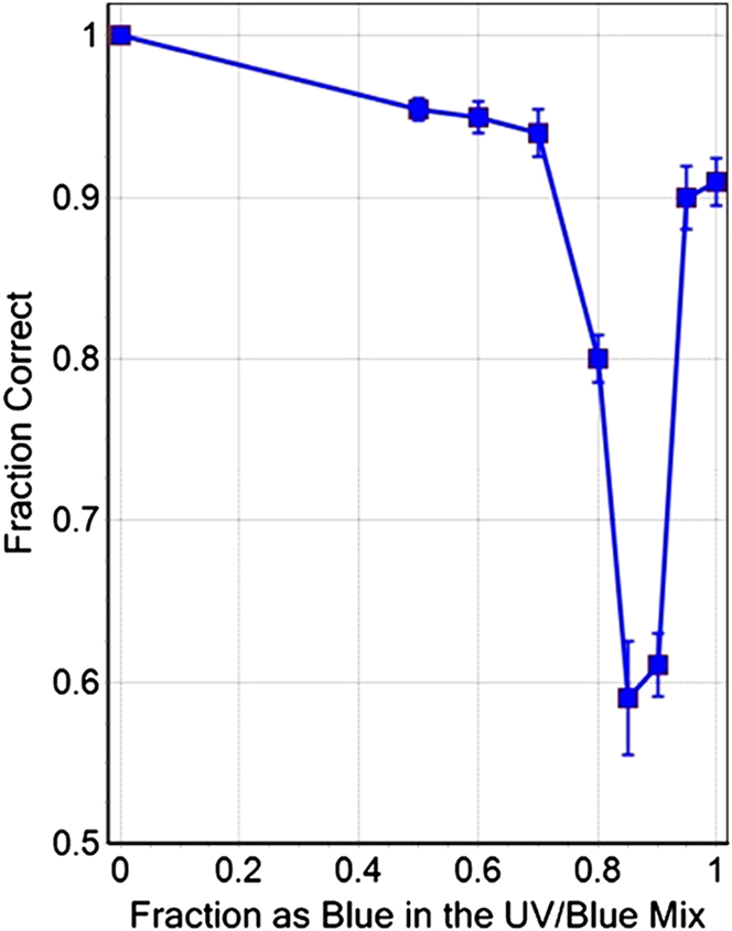
UV is seen by many birds as a distinct color. This figure shows the results of a behavioral experiment in which budgerigars were trained to discriminate 420 nm from mixtures of 440 nm (Blue) and 365 nm (UV). As the experiment ran, positions of the lights were interchanged and brightness altered in irregular fashion. Because red and yellow oil droplets screen the two cone pigments (MWS and LWS) active at longer wavelengths, the birds are dichromatic at wavelengths shorter than 440 nm. This experiment shows that UV and 440-nm lights were seen as different from the 420-nm training light with better than 90% accuracy in all mixtures except around 0.85–0.90 Blue, where there appears to be a color match. A match suggests the UVS and SWS cones project onto an opponent neural process.
Reflectance spectrum of the green back of a male painted bunting Passerina ciris. What appears green to our eye is reflectance from structural elements in the feathers, diminished by carotenoid absorption of blue wavelengths. Because all-trans isomers of C40 carotenoids have little absorbance in the near UV, there is a peak of reflectance in the UV that is not seen by us but is a distinct color for many birds. Inset shows regions of color of the plumage with varying amounts of UV reflectance plotted in Fig. 17.

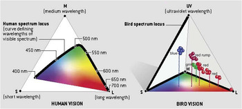
One way to represent color space is based on relative absorptions of the visual pigments, plotted for human color vision in Fig. 17 as a triangle. For birds, we need another dimension and can use a tetrahedron. The floor of the tetrahedron corresponds to the region of our visible spectrum but note how the spectrum locus follows the edges of the triangle, unlike the case for human color vision. This larger hypothetical color space is a result of the decreased overlap of the spectra of the cone pigments caused by the cone oil droplets.
Comparison of human and budgerigar color space, based on relative absorptions of the cone pigments. The fourth (UV) cone pigment of the bird requires an extra dimension, thus a tetrahedron rather than a triangle. The spectrum loci follow the heavy black lines. All colors for humans with normal color vision lie under the spectrum locus in the plane of the triangle, with nonspectral mixtures of red and blue across the bottom. For the budgerigar, the part of the spectrum without UV lies in the floor of the tetrahedron, and colors containing UV lie at various distances above the floor. The cone oil droplets reduce the spectral overlap of the cone pigments, the color space is expanded, and the spectrum locus follows two edges of the floor of the tetrahedron. The clusters of data points indicated by red, green, and blue are colors calculated from reflectance spectra from red, green, and blue regions of the plumage of male painted buntings. (Modified from Goldsmith, Reference Goldsmith2006.)

Colors above the floor contain UV wavelengths, and the points were calculated from reflectance spectra of different regions of the plumage of the painted bunting. This model of color space is based solely on the spectra of the cone pigments as filtered by the cone oil droplets, and it cannot tell us anything about the brain’s capacity to distinguish wavelength differences. That requires psychophysical experiments that are much easier to perform with high accuracy on humans than on any other animal. Even with the broad spectral overlap of the red- and green-sensitive visual pigments in human cones, wavelength differences less than 1 nm can be discriminated. Tetrachromacy and the presence of cone oil droplets likely have more to do with enriching the palate and increasing saturation than maximizing wavelength discrimination. Oil droplets may also play other roles not yet well understood. Retinas of birds that peer into bodies of water to catch prey are rich in red droplets, which are effective in reducing glare of short wavelengths.
Evolution as an irreversible process
This brief discussion of double cones, cone oil droplets, and avian color vision is also backdrop for an additional evolutionary point. We can think of evolutionary outcomes as peaks, scattered on an adaptive landscape of genetic possibilities. In this visual metaphor, adaptation consists of climbing peaks. But staying on a peak can be undone by environmental change too rapid for natural selection to keep pace. Crossing from one peak to another is an exceedingly low-probability event. Not only are peaks separated by realms of low fitness, but every climb is strewn with historical contingencies unlikely ever to be encountered again. And as reminded earlier, extinctions are regular events. Regular in the scale of geologic history, but whether humans are wise to generate extinctions through social and economic folly is a very different matter.
The color vision system of birds illustrates a quintessential adaptive peak for the diurnal vertebrate eye. Nothing quite like it is known among mammals. Placental mammals emerged from the Cretaceous having existed as small nocturnal creatures living in the shadows of larger reptiles for much of their entire existence, dating from the mid Jurassic, and they no longer possessed double cones or cone oil droplets. Moreover, they had only two of the four cone pigments present in so many reptiles and birds. The adaptive peak characterizing the placental mammalian eye had shifted with the change from diurnal to nocturnal habits, with more rods, fewer cones, and the loss of cone oil droplets and two cone pigments.
With the adaptive radiation of mammals dating from about 65 million years ago, mammals, particularly placental mammals, have diversified greatly, and many have become primarily diurnal. The evolution of color vision in mammals is nicely reviewed by Jacobs (Reference Jacobs2009). I will pursue a single point in what follows.
Most mammals remain with just the longest- and the shortest-wavelength pigment genes. The latter, however, has a pigment range from the near UV (e.g., rats and mice) to the violet (e.g., humans), a situation similar to birds. Trichromacy has been recovered among some primates, but the mid-range pigment gene is new and derived from the long-wave pigment gene (Yokoyama & Radlwimmer, Reference Yokoyama and Radlwimmer2001), a process made more probable with more than one copy of the parent gene adjacent on the X chromosome (Nathans et al., Reference Nathans, Piantanida, Eddy, Shows and Hogness1986). Thus, individual catarrhine (Old World) primates (including humans) have three cone pigments and trichromatic color vision. The outcome among the platyrrhine (New World) monkeys is more complex. Howler monkeys also have three cone pigments, but characteristically in other species, there is genetic polymorphism for three alleles of the long-wavelength pigment gene and correspondingly three different cone pigments. As these genes are on the X-chromosome, male monkeys and females homozygous for one of the long-wavelength pigment alleles are dichromatic, with three variants in the population. Females heterozygous for the long-wavelength pigment gene are trichromatic, again with three phenotypes possible.
Primates have managed an evolutionary reclamation. The outcome is not where the very early mammalian or reptilian ancestors started because during the Mesozoic, mammalian eyes relinquished some retinal components that were not sufficiently useful in their way of life for selection to be maintained. In evolution, what is not used is eventually lost. Species that live for generations in the total darkness of caves can lose their eyes. Complex features that are lost cannot be recovered in their original form, but if the loss is not too profound, evolutionary tinkering can sometimes produce an effective phenotypic replacement. Duplicated opsin genes have proved useful in the tinkerer’s toolbox, both among butterflies and some primates, and a small number of mutations have enabled selection for new cone pigments. Interestingly, the nervous system of primates has been sufficiently flexible to exploit the presence of an added cone pigment, and during development, neural connections are made to compare the quantum catches of a variable number of classes of cones containing spectrally different pigments. The result has been the evolutionary recovery of capacity for trichromatic color vision that functions impressively well, even in the case of species with genetic polymorphism for opsin genes and in which only some individual females have a third cone pigment.
Science, reason, and denial in the public square
At the Association for Research and Vision and Ophthalmology (ARVO) symposium at which this paper was presented, a discussant reminded the audience that the evolution of eyes played a special place in Darwin’s reasoning and that Darwin’s words have been misused by creationists in attacks on public education. This issue takes us in an unconventional direction, but for that very reason, it deserves our further attention.
In the Origin of Species, in a section on “Organs of extreme perfection and complications,” Darwin wrote:
“To suppose that the eye, with all its inimitable contrivances for adjusting the focus to different distances, for admitting different amounts of light, and for the correction of spherical and chromatic aberration, could have been formed by natural selection, seems, I freely confess, absurd in the highest possible degree” (Darwin, Reference Darwin1859).
Use of this passage by creationists and others to suggest Darwin had given up on eyes is more than troubling; it is intellectually dishonest in the tradition of much political discourse, for Darwin continued:
“Yet reason tells me, that if numerous gradations from a perfect and complex eye to one very imperfect and simple, each grade being useful to its possessor, can be shown to exist; if further, the eye does vary ever so slightly, and the variations be inherited, which is certainly the case; and if any variation or modification in the organ be ever useful to an animal under changing conditions of life, then the difficulty of believing that a perfect and complex eye could be formed by natural selection, though insuperable by our imagination, can hardly be considered real.”
Darwin’s inference has been amply confirmed, but this is not just a musty footnote to history. Denial of the reality of evolution permeates our culture, and attacks on science education and the role of science in understanding the world occur every day. In 1987, in Edwards vs. Aguillard, the Supreme Court of the United States decided 7:2 that the oxymoron “creation science” was religion and not science—“lacks a clear secular purpose” is the way the court put it—thus “violative of the Establishment Clause” and not appropriate to teach in a science classroom in public schools.
The capacity of the judicial mind to avoid the central issue is illustrated by Justice Scalia’s dissent, with Justice Rehnquist concurring: “. . . my views (and the views of this Court) about creation science and evolution are (or should be) beside the point. Our task is not to judge the debate about teaching the origins of life [sic], but to ascertain what the members of the Louisiana Legislature believed” (Edwards vs. Aguillard, 1987). Perhaps, it is a measure of scientific literacy in high places that these two Justices found the following legislative testimony compelling: “The body of scientific evidence supporting creation science is as strong as that supporting evolution. In fact, it may be stronger . . . Evolution is merely a scientific theory or ‘guess.’” One wonders what decision Justice Scalia would have reached if the Louisiana Legislature had insisted science classes teach about a flat earth.
In the antiscience world, the vernacular meaning of theory as tentative understanding or “guess” is timeless, and now, we also hear an effort to reposition global climate change as “theory not fact.” Although one might hope the recent decision in Kitzmiller vs. Dover disposed of intelligent design, issues of evolution and climate change remain alive in school boards, state legislatures, and among politicians, including several with presidential aspirations. “Teach the controversy” is the demand, but the controversy, where it exists, is not about science; it is about denial of evidence, injection of religious belief, displays of political and economic ideology, deliberate distortion, blaming the messenger, and heaps of wishful thinking.
Such obfuscation reflects a serious failure of education. Perhaps, this is not surprising; 13% of the biology teachers in public high schools in the United States are avowed creationists and a “cautious 60% . . . are neither strong advocates for evolutionary biology nor explicit endorsers of nonscientific alternatives” (Berkman & Plutzer, Reference Berkman and Plutzer2011). Berkman and Plutzer offer an insightful analysis of the causes at the high school scene as well as important suggestions for breaking the “cycle of ignorance.” The problem has political dimensions, and change will require active support and participation from the scientific community. This is the larger social issue that our discussant put in play and is the point I want to make in closing. Former Vice President Gore has concisely summarized the consequences of our current failure (Gore, Reference Gore2011):
“Dramatic changes in the way we communicate with one another about issues affecting the common good have diminished the role of reason and fact-based analysis, encouraging ideological extremists to construct their own alternative version of reality and defend it against fact-based reasoning.”
Acknowledgments
I am grateful to Gary Bernard, Nansi Colley, John Dowling, Roger Hardie, Mandyam Srinivasan, and an anonymous reviewer for useful discussions and helpful comments.