Introduction
Peritonsillar abscess is one of the commonest deep neck space infections. It is a suppurative complication of tonsillitis wherein the infection spreads to the peritonsillar space, an area between the tonsillar capsule and the superior constrictor muscle. Current management of peritonsillar abscess includes careful surgical drainage and appropriate antimicrobial therapy. Potential complications of peritonsillar abscess include spontaneous rupture of the abscess, aspiration of pus into the lower airways and extension of the infection to the lateral pharyngeal space.
The aetiology of peritonsillar abscess is often polymicrobial. Penicillins are used as empirical therapy. The definitive antibiotic management is usually selected after culture and sensitivity studies.
The aim of this study was to re-evaluate both the aerobic bacteriology of the peritonsillar abscess and the changing pattern of antibiotic sensitivity. Furthermore, these bacteriological data were compared with those of previous studies.
Materials and methods
Sixty patients who had suffered from peritonsillar abscess were included in this retrospective study. None of the patients in our study had suffered a previous episode of peritonsillar abscess or sought prior treatment for their complaints. Patients with systemic illnesses such as diabetes and hypertension, and those with immunodeficiency states, were excluded. The patients were managed at the Jawaharlal Institute of Postgraduate Medical Education and Research Hospital, a tertiary referral hospital, between May 2003 and June 2006. The clinical records of all these patients were reviewed regarding clinical features, diagnosis, bacteriological studies, treatment and outcome. The methods used in the study were approved by the institutional review board.
The medical history of all patients had been carefully elicited with regards to symptoms, duration, previous episodes of tonsillitis, peritonsillar abscess and antimicrobial therapy (if any). Follow-up data were recorded at successive visits at two, six and 12 weeks after discharge. In all patients, blood cell counts and blood sugar, urea and electrolyte concentrations were measured.
A sample was collected by needle aspiration of the abscess. The mucosal surface was sprayed with 5 per cent lignocaine and the aspiration performed using a 20 ml syringe with an 18 gauge needle, after disinfection of the surface with 70 per cent alcohol. All the air in the syringe and needle was expelled and the syringe plugged. The sample was then transported immediately, at room temperature, to the laboratory. Conventional bacteriological techniques were used to identify the aerobic bacteria in the abscess. The role of anaerobes has been proven by studies in the past; hence, anaerobic bacteriological assessment was not included in this present study.
Incision and drainage was performed for all patients and empirical antibiotic therapy with penicillin and metronidazole was commenced. Eighteen of the 60 patients showed either poor response or no improvement with empirical antibiotics; these patients required the addition of intravenous ciprofloxacin, based on their antibiograms.
At follow up, all the 60 patients showed complete resolution of symptoms with no complaints, and none required tonsillectomy.
Results
Sixty patients were included (32 males and 28 females). The youngest patient was aged eight years and the oldest 42 years. None of the patients had had a previous history of peritonsillar abscess, and none had sought any prior treatment for their present complaints.
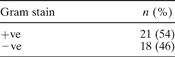
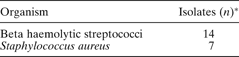
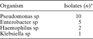
The blood sugar profiles of all the patients were normal, and the blood cell counts revealed only a mildly elevated neutrophil count. Pus was obtained from all 60 patients. Thirty-nine out of the 60 (65 per cent) samples showed a positive culture for aerobic bacteria. The aerobic infections were monomicrobial. Twenty-one out of these 39 (54 per cent) samples grew Gram-positive organisms (Table I). Gram-negative organisms were seen in the remaining 18 (46 per cent) samples (Table I). Of the Gram-positive organisms isolated, beta haemolytic streptococci were the commonest (Table II). Of the Gram-negative organisms, pseudomonas species were the commonest (Table III).

Gram-positive isolates

*Total = 21.
Gram-negative isolates

*Total = 18.
In the antibiogram, the Gram-positive organisms were uniformly sensitive to the conventionally tested antibiotics, whereas the Gram-negative organisms showed selective sensitivity to certain drugs, namely ciprofloxacin, gentamicin, amikacin, ceftazidime and ceftriaxone. The Gram-negative organisms were uniformly resistant to penicillins.
Discussion
Peritonsillar abscess is a suppurative complication of tonsillitis wherein the infection extends from within the tonsil into the peritonsillar space. It commonly occurs in adolescents and young adults, although any age can be affected. The patient usually complains of fever and a sore throat. There is difficulty in swallowing and dribbling of saliva, and the patient may have a muffled voice. Trismus may occur if the infection extends laterally. The abscess is usually unilateral.
The surface of the abscess is usually covered with normal oral flora type bacteria, wherein anaerobic bacteria far outnumber aerobic bacteria. Hence, careful aspiration after disinfection of the puncture site is essential in order to obtain a sample.
• Peritonsillar abscess is a common suppurative complication of tonsillitis
• Anaerobic bacteria and Gram-positive streptococci have been identified as the common aetiological agents
• In this study, Gram-negative aerobic bacteria were implicated in peritonsillar abscess, for the first time
• The presence of Gram-negative bacteria warrants the use of antibiotics other than penicillin (which hitherto has been the empirical therapy for peritonsillar abscess)
• In patients with peritonsillar abscess who do not improve with empirical therapy, Gram-negative organisms should be suspected and appropriate antibiotics added
The aetiology of peritonsillar abscess is often varied. Earlier studies indicated that anaerobic bacteria were the predominant species isolated from such abscesses, with peptostreptococcus species being the most common anaerobe isolated.Reference Sugita, Kawamura, Icikawa, Fujimaki, Oguri and Deguchi1 However, recent studies have found a predominately mixed isolate of aerobic and anaerobic bacteria in peritonsillar abscesses.Reference Sakae, Imamura, Sennes, Araujo Filho and Tsuji2 Of the Gram-positive organisms, beta haemolytic streptococci are most commonly isolated.Reference Sakae, Imamura, Sennes, Araujo Filho and Tsuji2–Reference Snow, Campbell and Morgan4 However, some studies have also indicated that other organisms, namely alpha haemolytic streptococci, may be implicated as the causative organism in peritonsillar abscess.Reference Matsuda, Tanaka, Kanaya, Kamata and Hasegawa5, Reference Fujiyoshi, Inaba, Udaka, Tanabe, Yoshida and Kakishima6
The role of Gram-negative aerobic bacteria in the aetiology of peritonsillar abscess has not been well defined. Gram-negative anaerobic bacteria have been isolated in previous studies.Reference Sugita, Kawamura, Icikawa, Fujimaki, Oguri and Deguchi1, Reference Savolainen, Jousimies-Somer and Makitie7, Reference Jousimies-Somer, Savolainen and Makitie8 However, Gram-negative aerobic organisms, namely pseudomonas sp, have not previously been reported as causative agents in peritonsillar abscess. The current study may represent the first report of Gram-negative aerobes, namely pseudomonas sp, being isolated from a peritonsillar abscess.
Conventional treatment of peritonsillar abscess includes incision and drainage of the abscess, along with empirical antibiotic therapy. Although some reports have indicated the use of culture sensitivity studies only in resistant cases,Reference Cherukuri and Beninger9 we advocate the routine use of bacteriological studies in order to ascertain the type of causative organism. Empirical antibiotic therapy usually consists of a penicillin plus metronidazole.Reference Kief, Bhattarcharyya and Siegel10, Reference Fairbanks11 Alternatives include ampicillin plus sulbactam or amoxicillin plus clavulanic acid.Reference Fairbanks11 However, this empirical therapy covers only Gram-positive bacteria and anaerobes. The presence of Gram-negative organisms in the isolate warrants the use of specific antibiotics, namely quinolones, aminoglycosides or cephalosporins. In our series, the Gram-negative organisms isolated were uniformly resistant to penicillins and sensitive to ciprofloxacin, ceftazidime, amikacin and ceftriaxone. None of our patients showing Gram-negative isolates completely improved until after the addition of intravenous ciprofloxacin.
Conclusion
The aetiology of peritonsillar abscess is often polymicrobial. Gram-positive organisms are usually implicated as causative organisms, especially group A beta haemolytic streptococci. However, there is an increasing trend toward Gram-negative aerobic bacteria being isolated from peritonsillar abscesses. When a patient on empirical antibiotic therapy fails to improve, Gram-negative organisms should be considered as possible causative agents, and an appropriate antibiotic with Gram-negative coverage should be included in the empirical therapy before the bacteriological studies are available.