It has been established that missense variations in the SCN5A gene encoding the alpha-subunit of the Nav1.5 cardiac Na+ channel lead to a multitude of arrhythmogenic and myopathic cardiac diseases.Reference Bezzina, Rook and Groenewegen1–Reference Savio-Galimberti, Argenziano and Antzelevitch10 SCN5A gene variants have been shown to cause conduction disease, polymorphic ventricular tachycardia syndromes, and cardiomyopathy, all of which can increase the risk of sudden cardiac death. Given the variable penetrance, the spectrum of clinical phenotypes is wide. Incomplete penetrance has been a rule for SCN5A variants, which can be associated with severe symptoms in some patients while presenting with apparently mild or sub-clinical symptoms in others. Given the heterogeneous phenotypes associated with SCN5A variants as a class, demonstrating a clear phenotypic association, when possible, is critical. Reference Ackerman, Priori and Willems11 We present a novel SCN5A variant, p. Asp197His that was originally found in a proband with progressive conduction disease, atrial and ventricular arrhythmia, and dilated cardiomyopathy as well as a paternal history of sudden cardiac death at an early age. Cardiac and disease phenotyping with extensive segregation analysis of the same variant in multiple family members demonstrated a tight relationship of this variant with the phenotypic cardiac disease. The aim of this work is to provide pathogenicity evidence of p. Asp197His and in doing so suggest that sound clinical phenotypic segregation analysis may be more important in doing so than current guidelines provide.
Methods
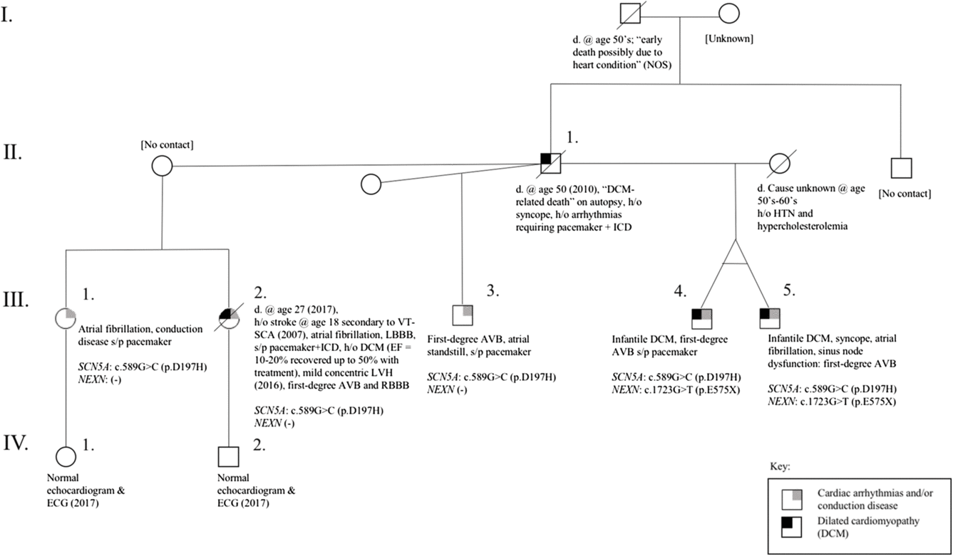
We completed a systematic chart review of the proband and all available family records, many of which are treated at our centre. This report was approved by the Indiana University Health Riley Children’s Hospital Institutional Review Board (IRB# 1811364611) with a waiver of consent. Clinical phenotypic disease was described by available data for each given family member and often included electrocardiogram, ambulatory electrocardiogram monitoring, exercise test, and echocardiogram with some individuals including pacemaker/implanted cardiac defibrillator data as well (refer to Fig 1 for the family pedigree). A phenotypic comparison of the clinical data was completed. Genotype data were available for four of the five offspring comprising all three parental pairs. No genotype data were available from the common father or grandparents.
Pedigree for the reported family. AVB = atrioventricular block; DCM = dilated cardiomyopathy; HTN = hypertension; ICD = implantable cardioverter difibrillator; LBBB = left bundle branch block; LVH = left ventricular hypertrophy; RBBB = right bundle branch block; SCA = sudden cardiac arrest; VT = ventricular tachycardia.

Cascade genetic testing was completed, and family segregation analysis of the variant was performed. The index affected male (Fig 1, individual III-4) in the family underwent next-generation sequencing of 58 genes associated with cardiomyopathies (see Supplementary material for the list of genes on this panel and relevant transcript identifier). All coding regions and splice sites were covered in this analysis. Testing was performed by the Clinical Laboratory Improvement Amendments-certified Indiana University Molecular Genetics Diagnostic Laboratory. Genomic DNA was processed by hybridisation-based target enrichment (Nextera library preparation technology and TruSight One Enrichment Kit, Illumina) followed by next-generation sequencing using sequencing by synthesis technology (Illumina MiSeq). Variant calls were generated using the Burrows-Wheeler Aligner followed by Genome Analysis Toolkit analysis. This test detects 100% of substitution variants (95% CI = 82−100) and 95% of small insertions and deletions (95% CI = 98.5−100). Sanger sequencing was used to provide data for bases with insufficient coverage (< 15× sequencing depth) and for bases in some known regions of genomic segmental duplications. All clinically significant and novel variants were confirmed by independent Sanger sequencing. Variants classified as likely benign or benign were not confirmed by Sanger sequencing. For familial cascade genetic testing, DNA was extracted from individuals’ buccal swab specimen and polymerase chain reaction amplified for relevant gene regions and then sequenced in forward and reverse directions. This provided a targeted analysis for the presence of the familial variants in other relatives.
Following genetic testing, a full literature and gene database review of all relevant areas was completed. Current interpretation and classification of genetic variants was based on guidelines from the American College of Medical Genetics and Genomics as well as a newer modified approach with more weight for family segregation studies. Reference Richards, Aziz and Bale12 , Reference Kelly, Caleshu and Morales13 A combined approach using the 2015 American College of Medical Genetics and Genomics guidelines and those of Kelly et al Reference Kelly, Caleshu and Morales13 provided the ability to better utilise information from the variant segregation testing and clinical phenotypic information completed and obtained in this study. While the approach by Kelly et al was used for assessing variants in a sarcomeric gene, MYH7, their method for weighting evidence for variant pathogenicity using family variant segregation can be applied to other genes of interest. Reference Kelly, Caleshu and Morales13 This addresses a limit of the American College of Medical Genetics and Genomics guidelines for variant interpretation, specifically, the lack of weighting variant classifications based on family variant segregation data. Criteria for pathogenicity specific to the family in this study were based on American College of Medical Genetics and Genomics strong, moderate, and supporting evidence categories including: PM2 (the variant is absent from controls in large databases), PP1 (the variant cosegregates with disease in multiple affected half-siblings with well-characterised phenotypes, and the strength of this evidence increased based on the half-siblingships), PP3 (concordance of multiple in silico variant interpretation tools with prediction of moderate effects on protein structure and function; software included: SIFT, PolyPhen2, PP2_HDIV, LRT, Mutation Assessor, and FATHMM), and PP4 (the patients’/family’s phenotype is consistent with a Mendelian genetic etiology). Criterion PP1 was modified using the approach of Kelly et al, Reference Kelly, Caleshu and Morales13 which weights evidence of pathogenicity more heavily with increasing numbers of affected relatives with the putative pathogenic variant of interest. The PP1 criterion can be converted to “PP1_Moderate” or “PP1_Strong” as variant segregations increase in affected relatives in a family.
Results
Clinical characteristics
A patient pedigree (Fig 1) was created following the clinical evaluation of three affected individuals, two monozygotic twins and a paternal half-brother, which lead to the evaluation of a paternal half-sister (four siblings with the same father and three different mothers) all of whom experienced varying degrees of atrial arrhythmias, conduction disease, and dilated cardiomyopathy in addition to a paternal history of unexplained sudden cardiac arrest in his 50s.
The initial presentation involved monozygotic (identical) twin males (Fig 1, individuals III-4 and III-5) originally born prematurely at 31 weeks estimated gestational age due to maternal pre-eclampsia in 1996. The twins’ genetic status was confirmed with zygosity testing several years prior to current ascertainment; the original testing records were obtained and verified. Despite birth-weights <1000 g, they developed well to discharge at 5 weeks of age with no described cardiovascular or arrhythmia concerns. They were discharged with an apnea monitor with a low heart rate set at 80 beats per minute. At home the twins had disparate trajectories, one thriving while the other had difficulty feeding and gaining weight. Following 5 weeks at home (10 weeks of age), the latter experienced a night of inconsolability and heart rates consistently bradycardic <80 beats per minute. He was brought to the local emergency department where he had a sinus tachycardia at 150 beats per minute and clinical evidence of severe dilated cardiomyopathy. An initial echocardiogram showed an ejection fraction of 15%. The larger twin was subsequently evaluated and shown to have evidence of mild dilation in the setting of intermittent second-degree AV block and left bundle branch block with heart rates in the 30s–40s. He underwent dual chamber pacemaker placement. No infectious or metabolic causes were identified, and both responded to anti-congestive medication, eventually discharged 1 month later with ejection fractions approximately 30% in the smaller and 55–60% with pacing in the larger.
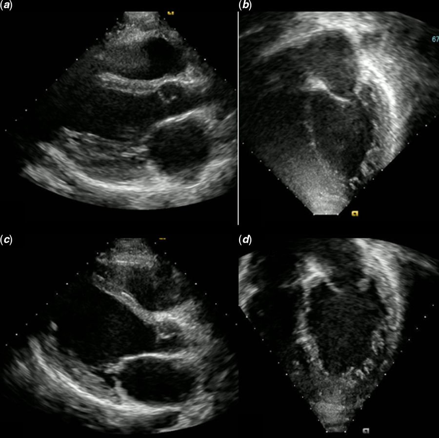
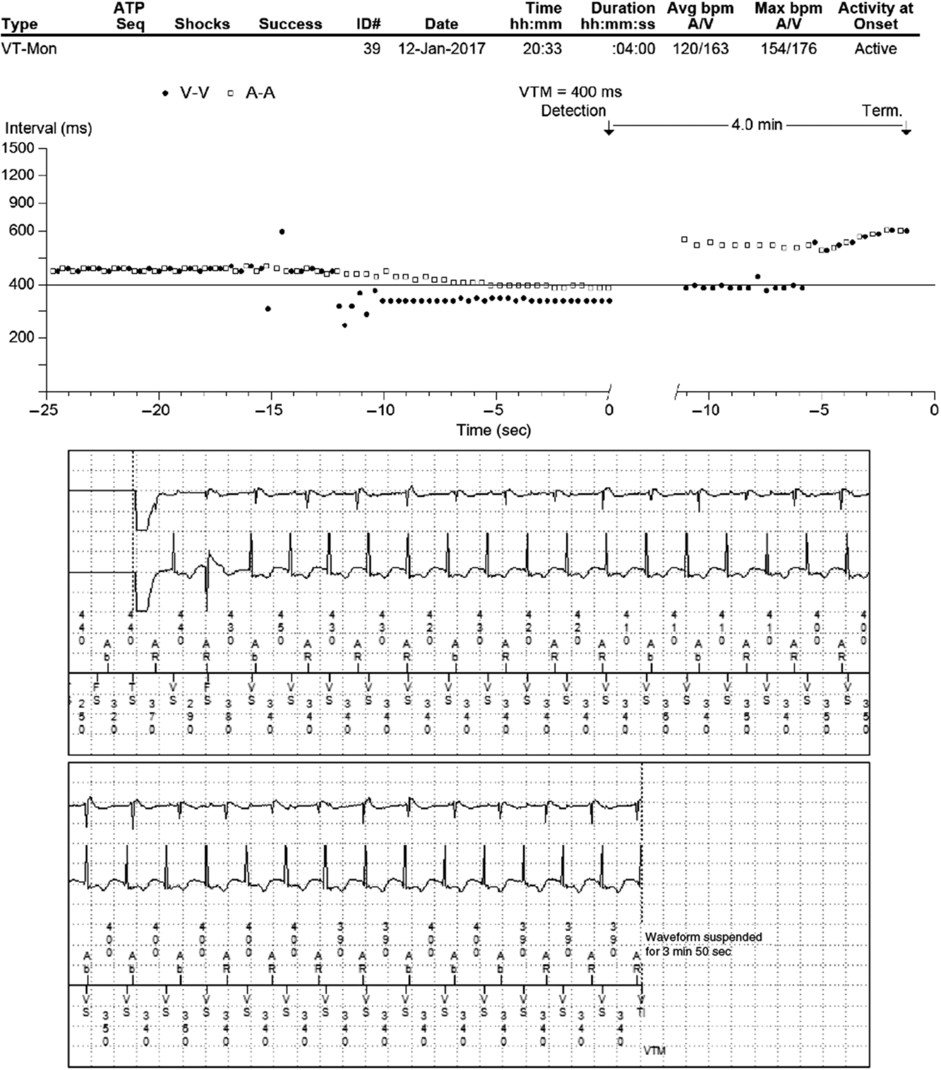
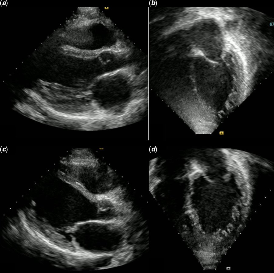
Despite repeat admissions during the winter in the smaller twin, by 9 months of age, systolic function was noted to be mildly depressed where it has been maintained with ejection fractions in the 50% range to 21 years (Fig 2). No significant atrial or ventricular arrhythmias were noted. The larger twin, in contrast, demonstrated only mildly diminished LV systolic function. As such, despite demonstrated periodic AV block, bradycardia, and progressive non-sustained atrial and ventricular tachycardia, his pacemaker was set to a backup rate only. At 19 years he underwent electrophysiology study, which demonstrated inducible sustained ventricular tachycardia with hemodynamic compromise. Pace-mapping resulted in successful radiofrequency ablation at the right ventricular apex though ventricular tachycardia was recurrent, and a dual chamber pacemaker/implantable cardiac defibrillator was placed. Spontaneous atrial activity and atrial capture with pacing has been variable. He had non-sustained runs of ventricular tachycardia on medication (Fig 3). At 20 years, he experienced a sustained ventricular tachycardia arrest off of medication, due to non-compliance, that failed implantable cardiac defibrillator cardioversion x6. He spontaneously reverted to sinus rhythm and recovered, which was followed by dual coil system upgrade.
Echo images of twin boys, individuals 4 and 5, at age 21 years. Images ( a ) and ( b ) are parasternal long and apical 4 chamber, respectively, of individual 4. Near-normalisation of size and function is present. Images ( c ) and ( d ) are parasternal long and apical 4 chamber, respectively, of individual 5. Near-normalisation of size and function is present.
Pacemaker/implanted cardiac defibrillator event interrogation demonstrating non-sustained ventricular tachycardia.

In this context, the twins’ mother noted that their father (Fig 1, individual II-1) was known to have a dilated cardiomyopathy by echocardiogram and had experienced unexplained syncope and palpitations. No specific cardiac diagnosis had been made prior to the family separation in 2001. The father was known to have had two daughters from a previous partner. The older sister (Fig 1, individual III-2) had progressive atrial and ventricular arrhythmias in the setting of dilated cardiomyopathy and underwent pacemaker/implantable cardiac defibrillator placement in 2007. A younger sister (Fig 1, individual III-1) has atrial arrhythmias, sinus node dysfunction, and intraventricular conduction delay. An additional son from a third partner was identified in February 2015 (Fig 1, individual III-3) due to an abnormal electrocardiogram consisting of sinus node dysfunction and a prolonged QRS duration consistent with non-specific intraventricular conduction delay.
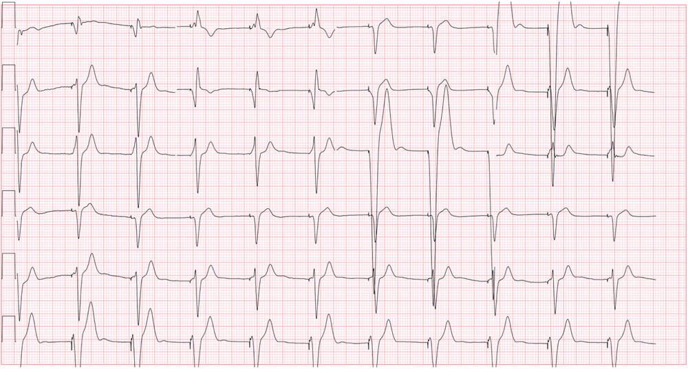
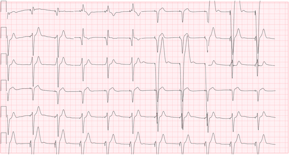
Genetic testing was initiated on the twins and younger half-brother in 2016 (Fig 1, individuals III-4, III-5, and III-3). The older half-sister was tested in 2017. Please refer to the testing results later in the article. Since these results, the oldest half-sister died from sudden cardiac arrest, not converted by her implantable cardioverter difibrillator. The youngest half-brother has a pacemaker due to symptomatic sinus node dysfunction and intraventricular conduction disease with recent onset of atrial tachycardia (Fig 4).
Resting 12-lead electrocardiogram of half-brother, individual 3 showing complete atrioventricular block with atrial standstill.
Molecular genetics
The genetic testing results demonstrated a missense variant in the SCN5A gene, SCN5A:NM_198056:exon5:c.589G>C (p. Asp197His, also known as p.D197H), in all affected siblings and half-siblings and a co-occurring non-sense variant in the NEXN gene, NEXN:NM_144573:exon13:c.1723G>T (p. Glu575*, also known as E575X), in the twin males with infant-onset cardiomyopathy. The SCN5A p. Asp197His variant was not detected in 202,186 control alleles from the gnomAD database (http://gnomad.broadinstitute.org) with sufficient coverage (50× for exome, 35× for genome), and it resides in the third transmembrane segment from the first transmembrane domain (DI-S3), indicating that this change does not directly affect the voltage sensor. Of note, a different variant affecting the same residue is reported in dbSNP (rs794728848), SCN5A:NM_198056.2:c.589G>T (p. Asp197Tyr). This latter variant may be defined as potentially deleterious, with no previous publication history or reference in other population databases. This SCN5A-Asp197Tyr variant is currently listed as likely pathogenic in the ClinVar database, though additional phenotypic information is needed for further characterisation of this variant (https://www.ncbi.nlm.nih.gov/clinvar/17242500/; accessed 27 September 2018). Notably, all affected relatives were tested for the NEXN variant, but only the monozygotic twins were found to have this variant in addition to the SCN5A variant.
Of interest, the neighbouring p. Thr187Ala Reference Makiyama, Akao and Tsuji14 , Reference Gui, Wang and Jones15 and the p. Trp193* Reference Kapplinger, Tester and Alders16 variants have been identified in Brugada syndrome subjects (associated with atrial fibrillation, conduction disease, and ventricular tachycardia). Reference Gui, Wang and Jones15 , Reference Francis and Antzelevitch17 – Reference Rodriguez-Manero, Namdar and Sarkozy19 While the p. Thr187Ala variant has evidence consistent with a likely pathogenic classification, the p. Trp193* variant does not currently have functional studies but is predicted to be deleterious.
With the extensive clinical phenotyping and family variant segregation across multiple half-siblings with overlapping disease, we classify the SCN5A p. Asp197His variant as likely pathogenic. This was based on meeting the American College of Medical Genetics and Genomics variant classification guideline categories of strong, moderate, and supporting criteria, including, PM2, PP1, PP3, and PP4 (see Methods section). When modifying the PP1 criterion using the approach of Kelly et al, Reference Kelly, Caleshu and Morales13 there are five meiotic segregations of the SCN5A variant in similarly affected relatives in the family, and this is considered moderate evidence of pathogenicity, converting PP1 to “PP1_Moderate” in this case. With a second moderate criterion, the American College of Medical Genetics and Genomics guidelines support upgrading the classification to likely pathogenic.
The NEXN variant p. Glu575* was observed in heterozygosity in one Non-Finnish European subject from the gnomAD database (http://gnomad.broadinstitute.org/variant/1-78408209-G-T; accessed September 2018). No truncating variant has unequivocally been demonstrated to be deleterious in heterozygosity in NEXN. The presence in gnomAD of radical variants throughout the gene suggests that the heterozygous loss of function of NEXN may not be sufficient to cause cardiomyopathy. This variant, present in the last exon, most likely skips nonsense mediated decay and leads to a truncated protein maintaining the most critical actin binding domains, and thus, at least partially, the main biological function. Reference Hassel, Dahme and Erdmann20 – Reference Aherrahrou, Schiossarek and Stoelting22 The p. Glu575* variant has been reported in one case of left ventricular non-compaction, Reference Pardun, Wenzel, Kramer, Berger, Geruli and Klaassen23 though given the presence of the variant in gnomAD, likely escape from nonsense mediated decay, and absence of ventricular non-compaction in the index cases in this study, this would be considered a variant of unclear significance at present.
Discussion
We have presented clinical and molecular genetic findings consistent with the SCN5A variant p. Asp197His as the most probable cause in a family with autosomal dominant dilated cardiomyopathy, atrial and ventricular arrhythmias, conduction disease, and sudden death. The primary findings indicate that p. Asp197His segregates with neonatal dilated cardiomyopathy of varying penetrance followed by progressive atrial, AV node, and His/Purkinje disease followed by later onset dilated cardiomyopathy and life-threatening ventricular tachycardia as early as the second decade of life. A second variant in NEXN may be contributory though segregation analysis could not confirm this finding in this family. To our knowledge, evidence (clinical or otherwise) supporting pathogenic effects of this variant has not yet been reported.
Based on the 2015 American College of Medical Genetics and Genomics variant interpretation guidelines, the SCN5A variant would meet criteria PM2, PP1, PP3, and PP4. Reference Richards, Aziz and Bale12 As such, based on these guidelines alone, the variant would be classified as a variant of unclear significance. However, there are reasonable critiques of the 2015 American College of Medical Genetics and Genomics variant classification guidelines, particularly when considering family segregation data. Reference Kelly, Caleshu and Morales13 , Reference Amendola, Jarvik and Leo24 – Reference Tavtigian, Greenblatt and Harrison26
One significant criticism is that the 2015 guidelines did not address the categorisation and weighting of evidence for variant interpretation, leading many laboratories to complement these guidelines with internal methods. Reference Amendola, Jarvik and Leo24 It is also important to note that these guidelines are not considered clinical standards, and that each assessed variant may have varying classification based on a team’s expert judgement and when evaluating the full body of evidence. Reference Richards, Aziz and Bale12
As described, the PP1 criterion converts to “PP1_Moderate” when ≥5 meioses are observed in a family (Logarithm of Odds 1.51). Reference Kelly, Caleshu and Morales13 Kelly et al Reference Kelly, Caleshu and Morales13 provide a quantitative approach to define family variant segregation thresholds not defined by the 2015 American College of Medical Genetics and Genomics guidelines.
In this family, there are five affected individuals all with the same SCN5A (p. Asp197His) variant, all of whom having the same/overlapping phenotypes consistent with previous reports of the clinical and cardiac disease heterogeneity of SCN5A-related disorders.
The clinical team agreed that PP1_Moderate was met. This was based on the family phenotype concordance and on pedigree analysis, and a reasonable conclusion can be made that the father of all affected cases in this family with dilated cardiomyopathy, arrhythmias, and sudden cardiac death has the SCN5A (p. Asp197His) variant as well (see Individual II-1 in Fig 1). Since this individual died with no available testable post-mortem samples before the family was ascertained, we were unable to confirm his genetic testing. Pedigree analysis combined with family variant segregation testing shows that there were at least five informative meiosis with the SCN5A variant, meeting the PP1_Moderate criterion. Reference Kelly, Caleshu and Morales13 Using the approach of Kelly et al, Reference Kelly, Caleshu and Morales13 the SCN5A variant here would meet two moderate criteria of PM2 and PP1_Moderate, as well as PP3 and PP4, meeting (v) “Likely pathogenic” as modified from the 2015 American College of Medical Genetics and Genomics guidelines.
A possible polygenic interaction between the SCN5A p. Asp197His variant and the NEXN p. Glu575* variant leading to variable penetrance and expression of cardiomyopathy in this family cannot be excluded, and additional functional studies to assess this are needed. Based on the clinical phenotypic and family segregation data, it is likely that the SCN5A p. Asp197His variant is driving the primary electrical disease in this family, with or without dilated cardiomyopathy. A polygenic influence (ie, the background genetic variation in individuals) leading to variable expression of overt cardiomyopathy with arrhythmias cannot be discounted and would require further studies. Reference Belkaya, Kontorovich and Byun27 – Reference Burkett and Hershberger30
Other genes have been well described to have significant concomitant dilated cardiomyopathy and rhythm disturbance, either or both of which can lead to significant clinical disease, sudden death, or the need for heart transplantation. Arguably the best described to date is lamin A/C defects. Reference Hasselberg, Haland and Saberniak31 There have been previous reports of SCN5A variants associated with atrial standstill. Reference Lehmann, Meltendorf and Klein32 , Reference Tan, Gando, Bu, Cecchin and Coetzee33 Interestingly, a relatively close c.664C>G (p. Arg222Gly) was reported in a family with atrial standstill without cardiomyopathy or sudden death. Reference Lehmann, Meltendorf and Klein32 Further investigation using well-designed in vitro studies are needed to determine how disruption of the SCN5A protein in this region may contribute to atrial standstill as well as cardiomyopathic and arrhythmic phenotypes in this family.
Conclusion
The SCN5A variant p. Asp197His, currently classified as a variant of unknown significance, may reasonably be re-classified as a likely pathogenic variant based on the segregation analysis of our family of interest. Despite the lack of molecular mechanism data, current evidence of pathogenicity seems sufficient based on variant interpretation guidelines and modified approaches. The associated clinical problems include cardiomyopathy, atrial brady-arrhythmias, atrioventricular nodal disease, ventricular tachy-arrhythmias, and sudden cardiac death.
Molecular mechanism studies are indicated to further describe these phenotypes and the possible role of the SCN5A variant as a Mendelian disease mechanism as well as a non-Mendelian contributor to complex cardiac disease. This family highlights an issue with genetic variant classification: interpretations are made on best available evidence at the clinical, molecular, individual, and family/population levels. Current guidelines leave room for some subjectivity due to underlying limitations of variant classification thresholds. Interdisciplinary approaches to variant classification are important to familial cardiac diseases and the need for careful individual and family phenotyping via clinical evaluation, detailed records review, and strategic use of genetic testing cannot be overemphasised. Certainly, judicious approaches to variant interpretation using multiple lines of evidence and best practices remain critical.
Supplementary material
To view supplementary material for this article, please visit https://doi.org/10.1017/S1047951119001860
Acknowledgements
None.
Financial support
This research received no specific grant from any funding agency, commercial or not-for-profit sectors.
Conflicts of interest
Adam C. Kean – none; Benjamin M. Helm – none; Matteo Vatta – primarily employed by Invitae Corporation; Mark Ayers – none; John J. Parent – none; Robert Darragh – none.
Ethical standard
The authors assert that all procedures contributing to this work comply with the ethical standards of the relevant United States guidelines on human experimentation and with the Helsinki Declaration of 1975, as revised in 2008, and the work has been approved by the Indiana University Health Riley Children’s Hospital Institutional Review Board (IRB-1811364611) with a waiver of consent.