Introduction
Malaria is an ancient disease with the earliest malaria reports dating back to more than 4000 years ago. According to the recent World Health Organisation (WHO) statistics (WHO, 2015), 91 countries reported ongoing malaria transmission with Africa experiencing disproportionately high malaria cases (90% of the total) and alone accounting for 92% of worldwide malaria deaths. The causative agent is an obligate intracellular, apicomplexan parasite called Plasmodium. Of the five human-infecting Plasmodium species (P. ovale, P. malariae, P. knowlesi, P. falciparum and P. vivax), P. falciparum and P. vivax contribute to the major burden of malaria in the world. Plasmodium falciparum is considered the deadliest species as it often leads to complicated malaria with clinical manifestations such as anaemia, renal failure, acidosis, cerebral malaria and multi-organ failure (Church et al., Reference Church, Le, Bryan, Gordon, Edelman, Fries, Davis, Herrington, Clyde, Shmuklarsky, Schneider, Mcgovern, Chulay, Ballou and Hoffman1997). In addition, there has been an emergence of multi-drug-resistant P. falciparum strains in South East Asia, Africa and some Central American provinces. Plasmodium vivax has recently received attention due to increasing severe and complicated cases causing mortality. Also, there is an emergence of drug resistance against P. vivax in endemic countries such as Papua New Guinea, Greece, Thailand, India and Indonesia (Klemola, Reference Klemola1988; Gething et al., Reference Gething, Elyazar, Moyes, Smith, Battle, Guerra, Patil, Tatem, Howes, Myers, George, Horby, Wertheim, Price, Mueller, Baird and Hay2012; Goncalves et al., Reference Goncalves, Cravo and Ferreira2014; Waheed et al., Reference Waheed, Ghanchi, Rehman, Raza, Mahmood and Beg2015). Although the mortality associated with P. vivax is lower than P. falciparum, its geographical reach is more diverse with about 2.9 billion people worldwide at a risk of P. vivax malaria (Bousema and Drakeley, Reference Bousema and Drakeley2011).
The malaria parasite requires two hosts to complete its life cycle; the primary host or the definitive host is the female Anopheles mosquito and the secondary host is a vertebrate (Bannister and Mitchell, Reference Bannister and Mitchell2003). A very small fraction of the cycling asexual parasites converts to the sexual stage called gametocytes. These are responsible for parasite transmission from human host to the mosquito. This phase of the life cycle provides the parasite with adequate possibilities for genetic recombination and clonal selection, under evolutionary as well as drug pressure, thereby acting as sentinels spreading drug resistance (Sinden, Reference Sinden1991). Recent epidemiological studies have emphasised the need for screening individuals for both gametocytaemia along with asexual parasitaemia. Asymptomatic populations in endemic areas often show high gametocytaemia and thus represent a potential reservoir of the parasite (Gouagna et al., Reference Gouagna, Ferguson, Okech, Killeen, Kabiru, Beier, Githure and Yan2004; Okell et al., Reference Okell, Bousema, Griffin, Ouedraogo, Ghani and Drakeley2012; Sumari et al., Reference Sumari, Mwingira, Selemani, Mugasa, Mugittu and Gwakisa2017). Gametocytes represent a rational target for transmission blocking antimalarial interventions. In spite of the central role of gametocytes in the parasite's life cycle, very little is known about their biology. Only recently we have begun to understand the physiological triggers and transcription factors important for differentiation from asexual to sexual stages. The study of gametocytes has revealed malaria diagnostic loopholes, the drawback of current frontline antimalarials and corresponding clinical challenges faced today.
Life cycle of Plasmodium: two hosts; completely different environmental niches
The life cycle of Plasmodium sp. (Josling and Llinas, Reference Josling and Llinas2015) is very complicated and intriguing; the parasite cycles between four very different niches – the salivary gland of the mosquito, liver cells in humans, red blood cells (RBCs) in humans and the mosquito midgut. During a blood meal, the mosquito salivary gland harbouring the parasite injects it into a healthy human host. These sporozoites now enter the liver cells and undergo several rounds of division. At this stage, there are no clinical symptoms. For P. vivax and P. ovale a quiescent stage called hypnozoites is responsible for relapse of infection weeks, or even years after the initial injection. Thousands of merozoites, each of which invades RBCs, are released from the liver. The parasite undergoes its asexual cycle within the RBCs. It progresses from a ring-shaped structure to the more metabolically active trophozoites and further into schizonts. On maturation, the schizonts burst and release 16–32 new merozoites, each of which enters new RBCs. This phase marks the symptomatic phase of malaria infection, which includes the characteristic recurrent febrile episodes.
A fraction of asexual parasites enters the sexual cycle to form male (microgametocytes) and female (macrogametocytes) gametocytes. The fact that both the male and female gametocytes can be generated from a single haploid clone, which indicates that sex is not determined by sex chromosomes. The mature gametocytes are present in the bloodstream of the parasitised human host and are picked up by the mosquito. In the mosquito midgut, a decrease in temperature, increase in pH and presence of a metabolite called xanthurenic acid induces the maturation of gametocytes into gametes (Billker et al., Reference Billker, Lindo, Panico, Etienne, Paxton, Dell, Rogers, Sinden and Morris1998; Arai et al., Reference Arai, Billker, Morris, Panico, Delcroix, Dixon, Ley and Sinden2001). The microgametocytes undergo three quick rounds of mitosis to produce eight male gametes in a process called exflagellation, while one macrogametocyte matures to form one female gamete. The male and female gametes fuse to form the diploid zygote, which further develops into the ookinete. The ookinetes are motile and travel to the midgut epithelial cells, where they develop to form the oocysts. These oocysts burst to release thousands of sporozoites, which travel to the salivary glands of the mosquito. In its next blood meal, the infected female Anopheles mosquito injects sporozoites into a healthy human and the cycle continues.
Zygote and ookinete are the only diploid stages of the parasite. At this stage, meiosis occurs and provides the parasite a chance for recombination (Sinden, Reference Sinden1991). It has been shown that a cross between two clones of P. falciparum differing in enzymes, drug sensitivity and chromosome patterns show recombination in high frequency (Walliker et al., Reference Walliker, Quakyi, Wellems, Mccutchan, Szarfman, London, Corcoran, Burkot and Carter1987). Clinically, this represents an important scenario; genetically different parasites infecting the same patient can undergo recombination events in the mosquito stage, leading to the development of parasites, which are genetically armed to face a myriad of stresses and unfavourable conditions. Several studies have shown that in natural populations of P. falciparum and P. vivax, high rates of meiotic recombination occur (Conway et al., Reference Conway, Roper, Oduola, Arnot, Kremsner, Grobusch, Curtis and Greenwood1999; Mu et al., Reference Mu, Awadalla, Duan, Mcgee, Joy, Mcvean and Su2005; Bright et al., Reference Bright, Manary, Tewhey, Arango, Wang, Schork, Yanow and Winzeler2014; Miles et al., Reference Miles, Iqbal, Vauterin, Pearson, Campino, Theron, Gould, Mead, Drury, O'brien, Ruano Rubio, Macinnis, Mwangi, Samarakoon, Ranford-Cartwright, Ferdig, Hayton, Su, Wellems, Rayner, Mcvean and Kwiatkowski2016). Meiosis, along with a number of mitotic cell divisions occurring throughout the life cycle of the parasite, provides a chance for the parasite to develop drug-resistant mutations.
Gametocyte biology and development
Plasmodium falciparum exhibits a unique set of stages during gametocyte development that are markedly different in shape, location and protein composition from the asexual stages (Josling and Llinas, Reference Josling and Llinas2015). The development of gametocytes in P. falciparum occurs over 10–12 days and is morphologically divided into five stages. Stage I resembles asexual stages. From stage II onwards, the parasite elongates, which is facilitated by the presence of subpellicular microtubules and an inner membrane complex (IMC) (Dearnley et al., Reference Dearnley, Yeoman, Hanssen, Kenny, Turnbull, Whitchurch, Tilley and Dixon2012; Kono et al., Reference Kono, Herrmann, Loughran, Cabrera, Engelberg, Lehmann, Sinha, Prinz, Ruch, Heussler, Spielmann, Parkinson and Gilberger2012). The IMC is a unifying feature in all Alveolates and is necessary for motility. However, in P. falciparum it is also known to play a vital role in the morphogenesis of gametocytes from trophozoite-like features in stage I to a crescent-shaped mature gametocyte in stage V. Stage II onwards, the parasite begins to elongate, acquiring a characteristic D-shape in stage III. Stage IV parasites have pointed ends, while stage V gametocytes acquire the characteristic crescent-shape. Immature gametocytes are sequestered in the bone marrow and re-enter circulation as mature stage V gametocytes, which are picked up by the mosquito blood meal and undergo further development (Aguilar et al., Reference Aguilar, Magallon-Tejada, Achtman, Moraleda, Joice, Cistero, Li, Suen, Nhabomba, Macete, Mueller, Marti, Alonso, Menendez, Schofield and Mayor2014; Joice et al., Reference Joice, Nilsson, Montgomery, Dankwa, Egan, Morahan, Seydel, Bertuccini, Alano, Williamson, Duraisingh, Taylor, Milner and Marti2014). A long-standing dilemma in gametocyte biology is the point of commitment to the sexual stage. It is well known that stages II–IV of gametocytes are completely absent from the bloodstream and are largely sequestered in the bone marrow, but the paucity of early gametocyte markers has restricted information regarding the point of commitment. Studies have shown that P. falciparum gametocyte formation is enhanced in reticulocyte-rich blood and in the presence of erythroid progenitors (Trager et al., Reference Trager, Gill, Lawrence and Nagel1999). In 2014, a study demonstrated that extravascular spaces rich in erythroid precursor cells in the human bone marrow favour gametocyte formation and development. Using an ex vivo erythropoiesis system and gametocyte favouring conditions, it was observed that P. falciparum schizonts could undergo sexual commitment in erythroid progenitor cells (Trager et al., Reference Trager, Gill, Lawrence and Nagel1999; Peatey et al., Reference Peatey, Watson, Trenholme, Brown, Nielson, Guenther, Timmins, Watson and Gardiner2013; Joice et al., Reference Joice, Nilsson, Montgomery, Dankwa, Egan, Morahan, Seydel, Bertuccini, Alano, Williamson, Duraisingh, Taylor, Milner and Marti2014). In another study, it was observed that in vitro P. falciparum growth leads to a decrease in the level of lysophosphatidylcholine (LPC) in the supernatant and that it is the major constituent of the sexual-stage triggering conditioned medium. The concentration of LPC was inversely related to the extent of gametocytogenesis. Further, the LPC level was lower in bone marrow than in the serum of mice (Brancucci et al., Reference Brancucci, Gerdt, Wang, de Niz, Philip, Adapa, Zhang, Hitz, Niederwieser, Boltryk, Laffitte, Clark, Gruring, Ravel, Blancke Soares, Demas, Bopp, Rubio-Ruiz, Conejo-Garcia, Wirth, Gendaszewska-Darmach, Duraisingh, Adams, Voss, Waters, Jiang, Clardy and Marti2017). These observations suggest that commitment occurs in the bone marrow, which is a rich source of erythroid progenitor cells and has reduced LPC concentration. On the contrary, a recent study has provided evidence for the presence of committed rings in the blood circulation and suggested that commitment perhaps occurs in the bloodstream (Farid et al., Reference Farid, Dixon, Tilley and Mccarthy2017). Thus, currently, there is a lack of consensus on the microenvironment that favours commitment of the parasite to the sexual stage development.
Recent studies have demonstrated that infected RBCs with mature gametocytes are highly deformable compared with immature gametocytes. This has been shown to be essential for preventing splenic clearance of mature gametocytes from the blood stream. Cyclic adenosine monophosphate (cAMP) signalling has been shown to be important in modulating the deformability of infected RBCs. Pharmacological agents interfering with this mechanical change in infected RBCs have been suggested as novel therapeutics (Ramdani et al., Reference Ramdani, Naissant, Thompson, Breil, Lorthiois, Dupuy, Cummings, Duffier, Corbett, Mercereau-Puijalon, Vernick, Taramelli, Baker, Langsley and Lavazec2015). Other studies have shown that xanthurenic acid in the mosquito midgut caused a dramatic increase in cytosolic calcium levels in the gametocyte via the calcium-dependent protein kinase 4 (Billker et al., Reference Billker, Dechamps, Tewari, Wenig, Franke-Fayard and Brinkmann2004). An inhibitor of cyclic guanosine monophosphate (cGMP) phosphodiesterase was capable of inducing exflagellation even in the absence of xanthurenic acid (McRobert et al., Reference Mcrobert, Taylor, Deng, Fivelman, Cummings, Polley, Billker and Baker2008). These observations have enhanced our understanding of the cAMP/cGMP-mediated signalling pathways operating in gametocytes and have proposed novel therapeutics capable of breaking the cycle of transmission. Some studies have hinted at the possibility that the key to the success of P. falciparum lies in a delay in its release of gametocytes. This enables them to avoid the febrile period, which is marked by the release of host immune factors that are known to disrupt transmission in other Plasmodium species (Naotunne et al., Reference Naotunne, Karunaweera, Del Giudice, Kularatne, Grau, Carter and Mendis1991; Drakeley et al., Reference Drakeley, Sutherland, Bousema, Sauerwein and Targett2006). The sequestration time of immature gametocytes is 12 days and circulation time estimated to be up to 22 days; thus, from the time of production, gametocytes can be present in the patient for up to 6 weeks. This provides enough time for transmission of the parasite from the host to the mosquito.
Sexual dimorphism is a hallmark of Plasmodium gametocytogenesis. From stage III, male microgametocytes and female macrogametocytes show ultrastructural differences. The female gametocyte shows rapid development of organelles such as mitochondria, endoplasmic reticulum, Golgi body and ribosomes. Microscopically, sexual dimorphism becomes discernible from stage IV onwards. Female gametocytes exhibit a concentrated nucleus and nucleolus and appear as violet cells when stained with Giemsa. Male gametocytes, on the other hand, exhibit a larger and more diffused nucleus with no nucleolus and appear pink when stained with Giemsa. Female gametocytes also exhibit the characteristic Golgi-derived small and ovoid structures called the osmiophilic bodies (de Koning-Ward et al., Reference de Koning-Ward, Olivieri, Bertuccini, Hood, Silvestrini, Charvalias, Berzosa Diaz, Camarda, Mcelwain, Papenfuss, Healer, Baldassarri, Crabb, Alano and Ranford-Cartwright2008). One male gametocyte leads to the development of eight male gametes, but one female gametocyte develops into one female gamete. Thus, the ratio of gametocytes is usually female biased (Guinet et al., Reference Guinet, Dvorak, Fujioka, Keister, Muratova, Kaslow, Aikawa, Vaidya and Wellems1996). In some cases, like increased asexual parasitaemia or low density of gametocytes, the sex ratio may be skewed towards male gametocytes (Schall, Reference Schall1989; Gbotosho et al., Reference Gbotosho, Sowunmi, Happi and Okuboyejo2011). The regulatory factors affecting this ratio remain unclear.
Gametocytes in P. vivax are large and oval or round and cover the whole area of the host RBC. Due to the absence of an in vitro P. vivax culturing system, most of our knowledge relies on clinical data. In most P. vivax cases, cytoadherence is not seen and gametocytes are detected from 3 days post-appearance of asexual parasites (Vallejo et al., Reference Vallejo, Garcia, Amado-Garavito, Arevalo-Herrera and Herrera2016). The other events of sexual development and sporogony are common between P. falciparum and P. vivax with the exception that P. vivax sexual stages can survive at 14.5°C as compared with 16°C for P. falciparum; thus, giving them a broader geographical presence (Guerra et al., Reference Guerra, Snow and Hay2006).
Epidemiology of Gametocytes
In the past two decades, The Roll Back Malaria programme has made global efforts to eradicate malaria and has had considerable success in eliminating malaria in 17 countries between 2010 and 2015. The combined use of artemisinin-based combination therapy with insecticide-treated nets has been at the heart of malaria control in endemic countries. However, there has been a surge in reports describing the emergence of submicroscopic and artemisinin-resistant malaria (Lindblade et al., Reference Lindblade, Steinhardt, Samuels, Kachur and Slutsker2013; Bousema et al., Reference Bousema, Okell, Felger and Drakeley2014; Chen et al., Reference Chen, Clarke, Gosling, Hamainza, Killeen, Magill, O'meara, Price and Riley2016). This is especially relevant in low endemic settings and areas of malaria pre-elimination or elimination phase, where submicroscopic carriers from 20 to 50% of the transmission pool (Okell et al., Reference Okell, Ghani, Lyons and Drakeley2009, Reference Okell, Bousema, Griffin, Ouedraogo, Ghani and Drakeley2012). Studies have indicated that gametocytes are present in most asymptomatic P. falciparum-infected individuals (Zhou et al., Reference Zhou, Mitchell, Kariuki, Odero, Otieno, Otieno, Onyona, Were, Wiegand, Gimnig, Walker, Desai and Shi2016; Rovira-Vallbona et al., Reference Rovira-Vallbona, Contreras-Mancilla, Ramirez, Guzman-Guzman, Carrasco-Escobar, Llanos-Cuentas, Vinetz, Gamboa and Rosanas-Urgell2017). Thus, these patients represent a potentially dangerous hidden parasite reservoir. From the limited data available on the relationship between gametocyte densities and mosquito infectivity, a log-sigmoid relationship between the two has been proposed (Jeffery and Eyles, Reference Jeffery and Eyles1955; Stepniewska et al., Reference Stepniewska, Price, Sutherland, Drakeley, von Seidlein, Nosten and White2008). However, host factors like humoral immunity and leucocyte counts are also known to play a role in determining mosquito infectivity (Stepniewska et al., Reference Stepniewska, Price, Sutherland, Drakeley, von Seidlein, Nosten and White2008).
Detection of gametocytes, like erythrocytic stages, is often performed by classical microscopy. The density of gametocytes in circulation is often close to the threshold of microscopy sensitivity and thus, densities as high as 10 gametocytes μl−1 can be missed (Schoone et al., Reference Schoone, Oskam, Kroon, Schallig and Omar2000). This problem becomes accentuated for detection of gametocytes of species other than P. falciparum in which the shape and characteristics of gametocytes often resemble asexual stages. Sensitive techniques such as real-time PCR (q-PCR), quantitative nucleic acid sequence-based amplification (QT-NASBA) and reverse transcription loop-mediated isothermal amplification (RT-LAMP) have sensitivities ranging from 0.02 to 10 gametocytes μl−1 of blood (Mens et al., Reference Mens, Spieker, Omar, Heijnen, Schallig and Kager2007; Rantala et al., Reference Rantala, Taylor, Trottman, Luntamo, Mbewe, Maleta, Kulmala, Ashorn and Meshnick2010; Wangai et al., Reference Wangai, Karau, Njiruh, Sabah, Kimani, Magoma and Kiambo2011; Pett et al., Reference Pett, Goncalves, Dicko, Nebie, Tiono, Lanke, Bradley, Chen, Diawara, Mahamar, Soumare, Traore, Baber, Sirima, Sauerwein, Brown, Gosling, Felger, Drakeley and Bousema2016; Roth et al., Reference Roth, Korevaar, Leeflang and Mens2016). Large cohort studies that have calculated gametocyte densities by both microscopy and molecular tools have exposed the pitfalls of using microscopy for epidemiological studies; in both symptomatic and asymptomatic as well as low and high endemic settings, gametocyte detection was 3–10-fold higher when more sensitive molecular tools were employed (Mawili-Mboumba et al., Reference Mawili-Mboumba, Nikiema, Bouyou-Akotet, Bahamontes-Rosa, Traore and Kombila2013; Zhou et al., Reference Zhou, Mitchell, Kariuki, Odero, Otieno, Otieno, Onyona, Were, Wiegand, Gimnig, Walker, Desai and Shi2016; Rovira-Vallbona et al., Reference Rovira-Vallbona, Contreras-Mancilla, Ramirez, Guzman-Guzman, Carrasco-Escobar, Llanos-Cuentas, Vinetz, Gamboa and Rosanas-Urgell2017). Thus, for an accurate and quantitative measure of gametocytes, molecular tools specific for gametocytes are essential.
The prevalence of gametocytes is higher in children, but gametocyte densities can increase with age (Coalson et al., Reference Coalson, Walldorf, Cohee, Ismail, Mathanga, Cordy, Marti, Taylor, Seydel, Laufer and Wilson2016). Interestingly, differences in gametocytogenesis occur in low and high transmission settings; gametocyte density is elevated in higher transmission areas, while the commitment to sexual forms is higher in low transmission settings – presumably to ensure maximal transmission from a rare host (Drakeley et al., Reference Drakeley, Akim, Sauerwein, Greenwood and Targett2000; Shekalaghe et al., Reference Shekalaghe, Bousema, Kunei, Lushino, Masokoto, Wolters, Mwakalinga, Mosha, Sauerwein and Drakeley2007; Ouedraogo et al., Reference Ouedraogo, De Vlas, Nebie, Ilboudo-Sanogo, Bousema, Ouattara, Verhave, Cuzin-Ouattara and Sauerwein2008). Several malaria eradication programmes have suffered a setback due to increased gametocytogenesis. Striking examples are the Garki project and the malaria control programme in New Guinea. Though cases of asexual parasitaemia initially dropped, there were increased cases of malaria due to increase in gametocytogenesis and subsequent transmission of P. falciparum (Metselaar, Reference Metselaar1960; Molineaux et al., Reference Molineaux, Storey, Cohen and Thomas1980).
Epidemiological study of P. vivax gametocytes is mainly focused on identification of the dormant hypnozoite stages that can be present in the liver of the host for a year and cause multiple relapses (Hulden, Reference Hulden2011). The rate of gametocyte production is faster in P. vivax, but their longevity in circulation is only 3 days (Moreira et al., Reference Moreira, Abo-Shehada, Price and Drakeley2015).
Gametocytes and drug resistance
Liver and sexual stages represent two bottlenecks in the lifecycle of the malaria parasite. Initially, only 100 sporozoites enter the host via the mosquito bite and thus drug treatment at this stage could eliminate the few parasites and the dormant hypnozoites in P. vivax and P. ovale infections that are responsible for relapses. The second bottleneck is the sexual stage as gametocyte numbers are low (<1% of total parasites). This results in fewer gametes and ookinetes that form oocysts that then undergo mitosis to form thousands of sporozoites. Thus, the fewer number of gametocytes compared with other stages and its critical link between the host and the vector provide a scientific rationale for targeting sexual stages of Plasmodium.
As discussed above, stage V gametocytes are present in the peripheral blood and are the only stage of the parasite capable of further transmission. These mature gametocytes are almost completely refractory to all known classes of antimalarials with the exception of primaquine (Peatey et al., Reference Peatey, Skinner-Adams, Dixon, Mccarthy, Gardiner and Trenholme2009). However, the use of primaquine is limited due to its haemolytic action in glucose-6-phosphate dehydrogenase-deficient individuals who are higher in number in malaria-endemic countries (Ganesan et al., Reference Ganesan, Chaurasiya, Sahu, Walker and Tekwani2012). Artemisinin-combination therapy has a potential to clear asexual as well as sexual forms; however, its gametocytocidal activity is restricted to the early and immature stages of gametocytes (Okell et al., Reference Okell, Drakeley, Ghani, Bousema and Sutherland2008). In fact, most frontline antimalarials are triggers for enhanced gametocytogenesis in vitro as well as in vivo (Puta and Manyando, Reference Puta and Manyando1997; Buckling et al., Reference Buckling, Ranford-Cartwright, Miles and Read1999; Peatey et al., Reference Peatey, Skinner-Adams, Dixon, Mccarthy, Gardiner and Trenholme2009). One study showed that male gametocytes are more susceptible to antimalarials than female gametocytes and this warrants the use of sex-specific assays for gametocyte sensitivity (Delves et al., Reference Delves, Ruecker, Straschil, Lelievre, Marques, Lopez-Barragan, Herreros and Sinden2013). Various clinical studies have pointed to the fact that gametocyte density is higher in drug-resistant P. falciparum patients compared with the drug-sensitive individuals (Handunnetti et al., Reference Handunnetti, Gunewardena, Pathirana, Ekanayake, Weerasinghe and Mendis1996; von Seidlein et al., Reference von Seidlein, Jawara, Coleman, Doherty, Walraven and Targett2001). One study found that antimalarials such as chloroquine (CQ) and sulphadoxine (SP) do not reduce gametocyte carriage (Hallett et al., Reference Hallett, Dunyo, Ord, Jawara, Pinder, Randall, Alloueche, Walraven, Targett, Alexander and Sutherland2006). On the contrary, a CQ/SP regimen leads to increased transmission of drug-resistant parasites. Interestingly, mutations in the Pfcrt and Pfmdr1 genes that are associated with chloroquine resistance leads to elevated gametocyte density and transmission after CQ or CQ/SP therapy. Thus, it is the higher potential of transmission that drives the spread of drug resistance in these parasites (Hallett et al., Reference Hallett, Dunyo, Ord, Jawara, Pinder, Randall, Alloueche, Walraven, Targett, Alexander and Sutherland2006).
There is strong clinical evidence that gametocyte rates can serve as an early warning of emerging drug resistance and gametocyte occurrence precedes changes in parasite clearance and a concomitant fall in cure rates (Greenwood, Reference Greenwood2008). A study in Kenya highlands identified increased gametocytogenesis as an indicator of an impending malaria epidemic (John et al., Reference John, Mchugh, Moormann, Sumba and Ofulla2005).
In clinical P. vivax malaria, gametocyte-specific markers have been detected as early as 3–7 days after infection (Moreira et al., Reference Moreira, Abo-Shehada, Price and Drakeley2015; Vallejo et al., Reference Vallejo, Garcia, Amado-Garavito, Arevalo-Herrera and Herrera2016). Fortunately, frontline antimalarial drugs show the efficient killing of P. vivax gametocytes (Pukrittayakamee et al., Reference Pukrittayakamee, Imwong, Singhasivanon, Stepniewska, Day and White2008).
Triggers of gametocytogenesis
During a P. falciparum infection, <1% of parasites commit to gametocytes (Eichner et al., Reference Eichner, Diebner, Molineaux, Collins, Jeffery and Dietz2001). The decision to commit to the sexual stage occurs during the asexual stage and all the merozoite progeny released from a schizont commit to form either male or female gametocytes (Smith et al., Reference Smith, Lourenco, Carter, Walliker and Ranford-Cartwright2000).
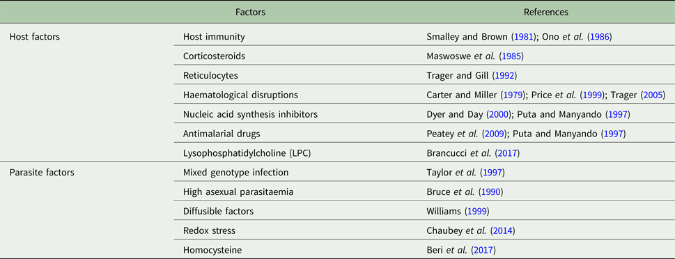
In P. falciparum, enhanced gametocyte counts have been achieved in vitro using a variety of molecules (Table 1). The process of gametocytogenesis has been linked to environmental stress. Factors that mimic stressful conditions have been used for in vitro production of gametocytes. For example, the corticosteroid is an important steroid and has been shown to enhance gametocytogenesis in vitro, exclusively in chloroquine-resistant strains (Maswoswe et al., Reference Maswoswe, Peters and Warhurst1985). Gametocytogenesis was markedly enhanced by host factors such as increased young RBCs in circulation, anaemia, haematological disruptions and by host-induced immune stress acting via lymphocytes and serum from P. falciparum-infected Gambian children (Carter and Miller, Reference Carter and Miller1979; Smalley and Brown, Reference Smalley and Brown1981; Ono et al., Reference Ono, Nakai and Nakabayashi1986; Bruce et al., Reference Bruce, Alano, Duthie and Carter1990; Trager and Gill, Reference Trager and Gill1992; Price et al., Reference Price, Nosten, Simpson, Luxemburger, Phaipun, ter kuile, van Vugt, Chongsuphajaisiddhi and White1999; Trager, Reference Trager2005). Pharmacological agents such as berenil, cholera toxin, antifolates, inhibitors of nucleic acid synthesis and most antimalarial drugs induce gametocytes (Ono et al., Reference Ono, Ohnishi, Nagamune and Kano1993; Puta and Manyando, Reference Puta and Manyando1997; Dyer and Day, Reference Dyer and Day2000; Peatey et al., Reference Peatey, Skinner-Adams, Dixon, Mccarthy, Gardiner and Trenholme2009). Interestingly, a study proposed that diffusible factor(s) from a parasite-conditioned medium-stimulated sexual stages in a paracrine or autocrine mechanism (Williams, Reference Williams1999). In vitro, conditioned medium is known to trigger sexual commitment in P. falciparum relative to fresh medium (Fivelman et al., Reference Fivelman, Mcrobert, Sharp, Taylor, Saeed, Swales, Sutherland and Baker2007; Brancucci et al., Reference Brancucci, Goldowitz, Buchholz, Werling and Marti2015). In the rodent malaria parasite P. chabaudi, mixed genotypic infections have been shown to result in greater transmission than infection by one clone of the parasite alone (Taylor et al., Reference Taylor, Walliker and Read1997). There have also been reports which suggest that exosome-like vesicles are released by parasitised RBCs as a tool to communicate with each other and trigger sexual differentiation (Del Portillo and Chitnis, Reference Del Portillo and Chitnis2013). However, a recent report suggests that vesicles are not essential for gametocyte formation (Brancucci et al., Reference Brancucci, Gerdt, Wang, de Niz, Philip, Adapa, Zhang, Hitz, Niederwieser, Boltryk, Laffitte, Clark, Gruring, Ravel, Blancke Soares, Demas, Bopp, Rubio-Ruiz, Conejo-Garcia, Wirth, Gendaszewska-Darmach, Duraisingh, Adams, Voss, Waters, Jiang, Clardy and Marti2017).
Factors contributing to sexual stage commitment in Plasmodium falciparum

The table summarises the host and parasite factors contributing to sexual stage commitment in the malaria parasite. The references for each of the triggers mentioned are listed.
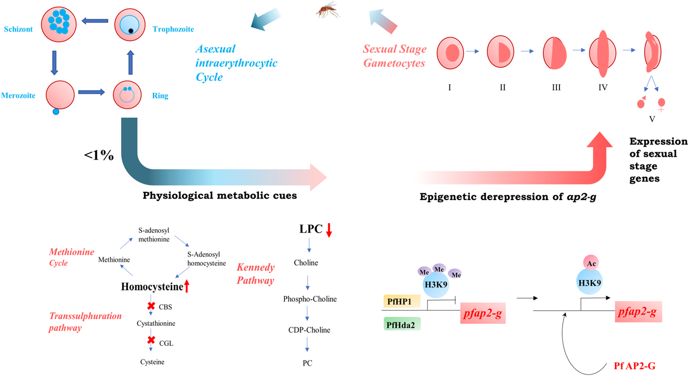
Recently, it has been shown that redox stress is capable of inducing gametocytogenesis in P. falciparum (Chaubey et al., Reference Chaubey, Grover and Tatu2014). Growth of the parasite in the human host is inherently prone to redox stress (Becker et al., Reference Becker, Tilley, Vennerstrom, Roberts, Rogerson and Ginsburg2004). Haemoglobin degradation and the host's immune response can both induce the formation of free radicals. However, the parasite has a compromised redox machinery and a crippled unfolded protein response machinery (Chaubey et al., Reference Chaubey, Grover and Tatu2014). Thus, in the face of an endoplasmic reticulum (ER) stress, the parasite cannot mount a robust stress response and forms terminally differentiated gametocytes. Our previous in vitro study as well as in vivo data revealed that the parasite has a truncated trans-sulphuration pathway (Beri et al., Reference Beri, Balan, Chaubey, Subramaniam, Surendra and Tatu2017). It lacks cystathionine beta-synthase and cystathionine gamma-lyase and consequently cannot convert homocysteine to cysteine. Homocysteine is an ER-stress-inducing molecule (Werstuck et al., Reference Werstuck, Lentz, Dayal, Hossain, Sood, Shi, Zhou, Maeda, Krisans, Malinow and Austin2001). The observations suggest that there is an accumulation of homocysteine in the host RBCs and surrounding medium/plasma. This was sufficient to induce the master transcriptional regulator of gametocytogenesis, ap2-g and in turn, trigger gametocytogenesis (Beri et al., Reference Beri, Balan, Chaubey, Subramaniam, Surendra and Tatu2017). This study proposed that the parasite times its own residence (as asexual stage parasites) in the host and once a threshold is reached, it prepares to exit the diseased host (as gametocytes). This was the first study to show a physiological metabolite causing gametocytogenesis in P. falciparum and P. berghei. Studies in clinical malaria have shown that hyper-homocysteinemia is associated with malaria infection (Chillemi et al., Reference Chillemi, Zappacosta, Simpore, Persichilli, Musumeci and Musumeci2004). Sexual stages are often linked to stress and it is unanimously agreed that increased stress conditions induce gametocytogenesis (Carter et al., Reference Carter, Kafsack, Llinas, Mideo, Pollitt and Reece2013). However, the presence of gametocytes in early infections and asymptomatic infections question the belief that induction of gametocytes is solely dependent on stress (Farid et al., Reference Farid, Dixon, Tilley and Mccarthy2017). Since homocysteine is a by-product of the parasite's own metabolism, it may therefore be one of the physiological triggers for gametocytogenesis (Beri et al., Reference Beri, Balan, Chaubey, Subramaniam, Surendra and Tatu2017). Another recent report suggests that the host-derived metabolite LPC controls sexual transition in the parasite (Brancucci et al., Reference Brancucci, Gerdt, Wang, de Niz, Philip, Adapa, Zhang, Hitz, Niederwieser, Boltryk, Laffitte, Clark, Gruring, Ravel, Blancke Soares, Demas, Bopp, Rubio-Ruiz, Conejo-Garcia, Wirth, Gendaszewska-Darmach, Duraisingh, Adams, Voss, Waters, Jiang, Clardy and Marti2017). It appears that depletion of LPC from the serum of the host is a cue for the parasite to undergo gametocytogenesis and suggests that the parasite takes up LPC from the host to synthesise choline and phosphatidylcholine via the Kennedy pathway. Comparative transcriptomics revealed 342 genes were induced by LPC depletion. Importantly, induction of ap2-g indicates that the lipid moiety acts upstream of the master regulator of sexual differentiation. However, LPC depletion did not affect sexual commitment in P. berghei, highlighting the differences in gametocytogenesis between Plasmodium species and indicating that the metabolite is not a genus-wide cue for sexual differentiation. These studies highlight the essentiality of understanding host–parasite metabolic pathways, which are critically linked to major life stage decisions made by the parasite (Fig. 1).
Metabolic triggers and epigenetic regulation of gametocytogenesis in Plasmodium: A small percentage of parasites exit the asexual intraerythrocytic cycle (shown in blue) to commit to the sexual stage (shown in red). Increase in homocysteine and decrease in the lysophosphatidylcholine level have been described as metabolic cues for sexual stage transition. This is followed by epigenetic regulation of ap2-g, the transcription factor associated with gametocytogenesis. Activation of ap2-g leads to expression of downstream genes which are essential for the development of gametocytes. The committed parasites undergo development from immature stage I gametocytes to banana-shaped, mature stage V gametocytes. During its blood meal, the mosquito vector ingests mature gametocytes that are circulating in the blood stream. Following development within the mosquito, parasites are injected into a healthy human host; thus, completing the life cycle of the parasite. (For more details, see text.)

Multi-OMICS approach to understand gametocytogenesis
In 2002, the first draft genome of a human isolate clone 3D7 of P. falciparum was obtained (Gardner et al., Reference Gardner, Hall, Fung, White, Berriman, Hyman, Carlton, Pain, Nelson, Bowman, Paulsen, James, Eisen, Rutherford, Salzberg, Craig, Kyes, Chan, Nene, Shallom, Suh, Peterson, Angiuoli, Pertea, Allen, Selengut, Haft, Mather, Vaidya, Martin, Fairlamb, Fraunholz, Roos, Ralph, Mcfadden, Cummings, Subramanian, Mungall, Venter, Carucci, Hoffman, Newbold, Davis, Fraser and Barrell2002) followed by genome sequencing of a P. vivax human isolate in 2008 (Carlton et al., Reference Carlton, Adams, Silva, Bidwell, Lorenzi, Caler, Crabtree, Angiuoli, Merino, Amedeo, Cheng, Coulson, Crabb, del Portillo, Essien, Feldblyum, Fernandez-Becerra, Gilson, Gueye, Guo, Kang'a, Kooij, Korsinczky, Meyer, Nene, Paulsen, White, Ralph, Ren, Sargeant, Salzberg, Stoeckert, Sullivan, Yamamoto, Hoffman, Wortman, Gardner, Galinski, Barnwell and Fraser-Liggett2008). Since then, the multi-OMICS approach has facilitated our understanding of various life stages of Plasmodium.
Forward genetic approaches like functional knockouts, as well as transposon-mediated insertional mutagenesis have been employed in delineating genetic control in Plasmodium (Ikadai et al., Reference Ikadai, Shaw Saliba, Kanzok, Mclean, Tanaka, Cao, Williamson and Jacobs-Lorena2013). The study identified 16 genes that are essential for gametocytogenesis in the commitment phase or developmental phase, with genetic perturbations leading to a functional arrest. Nine of these genes were reported to be involved in commitment, as well as early development. Of these, two coded for heat shock proteins (PF3D7_1253000, PF3D7_0527500), two were involved in RNA synthesis and metabolism (PF3D7_0416500, PF3D7_0924700), one was a transcription factor (PF3D7_1317200), one was involved in protein synthesis (PF3D7_0304400), one in phospholipid metabolism (PF3D7_1409900) and two of them were of unknown function (PF3D7_0532600, PF3D7_1455800). The remaining seven genes were involved in post-stage I development of the gametocyte. Except for PF3D7_0920000, which is involved in lipid metabolism, the functions of the other genes remain unknown (Ikadai et al., Reference Ikadai, Shaw Saliba, Kanzok, Mclean, Tanaka, Cao, Williamson and Jacobs-Lorena2013).
Other functional studies have shown a set of genes that are critical for the sexual development and now serve as canonical markers of gametocytogenesis [PF3D7_1302100 (Pfg27), PF3D7_0406200 (Pfs16), PF3D7_0935600 (Pfgig), PF3D7_1216500 (Pfmdv-1), PF3D7_0935400 (Pfgdv)] (Josling and Llinas, Reference Josling and Llinas2015). Recent studies have observed a transcription factor pfap2-g (PF3D7_1222600), belonging to the Apetala (Api2) family of transcription factors, as the master regulator of gametocytogenesis (Cai et al., Reference Cai, Zhou, Gu and Wang2012; Sinha et al., Reference Sinha, Hughes, Modrzynska, Otto, Pfander, Dickens, Religa, Bushell, Graham, Cameron, Kafsack, Williams, Llinas, Berriman, Billker and Waters2014). In P. falciparum, the DNA-binding protein PfAP2-G has been shown to have a strong correlation with gametocytogenesis. Forward and reverse genetic approaches have established the essentiality of ap2-g for gametocytogenesis. Using genome-wide ap2-g cognate motif occurrence, early gametocyte-related genes have been identified (Kafsack et al., Reference Kafsack, Rovira-Graells, Clark, Bancells, Crowley, Campino, Williams, Drought, Kwiatkowski, Baker, Cortes and Llinas2014). Similarly, in the rodent malaria parasite, P. berghei, it was found that Pbap2-g (PBANKA_143750) was essential for commitment to sexual stage. Other AP2 factors such as ap2-g2 were required for modulation, but not absolutely necessary to trigger gametocytogenesis. Further, ap2-g-specific binding motifs were found to be present upstream of Pbap2-g itself, suggesting an autoregulatory feedback loop in the regulation of gametocytogenesis (Sinha et al., Reference Sinha, Hughes, Modrzynska, Otto, Pfander, Dickens, Religa, Bushell, Graham, Cameron, Kafsack, Williams, Llinas, Berriman, Billker and Waters2014). Genome analyses of non-gametocyte producing lines have revealed mutations in ap2-g; thus, providing further evidence for its importance in the process of gametocytogenesis (Campino et al., Reference Campino, Benavente, Assefa, Thompson, Drought, Taylor, Gorvett, Carret, Flueck, Ivens, Kwiatkowski, Alano, Baker and Clark2016). Single-cell RNA-seq provided a deeper understanding on transcriptional changes induced by ap2-g and it identified 19 additional regulators (Poran et al., Reference Poran, Notzel, Aly, Mencia-Trinchant, Harris, Guzman, Hassane, Elemento and Kafsack2017). These included other AP2 members, SNF2 helicases, histone-modifying enzymes and putative histone lysine demethylases whose expression is temporally changed upon ap2-g induction. These results highlight a tight epigenetic control of ap2-g and gametocytogenesis.
Recent studies have shown that epigenetic mechanisms modulate gene expression in Plasmodium sp. (Fig. 1). The earliest of regulation decoded in Plasmodium was various histone post-translational modification (PTM) marks, cataloguing 232 distinct PTMs, 88 unique to Plasmodium (Doerig et al., Reference Doerig, Rayner, Scherf and Tobin2015). Of these, modifications in the above-mentioned ap2-g have been shown to be essential for gametocytogenesis in both P. falciparum and P. berghei (Modrzynska et al., Reference Modrzynska, Pfander, Chappell, Yu, Suarez, Dundas, Gomes, Goulding, Rayner, Choudhary and Billker2017). The ap2-g locus was shown to be regulated by the interplay between two epigenetic modifiers – PfHP1(PF3D7_1220900) and PfHDA2 (PF3D7_1008000). PfHP1 binds with H3K9me3, which is a hetero-chromatinisation signal and transcriptionally represses the Pfap2-g locus. PfHDA2, which is responsible for removing the acetylation mark, H3K9Ac3 is responsible for promoting the heterochromatinisation of the Pfap2-g locus. Knockout of PfHP1 or PfHDA2 enhances the gametocyte-forming ability of the parasite (Brancucci et al., Reference Brancucci, Bertschi, Zhu, Niederwieser, Chin, Wampfler, Freymond, Rottmann, Felger, Bozdech and Voss2014; Coleman et al., Reference Coleman, Skillman, Jiang, Childs, Altenhofen, Ganter, Leung, Goldowitz, Kafsack, Marti, Llinas, Buckee and Duraisingh2014), thus highlighting the role of ap2-g and its epigenetic regulation in gametocytogenesis. Recent studies have identified these epigenetic modulators in P. falciparum, as promising targets for antimalarial drugs (Gupta et al., Reference Gupta, Chin, Zhu, Mok, Luah, Lim and Bozdech2013).
Transcriptomic studies showed the presence of around 5500 protein-coding genes in Plasmodium. Suppression subtractive hybridisation studies have identified 126 genes which are differentially expressed in gametocytes, with functions spanning from the cell cycle, signalling, metabolism and cytoskeletal arrangement (Ngwa et al., Reference Ngwa, Scheuermayer, Mair, Kern, Brugl, Wirth, Aminake, Wiesner, Fischer, Vilcinskas and Pradel2013). Sex-specific RNA seq studies in P. falciparum showed upregulation of 1893 and 2078 transcripts in female and male gametocytes, respectively. This highlights the transcriptomic sexual dimorphism in microgametocytes and macrogametocytes. The study showed that more than 60% of the gametocyte transcriptome is differentially transcribed in the two sexes, which also strengthens the idea of a global transcript-based turnover in sex-specific gametocyte differentiation. Male gametocytes had enrichment of transcripts involved in flagellation of gametes, chromatin organisation and DNA replication, while female gametocytes expressed transcripts required for zygote formation, fertilisation, protein, lipid and energy metabolism. Thus, transcription-level sexual dimorphism occurs in the Plasmodium gametocytes (Lasonder et al., Reference Lasonder, Rijpma, van schaijk, Hoeijmakers, Kensche, Gresnigt, Italiaander, Vos, Woestenenk, Bousema, Mair, Khan, Janse, Bartfai and Sauerwein2016).
Proteomics analyses using high-resolution tandem mass spectrometry have identified proteins across different stages of P. falciparum (Florens et al., Reference Florens, Washburn, Raine, Anthony, Grainger, Haynes, Moch, Muster, Sacci, Tabb, Witney, Wolters, Wu, Gardner, Holder, Sinden, Yates and Carucci2002; Lasonder et al., Reference Lasonder, Ishihama, Andersen, Vermunt, Pain, Sauerwein, Eling, Hall, Waters, Stunnenberg and Mann2002). Recently, several separate studies have demonstrated differential protein signatures in gametocytes in comparison with asexual stages (Tao et al., Reference Tao, Ubaida-Mohien, Mathias, King, Pastrana-Mena, Tripathi, Goldowitz, Graham, Moss, Marti and Dinglasan2014; Lasonder et al., Reference Lasonder, Rijpma, van schaijk, Hoeijmakers, Kensche, Gresnigt, Italiaander, Vos, Woestenenk, Bousema, Mair, Khan, Janse, Bartfai and Sauerwein2016; Miao et al., Reference Miao, Chen, Wang, Shrestha, Li, Li and Cui2017). These revealed differences in the proteomes of the different life stages of the parasite reflecting their specialised functions. For example, the gametocyte proteome is highly enriched in ribosomal proteins. This occurs to accommodate the sudden surge in protein synthesis from previously repressed transcripts during gametogenesis and early zygote development. Commitment to gametocytes is accompanied by a shift in metabolic pathways of the parasite. Unlike the asexual stages, gametocytes have a fully functional TCA (tricarboxylic acid) cycle and fully developed mitochondria as a pre-adaptation for survival in the mosquito.
Another study examined the proteome at different stages of gametocytogenesis and discovered that stage I and II early gametocytes express 1427 proteins (Silvestrini et al., Reference Silvestrini, Lasonder, Olivieri, Camarda, van Schaijk, Sanchez, Younis younis, Sauerwein and Alano2010). Protein export was found to be highly prevalent in early gametocytogenesis. These exported molecules may have a role to play in the unknown sequestration mechanisms used by immature gametocytes to hide in the bone marrow. Another study identified an exported protein PfGEXP5, as early as 14 h post-invasion and suggested that it could be a marker of early gametocytes (Tiburcio et al., Reference Tiburcio, Dixon, Looker, Younis, Tilley and Alano2015).
The sex-specific proteomes showed significant differences that are consistent with the divergent functions of the two sexes (Khan et al., Reference Khan, Franke-Fayard, Mair, Lasonder, Janse, Mann and Waters2005). Both sexes shared around 1125 proteins (~74.7%), whereas 91 (7.3%) and 171 (12%) proteins were specific to male gametocyte and female gametocyte, respectively. Male gametocytes were enriched in proteins that play a crucial role in DNA replication and in flagellar proteins that guide the motility function in male gametocyte. In the female gametocytes, proteins of mitochondrial and apicoplast origin and proteins playing important roles in metabolic pathways, redox mechanisms and translation were highly expressed.
An example of successful integration of transcriptomic and proteomic data from male and female gametocytes is the discovery of translational repression in Plasmodium gametocytes (Mair et al., Reference Mair, Braks, Garver, Wiegant, Hall, Dirks, Khan, Dimopoulos, Janse and Waters2006; Lasonder et al., Reference Lasonder, Rijpma, van schaijk, Hoeijmakers, Kensche, Gresnigt, Italiaander, Vos, Woestenenk, Bousema, Mair, Khan, Janse, Bartfai and Sauerwein2016). The study revealed 512 highly expressed maternal transcripts with no corresponding protein expression. Certain key transcripts required for ookinete development are transcribed and stored preferentially in female gametocytes, but not translated until fertilisation in the mosquito midgut (Mair et al., Reference Mair, Braks, Garver, Wiegant, Hall, Dirks, Khan, Dimopoulos, Janse and Waters2006). This allows for temporal regulation of protein expression. The key proteins that guide these processes are Puf2 (PF3D7_0417100) and the P. berghei protein Pb DOZI (PBANKA_1217700), both of which are known to transcriptionally repress ~300 transcripts (Zhang et al., Reference Zhang, Joyce, Sullivan and Nussenzweig2013).
Discussion
Gametocytogenesis is a critical process in the life cycle of the malaria parasite as it underpins transmission of malaria. The advent of new technology has deepened our understanding of this dynamic process. The malaria parasite strategically adjusts between asexual reproduction and investment into gametocytes in response to changing environmental conditions. This so-called plasticity in the conversion rate enables the parasite to optimise between survival and transmission (Carter et al., Reference Carter, Kafsack, Llinas, Mideo, Pollitt and Reece2013).
The post-genomic era in gametocyte biology has seen exciting developments in gaining insights into transcriptomics, epigenomics and proteomics of different stages of P. falciparum gametocytes. Molecular tools have now established the transcription factor ap2-g as the master regulator of gametocytogenesis (Sinha et al., Reference Sinha, Hughes, Modrzynska, Otto, Pfander, Dickens, Religa, Bushell, Graham, Cameron, Kafsack, Williams, Llinas, Berriman, Billker and Waters2014). Elegant experiments have proven that depletion of PfHP1 or PfHDA2 leads to increase in ap2-g expression and a concomitant increase in gametocyte numbers (Brancucci et al., Reference Brancucci, Bertschi, Zhu, Niederwieser, Chin, Wampfler, Freymond, Rottmann, Felger, Bozdech and Voss2014; Coleman et al., Reference Coleman, Skillman, Jiang, Childs, Altenhofen, Ganter, Leung, Goldowitz, Kafsack, Marti, Llinas, Buckee and Duraisingh2014). Single-cell transcriptomics of parasites undergoing sexual commitment has provided clues to specific transcriptional sexual commitment programs that are executed by only a fraction of the parasites. The study emphasises the role of ap2-g and other ap2 transcription factors, histone-modifying enzymes and regulators of nucleosome positioning in facilitating sexual commitment in a fraction of the parasites. Until recently, induction of gametocytogenesis was primarily attributed to general stress conditions. However, recent studies have explored physiological metabolites associated with commitment. While homocysteine has been shown to trigger gametocytogenesis via ap2-g in both P. falciparum and P. berghei, LPC has been observed to upregulate ap2-g and gametocytogenesis exclusively in P. falciparum. This suggests that there might be multiple metabolic cues for the sexual commitment of the parasite and some of them could be species-specific. However, the mechanism by which the parasite converts a metabolic signal to an epigenetic/transcriptional change remains to be explored.
All parasites in a given patient are exposed to the same environmental conditions, yet only a small fraction (<1%) of these convert to gametocytes. Bet-hedging has been described to explain this phenomenon. It suggests in a small population of cells, certain regions of the genome are uniquely and variably transcribed within a genetically homogenous population (Alano, Reference Alano2014; Waters, Reference Waters2016). Such clonally variant gene expression ensures the presence of heterogenous transcripts within an otherwise homogenous genetic population. It is thought that ap2-g is bet hedged. This strategy aids modulation of gametocytogenesis in response to common fluctuations in the immediate environment. This allows the fittest parasite to survive, transmit and proliferate in the given environmental condition/stress condition. An in-depth understanding of the strategies used by the parasite to commit and regulate the process of gametocytogenesis will aid in the development of transmission-blocking therapeutics.
Gametocytes play a pivotal role in the parasite's life cycle yet their importance in clinical settings is undermined. Current methods of clinical malaria diagnosis are not well suited for gametocyte detection. Due to their low concentration, gametocytes can often be missed in the microscopic examination (Mawili-Mboumba et al., Reference Mawili-Mboumba, Nikiema, Bouyou-Akotet, Bahamontes-Rosa, Traore and Kombila2013; Rovira-Vallbona et al., Reference Rovira-Vallbona, Contreras-Mancilla, Ramirez, Guzman-Guzman, Carrasco-Escobar, Llanos-Cuentas, Vinetz, Gamboa and Rosanas-Urgell2017). Lateral Flow Assays used for malaria cannot differentiate between asexual and sexual stages (Krampa et al., Reference Krampa, Aniweh, Awandare and Kanyong2017). PCR-based diagnostic methods of gametocytes are available but seldom used in clinical settings. A recent study established that sub-microscopic and asymptomatic infections predominate in individuals and contribute significantly to the gametocyte reservoir in different communities in the Peruvian Amazon (Rovira-Vallbona et al., Reference Rovira-Vallbona, Contreras-Mancilla, Ramirez, Guzman-Guzman, Carrasco-Escobar, Llanos-Cuentas, Vinetz, Gamboa and Rosanas-Urgell2017). Further, most antimalarial drugs induce gametocytogenesis (Peatey et al., Reference Peatey, Skinner-Adams, Dixon, Mccarthy, Gardiner and Trenholme2009). In the last few years, more than 25 000 compounds have been identified that are active against asexual stages (Duffy and Avery, Reference Duffy and Avery2013). However, a recent study demonstrated that only 14% of these compounds have gametocytocidal activity (Duffy and Avery, Reference Duffy and Avery2013). Thus, there is an urgent need to develop methods specific for gametocyte detection and identify molecules that are active against both asexual and sexual stages of Plasmodium sp.
Future directions
Gametocyte biology is a striking example of the staggering complexity associated with a parasite. Even after decades of work, certain fundamental questions need to be addressed. We still do not completely understand the exact timing, precise point and the exact cue(s) that trigger gametocytogenesis. There is evidence that all merozoites released from a committed schizont form gametocytes, but we do not understand the mechanism(s) behind gender selection of gametocytes. The transcription factor ap2-g is considered the master regulator of gametocytogenesis. However, a recent study identified a protein, PfGEXP5, which has been described as the earliest gametocyte marker present in committed rings. Regulation of PfGEXP5 appears independent of ap2-g, since it was expressed even in ap2-g-mutated parasites (Tiburcio et al., Reference Tiburcio, Dixon, Looker, Younis, Tilley and Alano2015). This suggests that even before ap2-g induction, commitment to the sexual stage takes place and the molecular players operating upstream of ap2-g are yet to be identified. Time-lapse single-cell microscopy complemented with single-cell RNA-seq can be used as tools to examine fates of individual cells. Future research should be directed towards identifying the early players of gametocytogenesis. Two metabolites –homocysteine and LPC have been demonstrated to trigger gametocytogenesis. However, the mechanisms by which modulation in metabolite levels are perceived by the parasite remain unknown. Further research is needed to understand the signalling pathways that are responsible for the conversion of metabolic signals to genetic/transcript/protein read-outs. While some studies claim that gametocytes are formed in the bone marrow, a recent study used an early stage marker of commitment (PfGEXP5) to demonstrate the presence of committed rings in the bloodstream (Joice et al., Reference Joice, Nilsson, Montgomery, Dankwa, Egan, Morahan, Seydel, Bertuccini, Alano, Williamson, Duraisingh, Taylor, Milner and Marti2014; Farid et al., Reference Farid, Dixon, Tilley and Mccarthy2017). Also, a metabolomic study revealed major differences in the metabolomes of reticulocytes (present majorly in bone marrow) and mature RBCs (present in bloodstream) that may play an important role in commitment to the sexual stage (Srivastava et al., Reference Srivastava, Creek, Evans, de Souza, Schofield, Muller, Barrett, Mcconville and Waters2015). Immature gametocytes are sequestered in the bone marrow. However, the mechanism of sequestration of gametocytes in the bone marrow is not well understood but is known to be distinct from the asexual stages. Thus, there is a need to identify additional early markers of commitment, further to investigate the metabolic signatures of the host and parasite that may play a role in sexual stage transition and identify sequestration strategies used by immature gametocytes to evade the immune system.
PTMs are implicated in diverse and critical biological functions. Novel PTMs such as palmitoylation and glutathionylation are known to be present in Plasmodium, but their possible biological functions, especially in gametocytes, remain to be elucidated (Kehr et al., Reference Kehr, Jortzik, Delahunty, Yates, Rahlfs and Becker2011; Hodson et al., Reference Hodson, Invergo, Rayner and Choudhary2015). Using the multi-omics tools available to biologists today, robust and comprehensive information about sexual stage triggers, gametocyte development and host–parasite interactions can be obtained.
Currently, malariologists are aiming to control and ultimately eradicate malaria. Continuous and well-organised efforts from the WHO, Bill Melinda Gates Foundation and the Roll Back Malaria Programs have been successful in eradicating malaria from many countries. A recent study suggested that onset of gametocytogenesis occurs from the first round of asexual replication when the parasite load was <100 parasites ml−1 and individuals were completely asymptomatic. The study describes the potential presence of a pre-symptomatic pool of patients who are reservoirs for successful parasite transmission (Farid et al., Reference Farid, Dixon, Tilley and Mccarthy2017). There has been renewed interest in Transmission Blocking Vaccines (TBV) following the 2013 update to the Malaria Vaccine Technology Roadmap which urged researchers to focus on development of safe and effective vaccines against P. falciparum and P. vivax transmission stages (Carter et al., Reference Carter, Mendis, Miller, Molineaux and Saul2000; Carter, Reference Carter2001; Nunes et al., Reference Nunes, Woods, Carter, Raphael, Morin, Diallo, Leboulleux, Jain, Loucq, Kaslow and Birkett2014; Sauerwein and Bousema, Reference Sauerwein and Bousema2015; Lee et al., Reference Lee, Wu, Plieskatt, Miura, Hickey and King2017). The avian and mouse models of malaria have unequivocally established the highly effective nature of such vaccines, but the only sub-optimal reduction in infectivity had been observed with TBV25H used in human clinical trials. Thus, in the future, efforts must be directed to develop better candidates for TBVs (Carter, Reference Carter2001). The fundamental requirements of malaria research now are to understand the precise epidemiology of the disease, including the asymptomatic population harbouring gametocytes, which act as a hidden reservoir of the parasite, active surveillance to identify drug-resistant populations of the parasite, to develop a vaccine against the parasite and to identify existing/novel antimalarial drugs that target both the sexual and asexual stages of the parasite. Thus, understanding the various biological facets of its transmissible stage is of utmost importance in ultimately subverting the malaria parasite.
Financial support
Authors acknowledge funding for the study by the Department of Biotechnology (DBT), Department of Science and Technology (DST) and DBT–IISc partnership grant, India. D.B. acknowledges financial support from the Council for Scientific and Industrial Research, India.
Conflict of interest
The authors declare no competing financial interests.