Acute coronary syndrome is a general term for situations where the blood supplied to the heart muscle is suddenly blocked. It is uncommon in childhood and adolescence and can be a life-threatening situation. Acute coronary syndrome has clinical suspicion difficulties because of the rarity of cardiac causes and sudden onset among patients with chest pain complaint applied to paediatric emergency. Risk factors for myocardial infarction in children and adolescence are coronary artery abnormalities, CHD, and thromboembolic events. We present an adolescent with a history of Bentall operation who had acute coronary syndrome owing to coronary thrombosis as a result of inappropriate drug use.
Case report
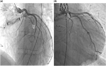
A 17-year-old boy presented to our emergency unit with the complaint of chest pain for the lasting 3 hours of the time of report, severity gradually increasing for minutes, feeling like pressure in his chest, no change with respiration nor position, radiating in his left shoulder, left arm and back. He was diagnosed with aortic coarctation, severe aortic stenosis, and bicuspid aorta and underwent Bentall procedure 2 years ago using 27-mm conduit (Medtronic, Inc., Minneapolis, Minnesota, United States of America) and coarctation stenting using 16-mm Z-MED balloon (NuMED, Inc., Hopkinton, New York, United States of America). There was no family history of hyperlipidaemia or early myocardial infarction. He had no history of fever for Kawasaki disease or other systemic diseases. Upon physical examination, arterial oxygen saturation was 96%, body temperature was 37°C, heart rate was 74 beat/minute, and arterial blood pressure was 130/80 mmHg. Laboratory investigations showed normal renal and liver function tests. Troponin I level was 0.01 ng/ml (normal value < 0.04 ng/ml) initially. Although the first cardiac marker levels were normal, there was a gradual increase in the control marker levels scheduled due to presentation with chest pain and history of heart surgery. Table 1 shows the increase in troponin I levels in hours. Troponin I level was obtained 36.6 ng/ml 8 hours after being admitted to the emergency unit. Activated prothrombin time was 31.5 seconds, and international normalised ratio was 1.08. Electrocardiogram showed T wave inversion in leads V4, V5, and V6. Echocardiography revealed opening and closing of leaflets and no abnormal densities for vegetation or thrombosis, both the left ventricular posterior wall thickness and interventricular septal thickness with a normal left ventricular ejection fraction, mild paravalvular regurgitation from prosthetic valve, a mean gradient of 14 mmHg with a 90 ms of acceleration time of prosthetic aortic valve and a mild residual aortic coarctation. Selective coronary angiography showed fresh thrombus in the mid left anterior descending artery at the level of second diagonal branch (Fig 1a). Prosthetic aortic valve was normal in function without any evidence of thrombus or restriction in leaflet mobility. The combination of anticoagulation with antiplatelet agents (salicylate; 100 mg/day orally and unfractionated heparin; an initial intraveous bolus of 60 IU/kg, maximum of 4000 U, followed by infusion of 12 IU/kg/h, maximum 1000 U/hour) was started immediately. The patient’s chest pain regressed about a day later. Control coronary angiography performed 2 days later showed no residual thrombus in coronary arteries (Fig 1b). Echocardiography performed before discharged showed normal systolic functions.
Troponin I levels during day. Although the first cardiac marker was normal, the troponin I level was restudied due to typical chest pain and high-risk patient for cardiac-induced chest pain.

(a) Left image shows fresh thrombus in the mid left anterior descending artery at the level of second diagonal branch; (b) Right image shows thrombus regression.

Discussion
Chest pain is a common finding in children and adolescents. Chest pain caused by heart disease is rarely seen among childhood, varies from 1 to 4%.Reference Selbst, Ruddy, Clark, Henretig and Santulli1,Reference Saleeb, Li, Warren and Lock2 Cardiac cause may be associated with life-threatening conditions and risk of sudden cardiac death.Reference Reddy and Singh3,Reference Friedman and Alexander4 Cardiac chest pain is seen in the precordial region, accompanied by palpitations or syncope, which can spread to the back, neck, jaw, or arms. In our patient, he had a chest pain suggesting cardiac-related chest pain which was no change with position nor respiration, described as a pressure in the chest and radiated in his left shoulder, left arm, and back. Cardiac causes of chest pain are cardiomyopathy, myo/pericarditis, pulmonary hypertension, arrhythmias, severe aortic/pulmonic stenosis, mitral valve prolapses, pulmonary embolus, cocaine uses, sympathomimetic overdose, and congenital/acquired coronary anomalies.Reference Selbst, Ruddy, Clark, Henretig and Santulli1–Reference Reddy and Singh3 The underlying cardiac disease should be suspected when sudden onset of pain or characteristics of chest pain with post-cardiac surgery and family members with sudden and early death histories. Our patient had a history of stenting due to aortic coarctation and aortic root replacement owing to aortic stenosis. Although troponin I levels taken after chest pain were normal, chest pain caused by cardiac reason was a high suspicion as a result of underlying cardiac history and characteristic of chest pain.
Acute coronary syndrome is extremely rare in childhood and adolescents. Etiologic factors for myocardial infarction in paediatric population are congenital/acquired coronary artery abnormalities (Kawasaki disease, myocardial bridging, anomalous origin of the coronary artery, fistulas, atresia, or stenosis of coronary artery), CHD, thrombosis susceptibility, poor coronary perfusion associated with perinatal asphyxia, hypoxia and sepsis, inflammatory conditions (myocarditis, systemic lupus erythematosus), genetic disorders (homocystinuria, progeria, mucopolysaccharidosis), and familial hypercholesterolemia.Reference Reddy and Singh3,Reference Osula, Bell and Hornung5
Because of the thrombogenicity of mechanical valves, patients with prosthetic mechanical valve may develop acute coronary syndrome. In aortic root replacements like Bentall operation, coronary steal complications such as kinking or pseudo aneurysms should be assessed. Mechanical heart valves are long-lived, and anticoagulant therapy is necessary and may cause bleeding or embolic complications if dose adjustment is not performed properly. In children, difficulties of dietary control and blood clotting tests need close monitoring.Reference Khitin, Sade, Bradley, Crawford, Widener and Stroud6,Reference Levis, Schultz and Lee7 Typical chest pain, ischaemic electrocardiogram findings with raised cardiac enzymes in patients with mechanical valves, and acute coronary syndrome should be suspected. Echocardiography showed normal systolic functions and no wall motion abnormality. When the patient’s history is deepened, it was learned that he was not using his medicine properly. It is important to take a detailed history of adolescent patients’ compliance with drug use. In addition, low international normalised ratio level led us to perform coronary angiography for coronary thrombus.
Management of acute coronary syndrome requires an urgent and careful approach. Close monitorisation of haemodynamic parameters (blood pressure, ECG and arrhythmia monitoring, pulse and saturation control) should be done. Oxygen treatment should be given if necessary and pain relief should be performed. Medical treatment with thrombolysis (streptokinase), antiplatelet drugs, and anticoagulation can be used. Percutaneous transluminal coronary angioplasty and stenting may be required in selected cases to reperfusion of coronary artery.Reference Nakazone, Tavares, Machado and Maia8,Reference Kiernan, Flynn and Kearney9 In our patient, the combination of anticoagulant and antiplatelet was sufficient in the treatment of thrombosis, and no additional interventional therapy was needed.
Conclusion
Identifying acute coronary syndrome in children presenting with chest pain is very important. Although it is rare in children, patients with history of heart disease, using anticoagulant treatment and having typical chest pain, should be urgently evaluated for acute coronary syndrome and need immediately coronary angiography for diagnosis and treatment.
Acknowledgements
None.
Financial support
This research received no specific grant from any funding agency, commercial or not-for-profit sectors.
Conflicts of interest
None.
Ethical standards
The authors assert that all procedures contributing to this work comply with the ethical standards of the relevant national guidelines on human medical regulations and with the Helsinki Declaration of 1975, as revised in 2008.