CHD is the most common congenital lesion in the term infant and is known to have long-lasting neurodevelopmental consequences during childhood and adolescence.Reference McQuillen and Miller 1 Brain abnormalities associated with CHD include delayed brain maturation and acute white matter injury.Reference McQuillen and Miller 1 Clinical neurodevelopmental abnormalities in these patients include motor, cognitive, and behavioural impairments.Reference McQuillen and Miller 1
The increase in the survival of newborns with CHD conferred by improvements in neonatal cardiac interventional techniques has allowed for neurodevelopmental assessment in these patients.Reference McQuillen and Miller 1 Recent studies have demonstrated delayed brain development in neonates diagnosed with CHD, which are due to delays in myelination, cortical development, maturation of the germinal matrix, and glial cell migration.Reference Andropoulos, Hunter and Nelson 2 Infants with CHD and immature brain development are at an increased risk for perinatal brain injury, specifically white matter injury and stroke.Reference Andropoulos, Hunter and Nelson 2
Approximately one-third of neonates with CHD demonstrate evidence of stroke and white matter injury on brain MRI before any surgical intervention.Reference McQuillen and Miller 1 The timing of brain injury is often unknown – for example, in utero versus peripartum versus postnatal – which is a hurdle for developing new therapeutics and care strategies to better protect the brain. MRI has been used extensively and valuably to identify perioperative brain injury in neonates with severe CHD;Reference McQuillen and Miller 1 however, it is challenging to implement MRI in a traditional care workflow, especially in ill neonates in whom MRI cannot be used to repeatedly monitor brain injury during care.
Glial fibrillary acidic protein is an abundant astrocyte/glial/ependymal cell cytoskeletal intermediate filament protein.Reference Pelinka, Kroepfl, Leixnering, Buchinger, Raabe and Redl 3 – Reference Bembea, Savage and Strouse 8 It is released from glial cells within the central nervous system as a result of cell necrosis.Reference Ennen, Huisman and Savage 9 According to Brunetti et al,Reference Brunetti, Jennings and Easley 10 an ideal biomarker for brain injury is specific to brain tissue and is released into the bloodstream immediately after neurological injury and accurately reflects the location and degree of injury. As the glial fibrillary acidic protein is released into the blood after astrocyte death, it is a suitable biomarker to predict neonatal brain injury.Reference Ennen, Huisman and Savage 9 Glial fibrillary acidic protein has been previously utilised as a diagnostic and prognostic tool in both children and adults; it has been shown to correspond with abnormal brain imaging in neonates,Reference Brunetti, Jennings and Easley 10 and to predict neurodevelopmental outcomes in neonates with hypoxic ischaemic encephalopathy, prematurity-related intracranial haemorrhage, extracorporeal membrane oxygenation, and cardiopulmonary bypass during repair of neonatal CHD.Reference Bembea, Savage and Strouse 8 , Reference Brunetti, Jennings and Easley 10
The aim of this study was to determine whether there is a difference in serum glial fibrillary acidic protein levels at birth and the immediate postnatal period between neonates with major CHD and two groups of controls: neonates with hypoxic ischaemic encephalopathy serving as positive controls, and neonates without brain injury serving as negative controls. The secondary objectives of this study were to determine whether glial fibrillary acidic protein levels can elucidate the timing of brain injury in neonates with CHD, and to evaluate the association between glial fibrillary acidic protein levels, abnormal head size, and brain structural abnormalities by head ultrasounds in the major CHD group.
Material and methods
Patients
The present study was a case–control study of neonates admitted to the neonatal intensive care unit at Johns Hopkins Hospital between October, 2009 and October, 2011. Cases were live-born infants, born between 30 and 41 weeks of gestation, with significant structural CHD defined as any cardiac defect requiring surgical intervention within the 1st year of life. Isolated patent arterial duct cases were not included in this study. We used two groups of controls. Positive controls had birth-related moderate-to-severe hypoxic ischaemic encephalopathy, as defined by Sarnat and Sarnat,Reference Sarnat and Sarnat 11 and were treated with therapeutic hypothermia. Negative controls were term or late preterm newborns admitted to the neonatal intensive care unit for reasons not related to cardiac or neurological concerns and without clinical brain injury at the time of discharge. The study was reviewed and approved by the Institutional Review Board at Johns Hopkins University. A consent waiver was granted by the Institutional Review Board at Johns Hopkins University School of Medicine for the study to collect discarded neonatal samples, no specific consent was deemed necessary to collect serum biomarker data on blood samples that would otherwise be discarded.
Specimens
Neonatal blood samples collected were remnant clinical samples. Neonatal waste blood was collected within 6 hours of birth, and then daily for the first 4 days of life from all cases and controls. Timing of sample collection was based on the clinical assessment of laboratory testing needs.
Glial fibrillary acidic protein assay
We utilised a glial fibrillary acidic protein electrochemiluminescent sandwich immunoassay (Meso Scale Discovery, Gaithersburg, MD, United States of America) as previously described.Reference Bembea, Savage and Strouse 8 – Reference Brunetti, Jennings and Easley 10 , Reference Savage, Everett and Casella 12 , Reference Savage, Barron‐Casella and Fu 13 Samples were assayed in duplicate and accepted for analysis with a CV% of <10%. The lower limit of quantitation was 0.04 ng/ml, and values below this level were recorded as 0. We have previously shown that glial fibrillary acidic protein is stable at room temperature for at least 72 hours, making use of remnant clinical blood specimens a feasible approach.Reference Savage, Barron‐Casella and Fu 13
Neonatal head ultrasound
Routine clinical neonatal head ultrasounds performed during initial admission to the neonatal intensive care unit were reviewed to assess for structural brain abnormalities in all the CHD patients. The timing of head ultrasound varied, and was not just limited to the 1st day of life. All the head ultrasound data obtained during the first 30 days of life, before cardiac surgery, were reviewed.
Cephalisation index
Neurodevelopment and brain maturity have been evaluated in newborns using the cephalisation index.Reference Harel, Tomer, Barak, Binderman and Yavin 14 This screening method utilises a ratio of head circumference to birth weight to predict possible brain vulnerability.Reference Harel, Tomer, Barak, Binderman and Yavin 14 This index has proven to be one of the most effective perinatal predictive parameters of abnormal neurodevelopment in cases of intrauterine growth restriction.Reference Leitner, Fattal-Valevski and Geva 15 Previous studies investigating neurodevelopment have concluded that a higher cephalisation index is associated with a greater risk for brain abnormality.Reference Harel, Tomer, Barak, Binderman and Yavin 14 A ratio of 0 indicates a healthy, normal-term, infant of 38–40 weeks of gestation, whereas negative values represent a prenatal stage of development, and positive values represent a postnatal stage of development.Reference Harel, Tomer, Barak, Binderman and Yavin 14
Statistical analysis
Our study population included 56 cases, 23 positive controls, and 23 negative controls who were all enrolled concurrently during the same study period. Summary statistics were generated to summarise the study population. Comparisons were made for non-parametric data, and medians were analysed using GraphPad Prism, version 6. The Kruskal–Wallis test with Dunn’s multiple comparisons were used to determine whether there was a difference in serum glial fibrillary acidic protein levels at birth between the CHD group, the positive controls, and the negative controls. Significance was set at p<0.05. Student’s t-test was performed to assess for differences between head size and glial fibrillary acidic protein levels. The two markers of head size included head circumference and the cephalisation index. The cephalisation index has not previously been validated in CHD patients. As part of this pilot study, we calculated the cephalisation index for the case and control groups to assess for trends. A χ2 test of independence was performed to examine the relation between single ventricle heart disease and glial fibrillary acidic protein levels.
Results
Study population and clinical characteristics
In the present study, we analysed a total of 102 neonates admitted to the neonatal intensive care unit. Cases consisted of 56 neonates diagnosed with CHD, who were compared with the two control groups of 23 neonates diagnosed with moderate-to-severe hypoxic ischaemic encephalopathy, qualifying for therapeutic hypothermia, and 23 neonates without CHD or brain injury. Of the 56 neonates with CHD, 39 were diagnosed with single ventricle heart disease.
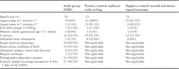
Neonatal clinical characteristics are summarised in Table 1. As shown, CHD cases – compared with negative controls – had lower 1- and 5-minute Apgar scores, with no difference in the number with low birth weight, preterm births, or caesarean section deliveries. As detailed below, three CHD cases had abnormal head ultrasounds.
Summary statistics for the study population.

C-section=caesarean section
Characteristics of the study group and two control groups (negative and positive control) are represented in this table, along with clinical variables that are known to be associated with abnormal neurological development, as well as haemodynamic risk factors that could theoretically be associated with abnormal neurological development
Glial fibrillary acidic protein as a biomarker
In the case group, 19 neonates had complete samples of neonatal serum at all time points during the study period, compared with only one negative control and seven positive controls with complete samples during all time points. The complete CHD group had the potential for 280 neonatal blood samples; 62 samples were not available. The complete control group was designed to include 115 neonatal blood samples from each of the two control groups; 25 positive control and 44 negative control samples were not available. Unavailability of samples was due to insufficient amount of remnant serum samples or due to the fact that clinical samples were not drawn at the chosen time points for the study. In addition, neonatal serum at birth was not available for outborn neonates who were transferred to Johns Hopkins Neonatal Intensive Care Unit.
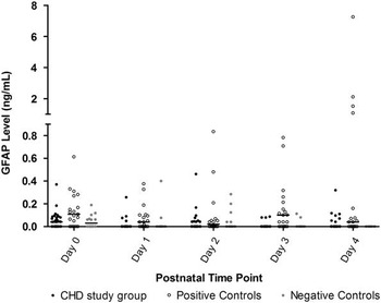
There were no significant differences in overall glial fibrillary acidic protein levels between the CHD group and the negative control group at any time point of the study (Fig 1). Negative controls demonstrated a pattern of highest levels of glial fibrillary acidic protein at birth with a progressive decline to unmeasurable levels by day 4 of life. On admission, defined as <6 hours of life, the CHD group had a median glial fibrillary acidic protein level of 0.042 ng/ml. The negative control group had a median glial fibrillary acidic protein level of 0.03 ng/ml, whereas the positive control group had a median glial fibrillary acidic protein level of 0.108 ng/ml. On the other hand, by day 4 of life, 7/56 (12.5%) CHD cases continued to have detectable or increasing glial fibrillary acidic protein levels, compared with 0/23 (0%) normal controls. The CHD cases differed significantly when compared with the positive control group on day 1, 3, and 4 of life (Kruskal–Wallis test with Dunn’s multiple comparisons; day 1 (p<0.0004), day 3 (p<0.0001), day 4 (p<0.0094)).
Median glial fibrillary acidic protein by postnatal time point: glial fibrillary acidic protein (GFAP) levels at each day of life of patients with CHD (study group) and the two control groups. This graph shows that the study group’s GFAP level at each time point is very similar to non-brain-injured neonates (negative controls). In comparison, patients with hypoxic ischaemic encephalopathy (positive controls) showed elevated GFAP levels over time.

In the CHD group, three patients with abnormal head ultrasounds had the following findings and glial fibrillary acidic protein levels: (1) increased echogenicity of brain parenchyma, and later by brain MRI on day 16 of life to have a subdural haemorrhage, with the highest glial fibrillary acidic protein level of 0.462 on day 2 of life; (2) absent corpus callosum with no oedema, bleeding, or other positive findings, with the highest glial fibrillary acidic protein level of 0.101 on day 0 of life; and (3) periventricular white matter cyst with glial fibrillary acidic protein levels below level of quantification, highest level detected on day 0 of life to be 0.02. Of the three CHD cases with abnormal head ultrasounds, only the CHD case with imaging evidence of acute brain injury had elevated glial fibrillary acidic protein levels and was the highest in the CHD group.
CHD subgroup analysis
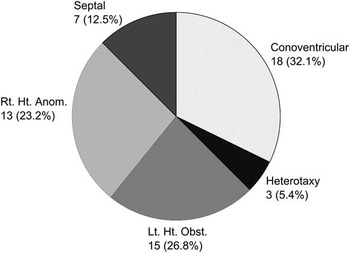
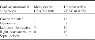
As shown in Figure 1, no negative controls and only a small group of CHD cases had persistent levels of glial fibrillary acidic protein by day 4. Therefore, we compared the glial fibrillary acidic protein levels on day 4 of life with cardiac anatomical subgroups to identify trends (Table 2). The CHD cases were divided into five anatomical categories: conoventricular, heterotaxy, septal defects, left heart obstruction, and right heart anomalies (Fig 2). The majority of postnatal time points for all five cardiac subgroups had glial fibrillary acidic protein levels of 0. The frequency of elevated glial fibrillary acidic protein levels was higher in patients with left heart obstruction on admission (day 0) and in patients with septal defects on admission (day 0) and day 2 of life. By day 4 of life, 5/8 (63%) patients in the CHD group with left heart obstruction continued to have detectable or increasing levels of glial fibrillary acidic protein. Half (5/10, 50%) of the left heart obstruction group versus 1/17 (6%) conoventricular, 0/13 (0%) right heart, and 1/6 (17%) septal defect patients continued to have elevated levels of glial fibrillary acidic protein at day 4 of life.
Study population by cardiac anatomical subgroup: breakdown of study group (patients with CHD) by cardiac anatomical subtype. Conoventricular heart disease represented the greatest portion of the study population (32.1%), followed by left heart obstruction (26.8%), right heart anomalies (23.2%), septal defects (12.5%), and heterotaxy syndrome (5.4%). Lt. Ht. Obst.=left heart obstruction; Rt. Ht. Anom.=right heart anomaly.

Glial fibrillary acidic protein (GFAP) levels at day 4 of life by cardiac anatomical subgroup.

This table demonstrates the number of patients by cardiac subtype with measurable and unmeasurable GFAP on day 4 of life. These data show that the left heart obstructive lesions were reported to have the highest number of cases with measureable GFAP levels on day 4 of life
These seven CHD cases with persistent glial fibrillary acidic protein levels are detailed in Table 3, and include two cases with hypoplastic left heart syndrome, two cases with aortic arch obstruction, one case with double-inlet left ventricle with pulmonary atresia, one case of acyanotic tetralogy of Fallot, and one case with atrioventricular canal; five out of these seven cases of CHD were prostaglandin dependent, and four of the seven had anatomy consistent with systemic outflow obstruction. Moreover, three out of the seven cases had an anatomical substrate for single ventricle palliation. Interestingly, five out of the seven cases exhibited the highest glial fibrillary acidic protein levels on day 4 of life. The medical record did not indicate any clinical hypotension, shock, or low cardiac output to explain these outliers at the corresponding time points.
Clinical characteristics of CHD cases with detectable glial fibrillary acidic protein (GFAP) within first 4 days of life.

VSD=ventricular septal defect
Head circumference and cephalisation index in CHD
Head circumference and cephalisation index data were obtained from CHD patients and were compared with normative data. We found that the mean head circumference (33.96+1.6 versus 33.78+2.0 cm) was not different between the CHD group and normative data (p=0.71). The mean cephalisation index (0.011+0.002 versus 0.011+0.002) was not different between the CHD group and normative data (p=0.4).
Discussion
Brain abnormalities in newborns with significant CHD have been well described in recent years, but the timing, aetiology, and significance of these abnormalities remain poorly understood. Many serum biomarkers have been evaluated for diagnosing, screening, and assessing cardiac function; however, this study represents one of the first studies evaluating brain injury reflected by glial fibrillary acidic protein in preoperative neonates at birth with CHD.
Negative (normal) control neonates demonstrated a pattern of glial fibrillary acidic protein elevation at birth reaching a nadir and being undetectable by day 4 of life. A subset of neonates with CHD, the majority of whom had left heart obstructive lesions, demonstrated a trend of persistent elevation of glial fibrillary acidic protein through day 4 of life. Glial fibrillary acidic protein levels at birth were not different from negative controls, suggesting that acute brain injury in neonates with CHD may not occur with the birth process. The marked elevation in patients with birth-related brain injury, such as that seen in the hypoxic ischaemic encephalopathy, the positive control group of this study, suggests a potential difference in both the severity and/or the timing of acute brain injury negatively impacting neurodevelopment in the CHD population.
There was a trend towards abnormal glial fibrillary acidic protein levels in certain subgroups of CHD by day 4 of life. Although not statistically significant, seven CHD patients had detectable glial fibrillary acidic protein levels on day 4 of life. Interestingly, the group that had the highest number of patients with elevated glial fibrillary acidic protein levels was left heart obstructive lesions. This subgroup has been hypothesised to be at increased risk for neurodevelopmental abnormalities secondary to anatomy and aortic arch flow characteristics. These findings are in agreement with previous studies reporting that microcephaly was most common in neonates with hypoplastic left heart syndrome.Reference Hinton, Andelfinger and Sekar 16 , Reference Shillingford, Ittenbach and Marino 17 This may reflect sub-clinical injury, but needs to be further studied in a larger patient sample and at longer time points to establish whether these glial fibrillary acidic protein trends are statistically significant in this population.
The timing of brain injury remains unknown within the population of CHD neonates. In an attempt to identify the period in which injury occurs, this study evaluated glial fibrillary acidic protein levels during the first 4 days of life. As a biomarker, glial fibrillary acidic protein has a short half-life of less than 24 hours. Therefore, this protein is unable to detect brain injury occurring in utero at a time point earlier than immediately after delivery. At the time of birth, the glial fibrillary acidic protein levels were not elevated in the CHD patients of this study, suggesting that no significant brain insult had occurred perinatally. Nevertheless, it is still possible that brain injury occurs at mid-gestation, but cannot be detected by assessment at birth. This study supports the idea that brain injury (stroke) observed by pre-surgical MRI in neonates with CHD does not commonly occur acutely in the immediate peripartum period, but likely during preoperative care. This is supported by the evidence from this report that five out of seven CHD neonates with circulating glial fibrillary acidic protein levels were prostaglandin dependent.
Patients with single ventricle physiology, particularly patients with a hypoplastic left heart, have significant cognitive and motor abnormalities at 1 year of age that persist at least until early school age.Reference Hoskoppal, Roberts, Kugler, Duncan and Needelman 18 , Reference Davidson, Gringras, Fairhurst and Simpson 19 Although our pilot study was not powered for CHD subgroup analysis, the finding of abnormal glial fibrillary acidic protein levels during preoperative care suggests this as a vulnerable window, and therefore a clinical opportunity to limit perioperative brain injury.
The aetiology of neurological abnormalities in newborns with CHD is not well understood, despite multiple studies describing the finding of white matter injury in a significant number of these neonates. The findings of this study demonstrate that the cephalisation index is not a good marker to predict abnormal brain development of CHD neonates in our study population. Newborns with CHD in this study had a normal head size. This underscores the complex nature of neurological abnormalities in this population. Other hypothesised aetiologies for neurological deficits in children with CHD include disturbances in brain metabolic function or developmental abnormalities, as opposed to an acute injury mechanism seen in hypoxic ischaemic encephalopathy.Reference Shedeed and Elfaytouri 20 This may parallel the type of neuromaturation defects described in premature babies.Reference Allen, Cristofalo and Kim 21 Quite possibly, the mechanism of brain injury in this fragile population is a combination of those listed above. Microcephaly has been noted in utero in a subset of CHD patients, supporting the notion that the abnormality may find its origin before birth.Reference Hinton, Andelfinger and Sekar 16 , Reference Shillingford, Ittenbach and Marino 17 As noted previously, one-third of CHD neonates have abnormal brain MRI before any cardiac intervention;Reference McQuillen and Miller 1 however, there is no clinically important brain injury in many of these cases. A combination of imaging and biomarker technologies that track normal fetal neurodevelopment would be the next step to understanding the nature of neurodevelopmental abnormalities in fetuses and newborns with CHD.
The present study is one of the first studies examining any serum biomarker of preoperative neonates with CHD. There are several limitations to this study. This study does not examine neonatal brain MRI as it is not part of clinical routine. Long-term neurodevelopmental follow-up data were also not available for this sample. In addition, as remnant clinical samples were used, there were missing samples from all cohorts. Another limitation of this study is the population of the negative control group. This group was comprised of non-hypoxic ischaemic encephalopathy and non-CHD neonates admitted to the neonatal intensive care unit. Our study was also not adequately powered for sub-study analysis of different CHD subgroups, which demonstrated interesting and possibly important trends.
Conclusions
Neonates with CHD as a group followed a glial fibrillary acidic protein pattern similar to the negative controls at birth and not the positive controls with clinical perinatal brain injury. This suggests that the nature of brain injury in CHD neonates is unlikely related to the birth process. In addition, glial fibrillary acidic protein levels suggested that certain high-risk CHD lesions may be at risk of acute injury before repair. Therefore, circulating biomarkers of brain injury may have utility as postnatal screening tools for brain injury to develop and test perioperative ICU protocols to reduce brain injury in CHD neonates. Additional investigation and discovery of biomarkers specific to prenatal brain development and injury will be important to further test the idea that brain injury in CHD neonates could also occur prenatally. Pinpointing the exact timing of brain injury and the high-risk groups in this population will help in timely intervention to prevent subsequent neurodevelopmental abnormalities.
Acknowledgements
The authors acknowledge partial support for the statistical analysis from the National Center for Research Resources and the National Center for Advancing Translational Sciences (NCATS) of the National Institutes of Health through Grant Number 1UL1TR001079. The authors thank their colleague, Jiangxia Wang, MS, MA, with the Johns Hopkins Institute for Clinical and Translational Research, who provided insight and expertise in biostatistics that greatly assisted the research. They also thank Michael V. Johnston, MD, Vice President and Chief Medical Officer at Kennedy Krieger Institute and Professor in the Department of Neurology and Pediatrics at Johns Hopkins University School of Medicine, for his assistance and feedback.
Financial Support
This research received no specific grant from any funding agency, commercial, or not-for-profit sectors.
Conflicts of Interest
None.
Ethical Standards
The authors assert that all the procedures contributing to this work comply with the ethical standards of the relevant national guidelines on human experimentation and with the Helsinki Declaration of 1975, as revised in 2008, and has been approved by the institutional committees, including the Institutional Review Board at Johns Hopkins University School of Medicine.