Tetralogy of Fallot with so-called “absent” pulmonary valve is a well-recognized variant of the classical form of tetralogy, occurring in up to one-twentieth of such patients.Reference Freedom, Yoo, Mikailian and Williams1 It is characterized by the absence or rudimentary formation of the pulmonary valvar leaflets, with gross dilation of the pulmonary trunk and its branches, usually with absence of the arterial duct in those having confluent intrapericardial pulmonary arteries. Anomalous pulmonary venous return is a rare finding in association with tetralogy of Fallot.Reference Redington, Raine, Shinebourne and Rigby2–Reference Vargas-Barron, Espinola-Zavaleta, Rijlaarsdam, Keirns and Romero-Cardenas4 We describe here a patient with tetralogy of Fallot, absent pulmonary valve syndrome, and supracardiac totally anomalous pulmonary venous connection, with the vertical vein obstructed between the left-sided descending aorta and the enlarged left pulmonary artery.
Case report
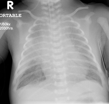
A full term Asian male born via spontaneous vaginal delivery was noted to be immediately cyanotic and in respiratory distress. The neonate was markedly acidotic with profound hypoxia, despite an infusion of prostaglandin E1 and inotropic support. The physical examination revealed no obvious dysmorphism, but there was cyanosis, a hyperactive precordium, and palpable femoral pulses. Auscultation revealed a single second heart sound, and a “to and fro” murmur best heard at the left sternal border. After intubation, a chest radiograph showed a heart of normal size, features of extensive oedema and atelectasis, with enlarged pulmonary arteries and pulmonary interstitial and alveolar oedema (Fig. 1). The child was immediately transported to our facility.
Supine, frontal chest radiograph. The lungs demonstrate marked pulmonary oedema with fluid tracking into the oblique fissure. There is patchy atelectasis, predominantly in the right upper lobes, reflecting the bronchial compression by the enlarged pulmonary arteries.

The transthoracic echocardiogram showed the usual arrangement of thoracic and abdominal contents, concordant atrioventricular and ventriculoarterial connections, an intact atrial septum, anterior deviation of the outlet septum, and an unrestricted ventricular septal defect. There was absence of the pulmonary valve, with grossly dilated pulmonary arteries measuring 2.8 centimetres in diameter. There was a left aortic arch with no arterial duct was identified. The relationship of the pulmonary venous confluence to the back of the atrial chambers was difficult to image, but a left vertical vein draining to the brachiocephalic vein, with continuous non-phasic flow consistent with obstruction, was noted by pulse wave and colour flow Doppler mapping.
A computed tomography angiogram (Brilliance Power 16CY Computed Tomography Scanner, Philips Medical Systems, Cleveland, Ohio, United States of America) of the chest was undertaken to define more clearly the anatomy of the obstructed pulmonary venous return. The study was performed with the child intubated and ventilated, following intravenous injection of 2 millilitres per kilogram, 300 milligrams Iodine per milliliter Iohexol contrast medium (Omnipaque, Amersham Health Inc, Oakville, Ontario, Canada) via a right transfemoral venous catheter. Breath-hold, non-gated, axial images were obtained from the apexes of the lungs to below the level of the hepatic inferior caval vein. Images were reviewed in axial and multi-planar reformatted planes. Additional maximum intensity projected and surface rendered three-dimensional images were reconstructed.
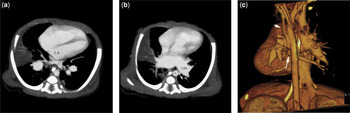
The computed tomography angiogram clearly demonstrated the isolated pulmonary venous confluence posterior to the small left atrium, with the draining vertical vein obstructed between the lateral aspect of the descending thoracic aorta and the grossly dilated left pulmonary artery, and connecting to the brachiocephalic vein (Fig. 2a). The bilaterally patent bronchuses likely reflected the images being acquired during end-inspiration. Surgical considerations were limited due the rapid deterioration of the clinical state. The child died 20 hours after birth.
Computed tomography angiogram (a) with breath held in inspiration. Axial image showing the pulmonary venous confluence (C) posterior to the left atrium, but with no connection to it. Note the enormous branch pulmonary artery and the right upper lobe atelectasis. LA=left atrium. Axial image (b) demonstrating compression of the vertical vein (arrow) between the hugely dilated left pulmonary artery and the lateral aspect of the descending aorta (*). LPA=left pulmonary artery. Three-dimensional surface rendered reconstruction image (c) showing the thin, obstructed, vertical vein (short arrow) connecting the pulmonary venous confluence (long arrow) with the left braciocephalic vein. The dilated branch pulmonary arteries are seen.

Discussion
The rare association of tetralogy of Fallot and anomalous pulmonary venous connection has been described by Redington et al.Reference Redington, Raine, Shinebourne and Rigby2 and Freedom et al.Reference Freedom, Yoo, Mikailian and Williams1 Two cases have been reported in patients with tetralogy of Fallot and absent pulmonary valve. Redington et al.Reference Redington, Raine, Shinebourne and Rigby2 described one neonate with unobstructed pulmonary veins to the coronary sinus, while Vargas-Barron et al.Reference Vargas-Barron, Espinola-Zavaleta, Rijlaarsdam, Keirns and Romero-Cardenas4 described an adult with similar anatomy. To the best of our knowledge, a case with supracardiac, obstructed, totally anomalous pulmonary venous return has not previously been reported in any patient with tetralogy of Fallot.
Neonates with absent pulmonary valve will often present with cyanosis and oligaemic lungs on chest radiography, likely due to the high pulmonary vascular resistance in the presence of pulmonary insufficiency complicated by impaired aeration due to bronchial compression. Delay in the clearance of retained fetal lung fluid, due to the bronchial compression, is well described.Reference Griscom, Harris, Wohl, Vawter and Eraklis5 In our neonate, the radiographic findings of persistent pulmonary oedema, despite aggressive ventilation, directed our attention towards an unexpected obstructed anomalous pulmonary venous return. Indeed, the clinical course was dominated by the pulmonary venous obstruction, rather than by compression of the airway, insufficiency of the pulmonary valve or the right-sided infundibular obstruction typical of Fallot’s tetralogy.Reference Zucker, Rozin, Aviva and Zalstein6
Further imaging was required better to delineate the venous anatomy prior to consideration of surgical options. Magnetic resonance imaging was not considered appropriate due to the time required to obtain the information, and due to the need to sequester the critically ill child in an environment not optimized for direct observation or resuscitation. Although the computed tomography angiogram delivered a significant radiation dose to the child, with its attendant risks,Reference Hall7 it did define the level and mechanism of obstruction of the vertical vein, as well as providing information on the quality of the obstruction to the airways (Fig. 2b, c).
Obstruction of the vertical vein may occur at a variety of levels, and be due to a variety of mechanisms.Reference Freedom, Mawson, Yoo and Benson8 Most commonly obstruction results from the vein being entrapped between the left pulmonary artery and left main bronchus. In this child, the vice formed by the descending aorta and the enlarged left pulmonary artery is unusual, but under the circumstances, not unexpected.
The association of totally anomalous pulmonary venous connection and tetralogy of Fallot is rare. This association in the setting of the variant of tetralogy with so-called absent pulmonary valve had a direct bearing on clinical course and outcome, as the enormous size and pulsatility of the left pulmonary artery contributed to venous obstruction. Our experience emphasizes the importance of cross-sectional modalities for imaging, including computed tomography angiography, as non-invasive modalities with which clearly to define cardiac anatomy in the critically ill neonate.
Acknowledgement
We acknowledge the help received from Dr. John B. Mawson, from the Department of Radiology, and Drs. Walter J. Duncan and Jim Potts from the Children’s Heart Centre at B.C. Children’s Hospital, in preparing this manuscript.