Introduction
Proliferating trichilemmal tumour (PTT) is a well-circumscribed dermal or subcutaneous neoplasm with trichilemmal-type of keratinisation.Reference Markal, Kurtay, Velidedeoglu and Hucumenoglu1 It is a benign tumour arising from hair follicles, which can rarely undergo malignant transformation.Reference Park, Yang and Cho2–Reference Mori, Hachisuka and Sasai6 It occurs most commonly in women over the age of 50 years.Reference Laing, Knipe, Flowers, Stoer and Ramos-Caro7, Reference López-Ríos, Rodríguez-Peralto, Aguilar, Hernández and Gallego8 Tumours of adnexal components of the skin are exceptionally rare and were first reported in 1966 by Wilson-Jones as a pathological entity. It can simulate squamous cell carcinoma.Reference Wilson-Jones9 These tumours occur most commonly in women as benign scalp lesions. Lanugo hair follicles of the bald scalp and follicles of other areas devoid of non-terminal hair are unlikely to produce these tumours.Reference Saida, Oohara, Hori and Tsuhiya10
Previous reports of benign trichilemmal tumours have been infrequent, that is, 0·1% of skin biopsies. The malignant variety is extremely rare. Despite the characteristic histological features of these tumours, earlier reports have emphasised the need to differentiate this pathology from a well-differentiated carcinoma. Confirmation of the diagnosis is made on the histological appearance of clear cells originating from the outer root sheath that exhibit marked pleomorphism and atypical mitosis. Characteristically, nodular trabecular formation of keratinocytes with a sharp border between parenchyma and stroma is seen. Malignant change in these tumours conforms to two growth patterns such as circumscribed nodular and diffuse spindle cell type. Malignant trichilemmal tumours need to be differentiated from metastases of spindle cell tumours with regions of squamous differentiation that tend to mimic their histopathological appearance. Differential diagnosis includes sebaceous carcinoma, clear-cell hidradenocarcinoma and cutaneous metastasis of renal cell carcinomas.Reference Siddha, Budrukkar and Shet11
Patients with aggressive tumours have been reviewed and none were addressed by any type of adjuvant treatment that lead to an unfavourable treatment outcome.Reference Sau, Graham and Helwig5, Reference Wilson-Jones9–Reference Poiares Baptista, Garcia, Silva and Born16 Similarly, in the series by Ye et al., four out of eight patients with malignant trichilemmal/pilar tumours recurred within a period ranging from 2 to 14 months. None of these patients were given any adjuvant treatment. Based on this evidence, we decided to give adjuvant radiotherapy to the operated tumour bed to achieve favourable locoregional control.
Case Description
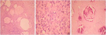
A 48-year-old woman presented with a solitary 4×3 cm-sized swelling over the parietal area of the scalp on the right side, increasing in size for the past 5 months. She reported a similar swelling in the same site 8 months ago, for which she underwent excision and skin grafting. She had no remarkable previous medical or family history. Histopathology was suggestive of PTT (Figure 1). The lesion recurred within 3 months of excision and grew to the present size in 5 months.
Histopathology: tumour cells arranged in varying size nodules. Notes: Nodules show basaloid cells (double arrow) palisading around areas of keratinisation (single arrow) with foci of micro-calcification (triple arrow).

On examination, a well-circumscribed swelling measuring 4×3 cm was observed on the right side of the scalp in the parietal region. An overlying scar from the previous excision and skin grafting was also noted. The swelling was ulcerated with foul-smelling discharge. It was also non-tender, mobile in all directions and not fixed to the bone. On palpation, bleeding was noted from the tumour. No palpable neck lymphadenopathy was observed.
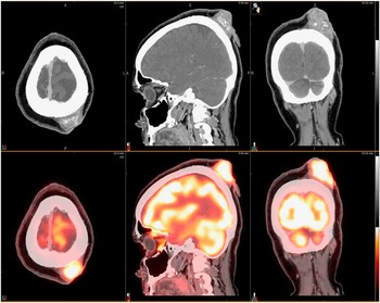
Complete metastatic workup was done. PET-CT showed a metabolically active lesion in left parietal region of scalp with a superior uptake value of 7 (Figure 2). No bony erosion was seen. Biopsy was suggestive of a malignant PTT of the scalp. Attributable to its aggressiveness, wide local excision of the tumour with primary skin grafting was done. Post-operative histopathology revealed a tumour measuring 4×3·6×2·8 cm with cells arranged in nodules of various sizes. These nodules showed basaloid cells with indistinct cell border, abundant eosinophilic cytoplasm and open chromatin palisading around areas of keratinisation with foci of micro-calcification and atypical cells. Features were suggestive of malignant trichilemmal tumour of the scalp.
PET-CT axial, sagittal and coronal views. Notes: PET-CT heterogeneously enhancing, metabolically active soft tissue mass is seen in the left parietal scalp abutting the outer surface of the calvarium. No evidence of underlying calvarial erosion. Abbreviation: PET-CT, positron emission tomography computed tomography.

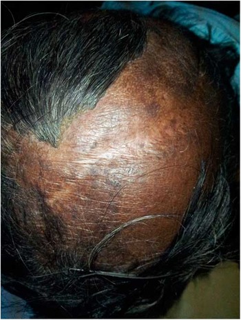
After complete healing of the surgical wound, adjuvant radiotherapy (electron therapy) was administered to a localised area of the scalp covering the tumour bed with 2 cm margin, using 9 MeV electrons to a dose of 60 Gy/30 fractions, 5 days a week for 6 weeks. Patient tolerated the treatment well. She had a mild hyperpigmentation over the treated area. Follow-up of 9 months post-therapy revealed no recurrence of the tumour (Figure 3).
Picture showing treated scalp.

Conclusion
Diagnosis of this patient was substantiated by identification of the characteristic histopathological features. However, this disease was unusual as the tumour is aggressive and it recurred in 3 months after the initial surgery. The role of adjuvant radiation therapy in malignant trichilemmal/pilar tumours, especially in the malignant variant is not clear. This is owing to the rarity of the disease. However, adjuvant radiotherapy is warranted for controlling belligerent malignant variant owing to high rates of locoregional as well as distant failures observed in previous series. Even though wide local excision with clear margins is the primary treatment, adjuvant treatment in the form of radiation and chemotherapy could be considered for aggressive and recurrent malignant PTTs. This could help in achieving a favourable local control and in preventing distant metastasis.
Acknowledgements
The authors would like to thank the surgical and radiation oncology teams of HCG Bangalore Institute of Oncology.