Implications
Lower levels of IgA are correlated with decreased mucosal immunity, which in turn can be followed by economic losses from poor performance. This work shows that horses under intense physical exercise and controlled sanitary management present lower levels of serum IgA when compared to animals kept in loose management. This might reflect in weaker mucosal responses by those animals, and thus how sanitary management and exercising influences the immune system of horses must be further investigated.
Introduction
Acquired immunity is characterized for its specificity and memory and it is usually classified into humoral and cellular branches. Cellular immunity is related to the destruction of intracellular microorganisms, while humoral immunity is associated to neutralization of toxins, opsonization of microbes and antibody-dependent cell death. Immunoglobulin A (IgA) is the main immunoglobulin isotype involved in neutralization of antigens of environmental origin (food- and air-borne) for being the predominant immunoglobulin isotype found in mucosae (Cerutti, Reference Cerutti2008). Therefore, IgA is essential for protection of the surfaces of respiratory, gastrointestinal and genitourinary tracts. Isotype switching to IgA occurs when B cells are stimulated in the presence of TGF-β1 produced by several cell types, including CD4+ T cells (Cerutti, Reference Cerutti2008).
Environmental antigenic load has direct influence in maturation and stimulation of the immune system and also in the increased IgA and IgG levels (Menezes et al., Reference Menezes, Mucida, Cara, Alvarez-Leite, Russo, Vaz and Faria2003 and Reference Menezes, Andrade, Senra, Rodrigues, Vaz and Faria2006). Furthermore, IgA switching is highly dependent on intestinal commensal flora and it has been shown that rodents kept under germ-free conditions or elemental diets present lower levels of both serum and mucosal IgA (Bos et al., Reference Bos, Meeuwsen, Wostmann, Pleasants and Benner1988; Menezes et al., Reference Menezes, Mucida, Cara, Alvarez-Leite, Russo, Vaz and Faria2003).
Among the known factors that decrease mucosal immunity is physical effort. In humans, intense exercising causes a significant decrease in IgA and IgM levels in serum (Mackinnon, Reference Mackinnon2000). As a consequence, elite athletes are more susceptible to respiratory and gastrointestinal tract disorders when compared to non-athletic individuals and are considered bearers of ‘chronic mild immunosuppression’ (Gleeson et al., Reference Gleeson, McDonald, Cripps, Pyne, Clancy and Fricker1995; Gleeson and Pyne, Reference Gleeson and Pyne2000). In horses, it has been shown that intensive exercise triggers an increase in cortisol and ACTH levels (Vara et al., Reference Vara, Marañon, Castejon, Sánchez de la Muela, Olabarri, Manley, Dominguez, Muñoz-Escassi and Cayado2006), a decrease in number of CD8+ T lymphocytes (Malinowski et al., Reference Malinowski, Kearns, Guirnalda, Roegner and McKeever2004), and is associated with higher incidence and morbidity of Babesia equi (al-Ani et al., Reference Al-Ani, al-Darraji, Lafi and Hailat1997).
The equine industry has reached a high level of specialization. Athlete horses are born and kept under controlled conditions such as concrete individual stalls, standardized industrialized food, multiple vaccination and vermifugation protocols, easy access to antimicrobial therapy and daily supervised physical training. It is conceivable that such conditions, which are much cleaner but physically demanding when compared to those found in a free-ranging farm, underlie the higher frequency of upper respiratory and digestive tracts infections found in athlete animals (Hines et al., Reference Hines, Schott, Bayly and Leroux1996). Herein, we worked with the hypothesis that intensive handling associated with chronic strenuous exercise results in mucosal humoral immunosuppression in horses.
Material and methods
Animals – management – design
Sixty male and female horses of various breeds and origins were included in this study. All horses were within the age range of 6 to 23 years. The ‘intensive’ group involved 35 animals under daily training at tracks or jumping Clubs (São Paulo Equestrian Club or São Paulo Jockey Club, São Paulo, Brazil) where they were subjected to at least 3 h of daily training at the tracks. This group lived under high sanitary standards: vermifugation with ivermectin paste every 6 months, vaccination against rabies and tetanus once a year, and against influenza and West Nile viruses every 6 months. This group was fully stabled in individual concrete boxes with concrete floor bedded with fresh straw. The animals were fed with commercial diet and fresh straw and had constant access to clean water. Stables were fully cleaned once daily and had manure removed as necessary. The ‘loose’ group was formed by 25 animals living in two farms at the countryside of São Paulo State (Brazil), under a partly stabled regimen. The animals were stabled in individual wooden boxes within a barn style complex during the night. The boxes had hard bare ground covered with fresh straw bedding and manure was removed only once a day. The animals received commercial food and had constant access to clean water inside the boxes. During the day the animals were kept together in the same large paddock with grass and access to water. This group received a loose protocol of vaccination/vermifugation where medication was administered only when the animal presented clinical symptoms of worm infestation. This group had no exercising routine.
Sampling
Venous blood samples were collected and allowed to clot at room temperature. Sera were stored at −20°C until analysis. All animals were clinically sound, with normal complete blood cell counts at the time of sampling, and no history of recurrent infections. Samples were divided into two groups according to sanitary standards and physical training. All procedures were carried out with the approval of the Animal Care and Use Committee of the University of São Paulo.
Laboratory analysis
IgA, IgG, IgG(T) and IgM isotypes were quantified using commercial radial immunodiffusion (RID) kits (VMRD, Pullman, WA, USA).
Statistical analysis
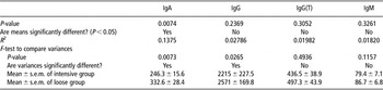
Normal distribution of the isotypes within each group (‘intensive’ and ‘loose’) was tested using the Kolmogorov–Smirnov test at a significance of P < 0.05 and GraphPrism software (GraphPad Software; data not shown). Since the data followed normal distribution, parametric Student’s t-test was used to compare means and s.e.m. of both groups (Tables 1 and 2). Power calculation was performed for all four immunoglobulin isotypes at a statistical power of 0.95 and α-level of 0.05. The obtained sample sizes were 38 for IgA, 329 for IgM, 215 for IgG and 330 for IgG(T).
Means and standard deviations of serum immunoglobulin isotypes concentrations obtained from ‘intensive’ and ‘loose’ groups (mg/dl)

Comparison of means and variances of serum isotypes IgA, IgG, IgG(T) and IgM between ‘intensive’ and ‘loose’ group (mg/dl)

Results and discussion
Table 1 shows means and standard deviations found in both ‘intensive’ and ‘loose’ groups. The ‘intensive’ group presented lower means for IgA, IgG and IgG(T), but only the IgA level was significantly (P < 0.0074) lower (Table 2). In studies carried out in mice, animals kept on elemental diet (Menezes et al., Reference Menezes, Mucida, Cara, Alvarez-Leite, Russo, Vaz and Faria2003; Menezes et al., Reference Menezes, Andrade, Senra, Rodrigues, Vaz and Faria2006) or under germ-free conditions (Bos et al., Reference Bos, Meeuwsen, Wostmann, Pleasants and Benner1988) presented significantly lower levels of secretory IgA and also lower serum IgA and IgG, while IgM levels remained within normality. In our case, variances between the two groups were significantly different for IgA (P = 0.0073) and IgG (P = 0.0265), also suggesting a tendency of IgG to be lower in the ‘intensive’ group. At this point, we believe that an increase in the number of animals per group could result in significantly different means for IgG as well since this study was underpowered for this immunoglobulin isotype. We analyzed 60 individuals and found the adequate sample size for IgG to be 215 (see Material and methods section for details).
Studies carried out in human elite athletes found lower levels of salivary IgA to be associated with increased incidence of respiratory illness (Gleeson et al., Reference Gleeson, McDonald, Cripps, Pyne, Clancy and Fricker1995; Gleeson and Pyne, Reference Gleeson and Pyne2000; Mackinnon, Reference Mackinnon2000). Our data show that IgA levels are lower in animals kept under intensive physical training and cleaner facilities, which are conditions that correlate with lower mucosal immunity in several species. It is important to point out that studies carried out on mice measured both secretory and serum IgA and the levels found in both compartments correlated to each other. In our study, we measured only serum IgA but there is no reason to believe that secretory IgA levels would not correlate with serum levels in horses. Studies carried out by others have validated measurement of serum IgA as a parameter to evaluate immunity of horses (Foote et al., Reference Foote, Love, Gilkerson, Rota, Trevor-Jones, Ruitenberg, Wellington and Whalley2005; Tallmadge et al., Reference Tallmadge, McLaughlin, Secor, Ruano, Matychak and Flaminio2009). Moreover, in a recent study involving horses with common variable immunodeficiency (CVID), the authors noticed a decline in serum IgA concentrations that correlated with worsening of the clinical symptoms. The study suggests that serum IgA be monitored as a prognostic tool to evaluate CVID progression (Flaminio et al., Reference Flaminio, Tallmadge, Salles-Gomes and Matychak2009).
It is expected that the higher the quantity and variety of environmental antigens that an animal is exposed to, the higher the concentrations of immunoglobulins will be, reflecting the expansion of repertoire activated by the antigenic load (Menezes et al., Reference Menezes, Mucida, Cara, Alvarez-Leite, Russo, Vaz and Faria2003 and Reference Menezes, Andrade, Senra, Rodrigues, Vaz and Faria2006; Cerutti, Reference Cerutti2008). In this sense, the higher levels of IgA found in animals raised under ‘loose’ management could be explained by the constant stimulation of mucosal lymphoid tissue by environmental and social contact antigens. Although one could argue that horses in the jumping clubs and racing tracks have plenty of social contacts, for example, from participating in sports competitions and from traveling, we are here arguing that such contacts are ‘antigenically irrelevant’ since they occur between animals that are unlikely to be carriers of infectious agents. In horses, it was shown that the presence of specific IgG against Streptococcus equi in the serum (and induced by vaccination) did not provide protection against strangles if IgA isotype was not induced, in which case specific IgA levels in mucosa remained low (Timoney et al., Reference Timoney, Holmes, Sponseller and Sheoran1997). By the same token, animals in our ‘loose’ group could be considered as better protected from air- and food-borne infections due to higher levels of IgA induced by their lighter working load and higher environmental antigenic load.
At this point, there is no epidemiological data to prove that horses kept under ‘intensive management’ are more affected by respiratory/digestive tract infections when compared to loose management horses. It would be interesting to perform a meta-analysis of epidemiological studies focusing on both groups of animals to test the hypothesis that intensive management produces less resistant animals.
The levels of immunoglobulins G and its subclasses are dependent on previous infections and vaccines responses. In our study, we found no difference on IgG and IgG(T) levels when we compared the means of both groups (Table 2), suggesting that environmental antigenic load and exercising routine play no important role over these isotypes on the ability of the animal to produce an IgG-dependent response. IgM is produced early in the course of an infection and does not re-emerge during secondary immune responses. This could explain the similarity of IgM levels in both groups since all samples were taken from clinically healthy animals (Table 2).
Taken together, our study results suggest that horses under intensive handling should not be considered as a loose horse in respect of its immunological status. Our data suggest that an athlete horse may be less poised to mount an effective mucosal immunity response to environmental challenges when compared to a horse that is subjected to loose management and lives within a free-ranging group. This potentially decreased immunity should be kept in mind when treating such animals for infectious diseases. But perhaps more importantly, the differences of immunity between athlete horses and their non-athlete counterparts must be further studied and better understood if we aim at developing more effective vaccines.
Acknowledgments
The authors thank all referring veterinarians for sending samples and providing information about the horses, Ms Claudia S. Cunha for technical assistance, Dr Ana Maria Caetano de Faria for critical input on the data and Dr Marcos Amaku for statistical advice. Sao Paulo State Council for Science supported this work (FAPESP 06/50543-7 and 08/54811-1 for M.M.C.). C.B.P. is an RUSP Scholarship recipient and B.A.M. is a CNPq/PIBIC Scholarship recipient.