Implications
Knowledge of sperm fertilizing potential has great economic impact for the breeding industry and facilitates the selection of high-fertility bulls. Reproductive success depends on the efficiency of several sperm functions. Correlations between fertility and individual sperm parameters are usually too weak to predict fertility. Backward elimination analysis provides a fertility prediction model that considers kinetic parameters and DNA integrity indicators, and accounts for almost half of the variation in fertility. The use of a reliable model of fertility prediction in artificial insemination (AI) centers would facilitate the rejection of low-potential fertility bulls and elimination of poor-fertility ejaculates.
Introduction
The main purpose of sperm evaluation is to study the relationship between semen quality and fertility in order to discover the potential fertility of samples and donors. The practice of freezing semen in cattle has become increasingly widespread, owing to its remarkable results, as it facilitates multiple aims, from biodiversity conservation to commercial purposes. The different steps in cryopreservation protocols can cause damage at various levels in the sperm cell, thereby reducing its ability to fertilize the oocyte. So it is crucial to implement the monitoring of cryopreserved semen quality to foresee its potential fertility. Reproductive success depends on the efficiency of several sperm functions. It would therefore be useful to have a system of fertility prediction, based on the evaluation of several semen quality parameters after freezing and thawing, to exclude low-potential fertility bulls from breeding programs, thus saving considerable time and economic resources. A number of studies have aimed to identify the most suitable parameters for semen fertility prediction. Correlations between fertility and semen quality have been reported in domestic species for various semen parameters, including membrane integrity (Januskauskas et al., Reference Januskauskas, Johannisson, Söderquist and Rodríguez-Martínez2000 and Reference Januskauskas, Johannisson and Rodríguez-Martínez2003), early (Del Olmo et al., Reference Del Olmo, García-Álvarez, Maroto-Morales, Ramón, Jiménez-Rabadán, Iniesta-Cuerda, Anel-Lopez, Martínez-Pastor, Soler, Garde and Fernández-Santos2016) and late stages of capacitation (Thundathil et al., Reference Thundathil, Gil, Januskauskas, Larsson, Söderquist, Mapletoft and Rodríguez-Martínez1999), and chromatin integrity (Evenson and Jost, Reference Evenson and Jost2000; Januskauskas et al., Reference Januskauskas, Johannisson and Rodríguez-Martínez2003; Evenson, Reference Evenson2016). To our knowledge, a combination of several parameters has rarely been analyzed by backward stepwise multiple regression analysis.
Motility assessment is a fundamental test for semen evaluation. Presently, subjective evaluation has been largely replaced by computer-assisted semen analysis (CASA) that can provide accurate and objective evaluation of semen motility and kinetic parameters. It is currently the most popular method, routinely used in AI centers. Sperm motility and kinetic parameters are correlated with field fertility in the bull (Farrell et al., Reference Farrell, Presicce, Brockett and Foote1998; Zhang et al., Reference Zhang, Larsson, Lundeheim and Rodríguez-Martínez1998; Januskauskas et al., Reference Januskauskas, Johannisson and Rodríguez-Martínez2003). Correlations between field fertility and DNA integrity, evaluated using the sperm chromatin structure assay have been detected in the bull and boar. The DNA fragmentation index (%DFI) has been proposed to identify subfertile individuals; however, this parameter usually presents a very narrow range of values in selected bulls (Januskauskas et al., Reference Januskauskas, Johannisson and Rodríguez-Martínez2003), thus reducing its predictive value. Generally, correlations between in vitro assays and fertility have shown high variability among various studies (Rodríguez-Martínez, Reference Rodríguez-Martínez2003). As fertilization is a phenomenon that requires a number of conditions, the identification of a specific set of parameters might facilitate improved predictions of fertility (Martínez-Pastor et al., Reference Martínez Pastor, Mata-Campuzano, Álvarez-Rodríguez, Álvarez, Anel and de Paz2010). Zhang et al. (Reference Zhang, Larsson, Lundeheim, Håård and Rodríguez-Martínez1999) observed that individual sperm parameters in the bull were not significantly correlated with field fertility; whereas when combined parameters were considered, the correlation was significant.
Fertility data are derived from many factors related to both male and female fertility, as well as several other sources of variation that contribute to the outcome of insemination (Amann and Hammerstedt, Reference Amann and Hammerstedt2002). Female fertility factors include among the others the ability to repair sperm DNA damage. The oocyte has this capability, although limited, whereas mature sperm lose their ability to repair DNA. Therefore, the biological effect of sperm DNA damage depends on both the extent of the damage and the ability of the oocyte to repair DNA (González-Marín et al., Reference González-Marín, Gosálvez and Roy2012).
Flow cytometry (FCM) is potentially an excellent tool for the evaluation of semen characteristics. Furthermore, the integration of several tests is a promising approach to achieve a better understanding of sperm functions. Flow cytometry enables the assessment of thousands of cells within a short time, and captures many features of each cell. In comparison to other techniques, it is characterized by very high reproducibility.
The aim of the present study was to evaluate the relationships among fertility and sperm parameters assessed by CASA (e.g. motility and kinetic parameters) and FCM (e.g. viability, acrosome integrity, membrane lipid disorder, lipid peroxidation, mitochondrial membrane potential (MMP), DNA integrity) in cryopreserved bull semen samples, in order to identify the most suitable parameters that would provide a reliable fertility prediction model.
Material and methods
Reagents and media
All chemicals used in the present study were of reagent grade. Unless otherwise indicated, reagents were purchased from Sigma-Aldrich Co. (St. Louis, MO, USA).
Semen source and fertility evaluation
Frozen semen samples from 18 Italian Holstein-Friesian bulls of known fertility were analyzed. The bulls were classified according to field fertility data expressed as the estimated relative conception rate (ERCR), provided by the Italian Holstein Breeders Association. To calculate the ERCR, the 56-day non-return rate (NRR) to service was adjusted for several factors, including farm, year and month of insemination, parity, days open of the cow at first insemination, bull, bull progeny and cow. The ERCR represents the effect of the bull on the NRR (expressed as a percentage) of cows inseminated in the herd, and is determined by the difference between that NRR value for a particular bull and the average NRR values obtained with the semen of other bulls. As its calculation is based on a large number of services from many different herds, the ERCR is considered a highly accurate measurement to identify high- and low-fertility bulls (Ibrahim et al., Reference Ibrahim, Gilbert, Loseth and Crabo2000; Clay and McDaniel, Reference Clay and McDaniel2001). Bulls with a reliability (Reliability=100×[1−(1−(Number of inseminations∕(Number of inseminations+200))½)×2.3]) of ERCR >60 were considered. According to the distribution of bulls based on the ERCR, nine high-fertility and nine low-fertility bulls were identified in the distribution tails.
The bulls included in the present study, routinely used in AI, displayed a very narrow fertility range, as ERCR values ranged from −1.84 to 1.91. On average, 1541 inseminations per bull (with a range of 106 to 11 036 inseminations) were used to estimate ERCR. The straws, collected from bulls between 10 months and 10 years of age, were provided by INSEME SpA, Modena, Italy. For each bull, eight to ten different batches were collected in different seasons and analyzed. The semen was packaged in 0.25 or 0.5 ml straws (each containing 20 106 spermatozoa), frozen with a commercial egg yolk-based extender, and stored in liquid nitrogen until further analysis. Three straws of the same batch were thawed in a water bath at 37°C for 2 min and pooled to reduce the straw’s variability effect.
Computer-assisted semen analysis
Sperm motility parameters were assessed using a CASA system (The Hobson sperm tracker 7V2B; Hobson Tracking Systems Ltd, Baslow, UK). Samples were diluted to ~30 106 cells/ml with EasyBuffer B® (EB; IMV Technologies, L’Aigle, France) and incubated for 10 min in a 37°C water bath. A pre-warmed (37°C) Makler counting chamber (10 µm depth) was loaded with 10 µl of each sample, at least three fields were acquired. Two technical replicates per sample were performed. The following parameters were recorded: total motility (MTOT (%)); average path velocity (VAP (µm/s): average velocity of the smoothed cell path); curvilinear velocity (VCL (µm/s): average velocity measured over the actual point-to-point track followed by the cell); straight line velocity (VSL (µm/s): average velocity measured in a straight line from the beginning to the end of the track); linearity (LIN (%): average value of the ratio VSL/VCL); straightness (STR (%): average value of the ratio VSL/VAP); amplitude of lateral head displacement (ALH (µm): mean width of head oscillation as sperm swim); active cells (ACT: percentage of sperm with VAP>25 µm/s and STR>75%); hyperactive cells (HYP: percentage of sperm with VCL>70 µm/s, ALH>5 µm and LIN<30%); and beat cross frequency (BCF (Hz): frequency with which sperm head cross the average path in either direction).
Flow cytometry
Measurements were recorded using a Guava easyCyteTM 5HT microcapillary flow cytometer with cytoSoft and IMV easySoft software (Merck KGaA, Darmstadt, Germany; distributed by IMV Technologies). The fluorescent probes were excited by a 20 mW argon ion laser (488 nm). Forward-scatter (FSC) v. side-scatter plots were used to separate sperm cells from debris. Non-sperm events were excluded from further analysis. Fluorescence detection was set with three photomultiplier tubes: detector FL-1 (green: 525/30 nm), detector FL-2 (yellow/orange: 583/26 nm) and detector FL-3 (red: 655/50 nm). Calibration was carried out using standard beads (Guava® Easy Check Kit; Merck Millipore, Milan, Italy). A total of 5000 sperm events per sample were analyzed at a flow rate 200 cells/s. Compensation for spectra overlap between fluorochromes was set according to the procedures outlined by Roederer (Reference Roederer2000).
Sperm viability
The LIVE-DEAD® Sperm Viability Kit (Life Technologies Italia, Monza, Italy) was used for analysis of plasma membrane integrity. The kit contained the membrane-permeant nucleic acid stain SYBR®14, and the conventional dead cell stain propidium iodide (PI). Both dyes can be used to label DNA. After staining, live sperm cells with intact cell membranes fluoresced bright green, whereas cells with damaged cell membranes fluoresced red. Aliquots of semen extended with EB (2.0 105 spermatozoa/ml) were supplemented with SYBR®14 0.1 µM and PI 12 µM (final dilutions) according to manufacturer instructions. After gentle mixing and 10 min incubation at 37°C in the dark, three replicates per sample were performed. Debris particles were gated out according to the intensity of green and red fluorescence. Three sperm populations were detected on the FL-1/FL-3 dot plot: viable (green), dead (red) and moribund (double stained) spermatozoa.
Acrosomal membrane integrity
The main challenge with FCM evaluation of frozen-thawed bovine semen processed in egg yolk-based extender is due to egg yolk particles having properties that are similar to those of sperm cells. Furthermore, removal of yolk particles is required to avoid overestimation of live acrosome-intact sperm. Therefore, the protocol of the present study considered the addition of an orange dye for nucleic acids (NAd) to identify egg yolk particles and non-sperm events (NAd negative), and exclude them from the analysis. The fluorescent stains fluorescein isothiocyanate-conjugated peanut agglutinin, PI and NAd at the appropriate concentrations and 2.0 105/ml frozen-thawed sperm cells were added to EB. Fluorescein isothiocyanate-conjugated peanut agglutinin labeled non-intact acrosomes (green), whereas PI-stained dead sperm cells (red). Samples were incubated for 45 min at 37°C in the dark. The protocol supplied by IMV Technologies was followed; the procedure, based on a confidentiality agreement, cannot be disclosed in detail. Three replicates were performed per sample. Compensations were set according to the procedures outlined by Roederer (Reference Roederer2000). After gating out non-DNA containing particles, four sperm populations were detected on the FL-1/FL-3 dot plot: live and dead spermatozoa, with intact and ruptured acrosomes.
Chromatin stability
Nuclear chromatin was analyzed using acridine orange, a fluorochrome that can turn from red to green, depending on the degree of chromatin compaction. The technique is based on the susceptibility of sperm DNA to acid-induced denaturation, as low-pH treatment causes partial DNA denaturation in sperm with altered chromatin structure. Acridine orange is a planar molecule which intercalates into double-stranded DNA but stacks on single-stranded DNA causing a metachromatic shift from green (double-stranded DNA) to red fluorescence (single-stranded DNA), when exposed to the 488 nm laser light of the flow cytometer. The assessments were performed using the sperm chromatin structure assay (Evenson and Jost, Reference Evenson and Jost2000): 3.0 105 cells were diluted in 200 µl of TNE buffer (0.01 M tris-HCl, 0.15 M NaCl, 1 mM ethylenediaminetetraacetic acid (EDTA), pH 7.4) and added to 400 µl of an acidic solution (Triton X-100 0.1%, 0.15 M NaCl, 0.08 N HCl; pH 1.2). After 30 s, cells were stained with 1.2 ml of acridine orange solution (6 µg/ml in 0.1 M citric acid, 0.2 M Na2HPO4, 1 mM EDTA, 0.15 M NaCl; pH 6). After 2.5 min, two replicates per sample were read using FCM. Data were analyzed using the EasyCompDNA software (IMV Technologies) with the following parameters: Alpha-T that is indicative of the shift from green to red fluorescence, is expressed as the ratio of red to total fluorescence intensity (red/(red+green)), and quantifies the degree of abnormal chromatin structure with an increased susceptibility to acid-induced denaturation; standard deviation of Alpha-T (ATSD) that shows the extent of abnormality in chromatin structure within a population; %DFI that indicates the percentage of sperm with fragmented DNA; and percentage of sperm with high green (%HG) fluorescence that is representative of the percentage of immature cells with reduced nuclear condensation (incomplete histone–protamine exchange).
Mitochondrial status
Mitochondria provide accessible energy to the cell that is required to reach and penetrate the oocyte. Functional mitochondria present high MMP that was evaluated using the lipophilic cationic probe 5,5',6,6'-tetrachloro-1,1',3,3'-tetraethylbenzimidazol-carbocyanine iodide (JC-1), that exhibits potential dependent accumulation in mitochondria. JC-1 aggregates in the presence of high MMP (~590 nm emission, orange) and assumes the form of a monomer in the presence of depolarization (~525 nm emission, green). The dye selectively enters mitochondria and reversibly turns from green to greenish-orange, as the membrane becomes more polarized. The Guava® Mitochondrial Depolarization Kit (Merck Millipore) was used to monitor the MMP. Sperm suspended in 1 ml of EB (2.0 105/ml) were incubated for 30 min in the dark at 37°C, with 10 µl of JC-1 100× solution prepared in dimethyl sulfoxide following the kit manufacturer instructions. Two replicates per sample were performed. The percentage of sperm with high MMP was evaluated on the FL-2/FL-1 dot plot.
Lipid peroxidation
Oxidative stress induced by cryopreservation is a major cause of defective sperm function. Oxidant attack on cell membranes results in lipid peroxidation, as evaluated by the fluorescent membrane probe 4,4-difluoro-5-(4-phenyl-1,3-butadienyl)-4-bora-3a,4a-diaza-s-indacene-3-undecanoic acid, C11-BODIPY581/591, in bovine sperm (Brouwers and Gadella, Reference Brouwers and Gadella2003). C11-BODIPY permeates cells, becomes incorporated into membranes, and turns from orange to green in the presence of oxidation. Semen samples were washed in EB (semen/washing buffer rate: 1/5) at 500 g for 10 min, resuspended at 2.0 107/ml in EB, and incubated with 2 µM C11-BODIPY (Life Technologies Italia) for 30 min at 37°C in the dark. After centrifugation at 500×g for 10 min to remove the unbound probe, pellets were resuspended in EB, and incubated at 37°C for 10 min, following which 12 µM PI was added and samples were incubated for 5 min. Samples were then diluted 1 : 50 in EB and read with the aid of FCM (three replicates per sample). Four sperm populations were detected on the FL-1/FL-3 dot plot: viable and dead spermatozoa, with presence and absence of lipid peroxidation. In preliminary investigations, lipid peroxidation levels in our samples were low, with limited differences among individuals. According to Aitken et al. (Reference Aitken, Wingate, De Iuliis and McLaughlin2007), the differences observed could be enhanced by the addition of a ferrous ion promoter. For this purpose, following centrifugation to remove the unbound probe, additional sperm suspensions were added to 120 µM FeSO4 and processed as the non-promoted samples. In order to distinguish the cell population from the egg yolk and other alien particles of similar size to spermatozoa and to avoid overestimation of the proportion of live sperm, a mathematical correction was applied according to procedures previously described by Petrunkina et al. (Reference Petrunkina, Waberski, Bollwein and Sieme2010).
Plasma membrane stability
Lipid disorder is indicative of membrane destabilization, similar to that which occurs during the first steps of capacitation. The lipid dye merocyanine 540 binds preferentially to membranes with loosely packed lipids and increases in yellow–orange fluorescence intensity depending on the extent of the lipid disorder. Samples were stained with fluorochromes specific for nucleic acids, in order to identify sperm cells and avoid egg yolk interference. The protocol described by Fernández-Santos et al. (Reference Fernández-Santos, Martínez-Pastor, García-Macías, Esteso, Soler, de Paz, Anel and Garde2007) was followed with some modifications. Following incubation with 12 µM PI and 0.1 µM SYBR-14 (LIVE-DEAD® Sperm Viability Kit) for 10 min at 37°C, samples (2.0 105 cells/ml) were stained with 2.7 µM merocyanine 540 (Life Technologies Italia) and immediately analyzed. Three replicates per sample were performed. Compensations were set according to the procedures outlined by Roederer (Reference Roederer2000). After gating out non-sperm events, based on SYBR-14 and PI fluorescence (DNA content), two dot plots (FL-2/FSC) representing viable and dead sperm were set to identify four populations: live and dead cells, with high and low membrane lipid disorder.
Statistical analysis
Data obtained from CASA and FCM measurements were analyzed using the SASTM package v 9.2 (SAS Institute Inc., Cary, NC, USA). In particular, the PROC MIXED procedure was used to perform analysis of covariance on in vitro sperm parameters. The mixed model included the fixed effects of fertility rank, categorized as low fertility (ERCR ⩽1.0) and high fertility (ERCR⩾1) and season of semen collection (four levels as follows: winter, December to February; spring, March to May; summer, June to August; autumn, September to November). The age of bulls was included in the model as covariate, and the bull effect was considered as random. PROC CORR was applied to calculate Spearman’s correlation coefficients within in vitro parameters, and between in vitro sperm quality parameters and ERCR.
A backward stepwise multiple regression analysis (PROC REG) was used to obtain a model with high relation between sperm quality parameters and ERCR. The redundant parameters were excluded from analysis. The procedure started with a model that contained all in vitro sperm parameters and repeatedly eliminated non-significant variables, using a significance level of P<0.05 to retain variables. The real values of the significant variables obtained from each bull, as determined by backward elimination, were included in an equation (real values were multiplied for the estimated parameters, calculated by backward elimination) to calculate the predicted fertility values. Correlation between real and predicted fertility values was then assessed.
Results
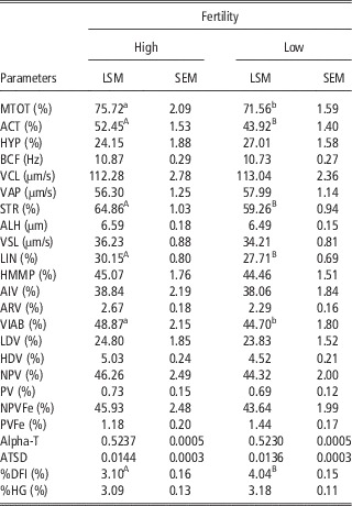
In vitro sperm quality parameters based on the fertility level are presented in Table 1. Computer-assisted semen analysis sperm kinetic parameters MTOT, ACT, STR and LIN were significantly greater in high-fertility bulls, whereas among the other semen parameters, significant differences were observed only for viability and %DFI. Regarding sperm susceptibility to oxidative stress, a significant increase in the number of viable cells with lipid peroxidation was observed in both fertility groups, following treatment with FeSO4. However, this increase failed to highlight any significant differences between the two groups (Supplementary Table S1).
Computer-assisted semen analysis and flow cytometry semen quality parameters of 18 Holstein bulls based on fertility ranking (LSM±SEM)

LSM=Least square mean; MTOT=total motility; ACT=active cells; HYP=hyperactive cells; BCF=beat cross frequency; VCL=curvilinear velocity; VAP=average path velocity; STR=straightness; ALH=amplitude of lateral head displacement; VSL=straight line velocity; LIN=linearity; HMMP=high mitochondrial membrane potential sperm; AIV=intact acrosome viable sperm; ARV=ruptured acrosome viable sperm; VIAB=viability; LDV=viable sperm with low membrane lipid disorder; HDV=viable sperm with high membrane lipid disorder; NPV=viable sperm without lipid peroxidation; PV=viable sperm with lipid peroxidation; NPVFe=viable sperm without lipid peroxidation, Fe induction; PVFe=viable sperm with lipid peroxidation, Fe induction; Alpha-T=red/(red+green) fluorescence intensity; ATSD=Alpha-T standard deviation; %DFI=fragmented DNA sperm; %HG=high green fluorescence sperm.
a,b,A,BValues within a row with different superscript letters differ significantly at P<0.05 and P<0.01, respectively.
Correlations among CASA and FCM semen parameters are given in Supplementary Table S2. Motility descriptors, particularly total motility, displayed considerably significant correlations with parameters that were indicators of good semen quality.
Relations between sperm parameters and fertility prediction models
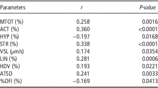
Table 2 shows significant Spearman correlation coefficients between sperm quality parameters and fertility. The kinetic parameters MTOT, ACT, STR, VSL, LIN and HYP and the FCM parameters, such as viable cells with high membrane disorder, ATSD and %DFI all showed correlations with ERCR.
Spearman correlation coefficients between computer-assisted semen analysis and flow cytometry semen parameters and fertility in 18 Holstein bulls

MTOT=total motility; ACT=active cells; HYP=hyperactive cells; STR=straightness; VSL=straight line velocity; LIN=linearity; HDV=viable sperm with high membrane lipid disorder; ATSD=Alpha-T standard deviation; %DFI=fragmented DNA sperm.
Simple correlations between fertility and sperm quality parameters were too weak to predict fertility. Although individual tests on semen samples were partially related to fertility, when those tests were combined in a model, they could account for a substantial proportion of variability. Thus, backward elimination analysis was assessed to obtain a fertility prediction model. The backward elimination analysis produced a final model that accounted for almost half of the variation of ERCR (R 2=0.47). The predictive equation included nine variables: five kinetic parameters (MTOT, ACT, BCF, VCL and ALH) and four DNA integrity indicators (Alpha-T, ATSD, %DFI and %HG). Figure 1 highlights the positive relationship (R 2=0.84, P<0.05) observed between real and predicted fertility.
Relationship between fertility rates predicted by an equation based on the combination of nine sperm quality parameters and field fertility (expressed as the estimated relative conception rate) observed in 18 Holstein bulls (R 2=0.84, P<0.05). The line indicates the trend in the data.

Discussion
In order to identify the most suitable parameters and provide a reliable prediction of fertility, we determined whether a number of semen characteristics, assessed by FCM and CASA in cryopreserved bull semen, were associated with ERCR. In the present study sperm numbers contained in the semen doses were right over the threshold amount of post-thaw viable sperm per AI dose. According to our results, some of the descriptors of motility, viability and DNA integrity were significantly different between bulls with different fertility levels. García-Macías et al. (Reference García-Macías, de Paz, Martínez-Pastor, Álvarez, Gomes-Alves, Bernardo, Anel and Anel2007) observed no significant differences in bull kinetic parameters between fertility groups or most of the FCM parameters; however, significant differences were noted only in morphological abnormalities and DNA integrity. Motility is the most commonly used parameter in routine assessments performed in AI centers, but its correlation with fertility is not universally accepted. We found that some CASA parameters were related to fertility, as previously reported in the bull (Zhang et al., Reference Zhang, Larsson, Lundeheim and Rodríguez-Martínez1998; Januskauskas et al., Reference Januskauskas, Johannisson and Rodríguez-Martínez2003).
The literature reports conflicting data concerning the association between fertility and viability, from a lack of correlation (García-Macías et al., Reference García-Macías, de Paz, Martínez-Pastor, Álvarez, Gomes-Alves, Bernardo, Anel and Anel2007), to positive correlation (Januskauskas et al., Reference Januskauskas, Johannisson, Söderquist and Rodríguez-Martínez2000 and Reference Januskauskas, Johannisson and Rodríguez-Martínez2003). We observed no correlation between viability and fertility rates. The limited differences observed between fertility groups in the present study could be attributed to the narrow fertility range, although all bulls were chosen at the extremes of the fertility distribution curve.
Levels of viable cells with low-lipid disorder were consistent with those reported in previous articles on cryopreserved semen of bulls (García-Macías et al., Reference García-Macías, de Paz, Martínez-Pastor, Álvarez, Gomes-Alves, Bernardo, Anel and Anel2007) and boars (Guthrie and Welch, Reference Guthrie and Welch2005). Moreover, we found no significant differences in membrane stability between high- and low-fertility bulls, in agreement with García-Macías et al. (Reference García-Macías, de Paz, Martínez-Pastor, Álvarez, Gomes-Alves, Bernardo, Anel and Anel2007).
At low and controlled concentrations, reactive oxygen species (ROS) have a regulatory role in specific sperm functions, such as hyperactivation, capacitation, the acrosomal reaction and zona binding (Bailey et al., Reference Bailey, Bilodeau and Cormier2000). However, oxidative stress is recognized as one of the contributing factors to male infertility, as high levels of polyunsaturated fatty acids make sperm particularly vulnerable to peroxidative damage (Aitken et al., Reference Aitken, Wingate, De Iuliis and McLaughlin2007). It is important to establish a balance between ROS production and antioxidant activity, to minimize oxidative stress created by excess ROS and its numerous damaging consequences (Bailey et al., Reference Bailey, Bilodeau and Cormier2000). In the present study, we found low levels of peroxidation and no significant difference between fertility groups. The peroxidation values generally reported in the literature are quite low for frozen-thawed spermatozoa from the bull (Gürler et al., Reference Gürler, Calisici, Calisici and Bollwein2015) and boar (Guthrie and Welch, Reference Guthrie and Welch2007). In a study on horses, Ortega Ferrusola et al. (Reference Ortega Ferrusola, González Fernández, Morrell, Salazar Sandoval, Macías García, Rodríguez-Martínez, Tapia and Peña2009) found large individual variability in peroxidative damage after thawing, that can possibly be attributed to the selection of most stallions based on performance, as opposed to semen freezability.
The proportion of DNA damaged sperm was considerably low, confirming the limited vulnerability of DNA to freezing and thawing, compared with other sperm cell components, such as plasma membranes and mitochondria (Bollwein et al., Reference Bollwein, Fuchs and Koess2008). Consistent with the reports of Ballachey et al. (Reference Ballachey, Hohenboken and Evenson1987) and Waterhouse et al. (Reference Waterhouse, Haugan, Kommisrud, Tverdal, Flatberg, Farstad, Evenson and De Angelis2006), we found a correlation, although lower, between the %DFI and fertility. In the bull, the threshold %DFI for subfertility was defined within the 10% to 20% range (Evenson, Reference Evenson2016). The %DFI recorded in the two fertility groups, despite being significantly different, were both considerably lower than the subfertility range, making it difficult to highlight relationships with fertility. In the present study, the extent of chromatin structure abnormality (ATSD) was positively correlated with fertility, a finding that is inconsistent with those of other authors, who have reported negative correlations (Ballachey et al., Reference Ballachey, Hohenboken and Evenson1987; García-Macías et al., Reference García-Macías, de Paz, Martínez-Pastor, Álvarez, Gomes-Alves, Bernardo, Anel and Anel2007). Martínez-Pastor et al. (Reference Martínez-Pastor, Johannisson, Gil, Kaabi, Anel, Paz and Rodríguez-Martínez2004) observed positive correlations between DNA damage and motility parameters, and suggested that this could be attributed to individual differences between males, rather than a relationship between motility and chromatin status. The unexpected trend of ATSD in our population could be attributed to the low levels of DNA damage that were below the threshold for subfertility in bulls.
Our results regarding acrosome integrity were consistent with those of previous reports on bovine sperm (Gürler et al., Reference Gürler, Calisici, Calisici and Bollwein2015; Sellem et al., Reference Sellem, Broekhuijse, Chevrier, Camugli, Schmitt, Schibler and Koenen2015). This proves the favorable stability of this parameter, as a low percentage of viable acrosome-exocytosed sperm was evident. The acrosomal state caused no significant differences in bull fertility, in agreement with García-Macías et al. (Reference García-Macías, de Paz, Martínez-Pastor, Álvarez, Gomes-Alves, Bernardo, Anel and Anel2007), but in disagreement with Sellem et al. (Reference Sellem, Broekhuijse, Chevrier, Camugli, Schmitt, Schibler and Koenen2015). Levels of mitochondrial functionality observed in the present study were also consistent with previous reports on frozen-thawed semen in the bull (García-Macías et al., Reference García-Macías, de Paz, Martínez-Pastor, Álvarez, Gomes-Alves, Bernardo, Anel and Anel2007; Bollwein et al., Reference Bollwein, Fuchs and Koess2008; Sellem et al., Reference Sellem, Broekhuijse, Chevrier, Camugli, Schmitt, Schibler and Koenen2015) and boar (Guthrie and Welch, Reference Guthrie and Welch2006).
Data processing by the stepwise backward selection procedure facilitated the development of a model that included variables accounting for most of the differences observed in bull fertility, comprising five kinetic parameters and four DNA integrity indicators. This statistical approach, which proceeds by fitting a model with all the variables following by the removal of the least significant variables (the opposite respect to the forward selection), has the advantage of increasing the joint predictive capability of all the variables retained together.
Sperm motility traits and parameters related to chromatin structure have been previously identified as variables highly related with fertility and useful in the development of fertility predictive models (Gillan et al., Reference Gillan, Kroetsch, Maxwell and Evans2008; Oliveira et al., Reference Oliveira, de Arruda, de Andrade, Celeghini, Reeb, Martins, dos Santos, Beletti, Peres, Monteiro and Hossepian de Lima2013). Farrell et al. (Reference Farrell, Presicce, Brockett and Foote1998) indicated that total motility alone had modest correlation with field fertility, whereas if combined with other kinetic parameters (e.g. BCF, VCL and ALH as in our model) it yielded higher levels of correlation. Generally, although regression of the developed model was notable, it was not very high. This could be partially attributed to the inclusion of proven preselected fertile bulls, based on their performance in a progeny test. The inclusion of subfertile subjects in trials might facilitate the quest for a good prediction model (Sellem et al., Reference Sellem, Broekhuijse, Chevrier, Camugli, Schmitt, Schibler and Koenen2015). The use of doses with a limited number of sperm could also highlight differences between bulls for compensable parameters that might otherwise be masked (Waterhouse et al., Reference Waterhouse, Haugan, Kommisrud, Tverdal, Flatberg, Farstad, Evenson and De Angelis2006). However, both these approaches are expensive and impractical in the AI industry.
Our results suggest that sperm characteristics, including motility and chromatin integrity, are important to ensure the fertility potential and play a crucial role in the prediction of fertility. The combined aspects of sperm function are prerequisites for normal in vivo fertilization and early embryonic development. In several previous reports (Gillan et al., Reference Gillan, Kroetsch, Maxwell and Evans2008; Oliveira et al., Reference Oliveira, de Arruda, de Andrade, Celeghini, Reeb, Martins, dos Santos, Beletti, Peres, Monteiro and Hossepian de Lima2013) and in the present study, both these aspects, motility and chromatin integrity, were related to field fertility, thus confirming their relevance to the development of models for predicting fertility. Estimated relative conception rate data account for these aspects that not only indicate true sperm fertility potential (contribution of various cellular functions that allow proper progression of the steps that culminate in fusion of the gametes), but also the possible inadequacy of DNA with consequences on the early stages of embryonic development (Puglisi et al., Reference Puglisi, Pozzi, Foglio, Spanò, Eleuteri, Grollino, Bongioni and Galli2012).
To our knowledge, limited information is presently available in the literature, regarding the use of stepwise backward selection procedures to develop models that associate semen attributes with fertility prediction. Waterhouse et al. (Reference Waterhouse, Haugan, Kommisrud, Tverdal, Flatberg, Farstad, Evenson and De Angelis2006) tested the relationship between multiple sperm parameters and field fertility with a stepwise backward selection procedure, and considered only variables related to DNA status to develop multiple significant models. They found that none of the other parameters evaluated (acrosomal status, viability and mitochondrial potential) were associated with field fertility, in agreement with our findings. Waterhouse et al. (Reference Waterhouse, Haugan, Kommisrud, Tverdal, Flatberg, Farstad, Evenson and De Angelis2006) sought to discriminate between fertility levels of selected high-fertility subjects, and found significant association between the %DFI and field fertility. Similarly, despite the narrow fertility range, we identified an association between DNA parameters and fertility. The model developed in the present study combined different variables that accounted for almost half (R 2=0.47) of the total variability of ERCR and could be easily implemented in AI centers, where motility and kinetic parameters are routinely analyzed and DNA analysis could be readily performed by an external laboratory equipped with FCM, at reasonable costs. Once the accuracy has been confirmed, the prediction of fertility with this model might be beneficial to AI centers in the selection of bulls, and facilitate the elimination of subfertile subjects or ejaculates with low fertilizing capacity.
In conclusion, the present study confirms that a single laboratory test is inadequate to make reliable fertility predictions, whereas the combination of several sperm attributes provides a more accurate and effective predictive tool. In particular, the combination of kinetic semen parameters originating from CASA and DNA analysis based on FCM seem able to distinguish between fertility levels in bulls even in high-fertility range.
Acknowledgment
Research activities were supported by MIUR GenHome project “Technological Resort for the advancement of animal genomic research.”
Supplementary Material
To view supplementary material for this article, please visit https://doi.org/10.1017/S1751731117000684