First described by Zipes et al, fascicular ventricular tachycardia constitutes 10–15% of all idiopathic ventricular tachycardias and is a common form of idiopathic left ventricular tachycardia substrates.Reference Zipes, Foster, Troup and Pedersen1 The literature has established that these arrhythmias tend to be benign, especially in patients with structurally normal hearts. However, tachycardia episodes may be frequent and incessant, rarely leading to cardiomyopathy, haemodynamic collapse, or even sudden cardiac death.Reference Ohe, Aihara, Kamakura, Kurita, Shimizu and Shimomura2
Patients with fascicular tachycardia may respond well to acute and chronic antiarrhythmic therapy, especially to verapamil.Reference Ohe, Aihara, Kamakura, Kurita, Shimizu and Shimomura2 Nonetheless, paediatric patients in need of long-term oral antiarrhythmic therapy may suffer from significant adverse effects, including negative inotropism and extracardiac toxicities.Reference Tondo, Carbucicchio and Russo3 Therefore, for this subgroup of patients, transcatheter ablation has become a preferable treatment option. Research has documented that the procedure is a safe, effective, and curative method, making it a favourable option in drug resistant and non-compliant patients with fascicular tachycardia.Reference Alahmad, Asaad, Arafa, Ahmad Khan and Mahmoud4
In a recent multicentre study, Collins et al reported on the procedural outcomes of catheter ablation in paediatric patients with fascicular tachycardia; the median fluoroscopy time was 24 (1–79) minutes.Reference Collins, Schaffer and Liberman5 Each hour of fluoroscopy increases the lifetime risk of fatal cancer by up to 1%, and children are more susceptible to ionising radiation; therefore, radiation exposure should be minimised to reduce the long-term cancer risk in this population.Reference Pearce, Salotti and Little6
This study aimed to demonstrate the outcomes of transcatheter radiofrequency ablation procedures performed via three-dimensional electroanatomic mapping systems using near-zero fluoroscopy in a paediatric patient cohort with fascicular tachycardia.
Materials and methods
In this study, we included consecutive patients with fascicular tachycardia who underwent an electrophysiologic study and a transcatheter radiofrequency ablation in our hospital from January 2014 to June 2019. The study protocol was approved by the local ethical committee, and written informed consent was obtained from the legal guardians of all patients in the study.
Procedural data
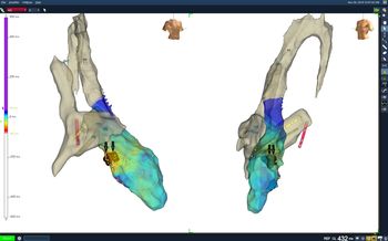
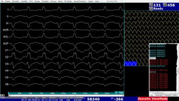
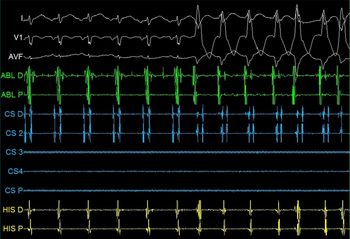
For all patients, the indication for ablations was fascicular tachycardia. Antiarrhythmic medications, if any, were ceased for a period of at least five half-lives prior to procedures. All procedures were performed under deep sedation in a fasting state. The right femoral artery and both femoral veins were cannulated in all patients. Six French quadripolar catheters were placed at the His bundle and right ventricular apex. The anatomy of the right atrium was mapped, and the inferior and superior caval veins as well as the His bundle were marked using a three-dimensional mapping system (Ensite NavX System; St Jude Medical Inc, St Paul, MN) (Fig 1). Basic measurements were made at the beginning of the procedures. Then, we performed a programmed stimulation implementing ≤3 extra stimuli with two different cycle lengths from the right ventricular apex, right ventricular outflow tract, and right atrium. We also conducted incremental burst pacing from the right ventricle and right atrium (up to cycle lengths of 280 and 200 ms, respectively) whenever needed to induce ventricular tachycardia. We used theophylline ethylenediamine, a non-selective adenosine receptor antagonist and a non-selective phosphodiesterase inhibitor, to induce fascicular tachycardia when needed. Fascicular tachycardia was inducible in all patients with or without the use of intravenous bolus of theophylline ethylenediamine (up to 6 mg/kg) (Figs 2 and 3). We excluded differential diagnoses, such as atrioventricular reentrant tachycardia and bundle branch reentry, using established deductive criteria during the electrophysiologic study in combination with diagnostic maneuvers, activation mapping, and entrainment.
Three-dimensional anatomy of the right atrium, superior caval vein inferior caval vein, coronary sinus, left ventricle, ascending aorta, and his bundle region are demonstrated in left anterior oblique and right anterior oblique views. Radiofrequency lesions given are shown on the three-dimensional electroanatomic mapping system.

Twelve channel electrocardiogram obtained during tachycardia is shown.

Fascicular tachycardia induced by programmed stimulation is demonstrated.

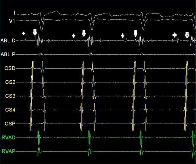
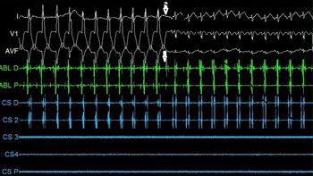
The procedures were performed without using any fluoroscopy in all but three patients. In three patients, limited fluoroscopy was employed while passing through the aortic arch and the aortic valve in order to avoid any damage to these structures. The left ventricle was accessed retrogradely with a Marin® 7 Fr 4-mm deflectable tip catheter for ablation and left ventricular mapping (Medtronic Inc., Minneapolis, MN, USA). To pass through the aortic arch, we gave the ablation catheter a nice curve once it reached the level of the pre-marked superior caval vein in the right and left anterior oblique positions on the electroanatomic map. Then, we paid close attention to giving the ablation catheter a maximum curve in the ascending aorta to easily access the left ventricle through the aortic valve. We created an endocardial left ventricle geometry on the electroanatomic mapping system and investigated the earliest prepurkinje potentials during activation mapping performed during ventricular tachycardia (Figs 4 and 5). We took the utmost care to position the ablation catheter at a safe distance from His structures. Whenever possible, entrainment was performed to confirm the targeted site’s participation in the arrhythmia circuit.
Prepurkinje (cross) and purkinje potentials (arrow) during sinus rhythm are shown on the electrocardiogram screen.

Fascicular ventricular tachycardia has been stopped by radiofrequency ablation in the earliest activation site.

Ablation procedure
Temperature-controlled radiofrequency energy was delivered at the site with the earliest prepurkinje potentials during ventricular tachycardia. The power output was titrated to as high as 30–35 W to achieve a target temperature of 45–60°C for 60–90 s. The ablation procedure was considered successful if the fascicular ventricular tachycardia terminated during ablation or if any ventricular tachycardia was not inducible 45 minutes after ablation despite repeat programmed stimulations and an intravenous bolus of theophylline ethylenediamine.
Statistical analysis
Statistical Package for the Social Sciences 22.0 (IBM Corporation, Armonk, New York, United States) was used to analyse all data. Quantitative data were expressed as mean ± SD (standard deviation) (maximum–minimum) values. Categorical values were stated as n (number) and percentage (percentage).
Results
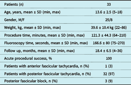
Thirty-three paediatric patients with fascicular tachycardia were included in the study (Table 1). Twenty-five patients were male, and eight were female. The mean weight of the patients was 39.6 ± 10.4 kg (22–86), and the mean age was 13.6 ± 2.5 years (5–18).
Demographic, procedural, and follow-up data.

The indication for ablation was decreased left ventricular systolic function, revealed by echocardiography (EF: 45%) in one patient due to incessant fascicular tachycardia. Eight patients refused to use antiarrhythmic medications, nine patients were on monotherapy, and 15 patients received dual antiarrhythmic therapy. Among patients on medication, 6 were symptomatic, and 4 had received direct cardioversion due to the recurrence of fascicular tachycardia and haemodynamic instability. The decision to undergo the procedure was made based on patient preferences, symptomatology, and possible drug side effects. A mitral valve prolapse was observed in one patient, and three patients had undergone unsuccessful ablation procedures previously (one anterior fascicular, two posterior fascicular).
The mean procedure time was 121.3 ± 44.3 minutes (64–210) (Table 1). We were able to induce fascicular tachycardia in all patients, and left ventricular mapping was performed during the tachycardia in all. A 7-Fr, 4 mm, non-irrigated radiofrequency ablation catheter was employed in all patients. The radiofrequency energy (limit 60°C 35 W) was delivered for 60 seconds when the tachycardia ceased within 10 seconds of ablation. The application of ablation induced rapid firing of fascicular tachycardia in all patients. The ablation was terminated when the tachycardia persisted after the first 10 seconds of ablation. The mean number of effective lesions was 2.8 ± 1.2 (2–7). All tachycardias stemmed from the posterior fascicle except in one patient where the site of tachycardia was the left anterior fascicle. No fluoroscopy was needed in 30 patients. The mean fluoroscopy time in the remaining patients was 166.6 ± 80 seconds (75–270).
The procedural acute success rate was perfect (100%) without any major complications. No patients developed left bundle branch block or complete atrioventricular block. A posterior fascicular block was observed in three patients after the ablation procedures.
The mean follow-up period was 18.4 ± 6.5 months (range: 4–36). During the study period, nine patients complained of palpitation after their procedures. The rhythm documented on electrocardiogram, Holter monitoring, or event recorder during palpitation was sinus tachycardia in eight patients. Recurrence was noted in only one patient 12 months after the index procedure. An electrocardiogram performed during palpitation revealed recurrent fascicular ventricular tachycardia. In response, the patient received a repeat radiofrequency ablation procedure. The initial procedure was performed via three-dimensional electroanatomic mapping without using fluoroscopy, and the site of ablation in the second procedure was the same on the three-dimensional map as in the first procedure. The patient has been symptom-free for the last 14 months, and no permanent ablation-related complications occurred.
Discussion
In this study, we demonstrated the outcomes of radiofrequency ablation procedures performed with the EnSite® system using near-zero fluoroscopy in a paediatric cohort with fascicular tachycardia.
The acute success rate of our procedures was excellent with a very satisfying recurrence rate in the follow-up period. In a recent review analysing 953 patients from 38 studies, the success rate of radiofrequency ablation in patients with fascicular tachycardia was very high, reaching up to 100% in some studies. However, compared to adult patients, the success rate was significantly lower in paediatric studies (90% versus 94.3%).Reference Creta, Chow and Sporton7 Use of a three-dimensional electroanatomic mapping system varied significantly among studies included in the review. In our procedures, three-dimensional electroanatomic mapping systems helped confirm the exact catheter position prior to ablations, allowing us to label the location of ablation lesions on the three-dimensional map. We thought that the three-dimensional mapping system rendered our ablation lesions more precise and effective, compared to fluoroscopy-only procedures.
Three-dimensional electroanatomic mapping systems have proven useful in the ablation procedures of paediatric patients with supraventricular and ventricular outflow tract tachycardia. However, there is limited experience in the use of mapping systems in paediatric patients with fascicular tachycardia, and conventionally, X-rays have been used for catheter navigation during these procedures.Reference Balli, Kucuk, Orhan Bulut, Kemal Yucel and Celebi8,Reference Koca, Akdeniz, Karacan and Tuzcu9 It is known that children are very susceptible to ionising radiation beginning from fetal life.Reference Liu, Fang and Yang10–Reference Lyan, Tsyganov and Abdrahmanov13 Therefore, radiation exposure should be minimised in paediatric patients. Recently, Koca et al showed that the ablation of fascicular tachycardia using electroanatomic mapping systems with limited fluoroscopy is feasible and effective in paediatric patients. In their study, fluoroscopy was used only for the introduction of radiofrequency ablation and diagnostic catheters, retrogradely into the left ventricle.Reference Koca, Akdeniz, Karacan and Tuzcu9 Likewise, in our study, we used limited fluoroscopy in some patients when we had difficulty advancing our catheters through the aortic arch and the aortic valve. We believe that, with increasing knowledge and experience with electroanatomic mapping systems, physicians will be able to decrease significantly the use of fluoroscopy in paediatric patients undergoing ablation procedures for fascicular tachycardia.
The technique employed to find and confirm the exact site of a tachycardia circuit significantly affects the success rate of procedures in patients with fascicular tachycardia. In some studies, ablation of the fragmented purkinje potentials has been successful in patients with non-inducible or nonsustained ventricular tachycardia. However, these techniques are associated with lower success rates and higher recurrence rates when compared to those performed during tachycardia.Reference Liu, Fang and Yang10,Reference Talib and Nogami14–Reference Luo, Zhou and Guo16 In our study, we tried to map the earliest fascicular and purkinje potentials during ventricular tachycardia. Fascicular entrainment was also performed whenever possible. These techniques are highly useful in determining the exact location of the arrhythmia circuit and in increasing the success rate of ablation procedures in patients with fascicular tachycardia.
There have been several possible mechanisms of fascicular tachycardia recurrence reported. Nishiuchi et al have reported that upper septal VT may be responsible for fascicular tachycardia recurrence in patients who had previously undergone successful ablation of posterior fascicular tachycardia.Reference Nishiuchi, Nogami and Naito17 The secondary exit point of the arrhythmia circuit may change after the ablation, moving to the upper septal region or papillary muscles.Reference Nair, Thomas, Stoyanov, Nery, Sadek and Green18 Nonetheless, in our patient with recurrence, the arrhythmmia circuit was found to be in the exact same location as detected in the first procedure.
Researchers have used isoproterenol and isoprenaline infusions when the induction of tachycardia is not possible through extrastimulation and burst pacing maneuvers.Reference Gopi, Nair and Shelke19 In our study, we used theophylline ethylenediamine when tachycardia could not be induced through programmed stimulation and burst pacing. Theophylline ethylenediamine (aminophylline) is a short-acting methylxanthine that shortens sinoatrial, atriventricular, and His-purkinje conduction times by increasing intracellular cAMP. Theophylline ethylenediamine is known to enhance the triggered activity causing ventricular tachycardia.Reference Belardinelli, Fenton, West, Linden, Althaus and Berne20–Reference Aliot, Stevenson, Almendral-Garrote, Bogun, Calkins and Delacretaz22 An intravenous bolus of theophylline ethylenediamine proved useful in helping to induce the tachycardia in all 15 patients when it was not possible through standard protocols.
Conclusions
In this study, we demonstrated the outcomes of radiofrequency ablation procedures performed through the EnSite system using near-zero fluoroscopy in a paediatric cohort with fascicular tachycardia. We suggest that radiofrequency ablation procedures, performed via an electroanatomic mapping system, are quite safe and effective, demonstrating high success rates in paediatric patients with fascicular tachycardia. This technique has the benefit of avoiding ionising radiation exposure for both the patient and operator, thus reducing the lifetime risk of malignancy for this population.
Limitations
The relatively short follow-up period is the main limitation of this study. However, the study presents valuable information on the feasibility of catheter ablation procedures using near-zero fluoroscopy in paediatric patients with sustained fascicular tachycardia.
Acknowledgements
None.
Financial support
This research received no specific grant from any funding agency, commercial, or not-for-profit sectors.
Conflicts of interest
None.
Ethical standards
The authors assert that all procedures contributing to this work comply with the ethical standards of the Office for Human Research Protections of the Department of Health and Human Services and with the Helsinki Declaration of 1975, as revised in 2008, and has been approved by the University of Maryland Institutional Review Board.