Coarctation of the aorta is present in 5–8% of all patients with congenital heart disease and is seen in 0.04% of all live births. In 1981, balloon angioplasty was successfully administered to a neonate for the first time.Reference Singer, Rowen and Dorsey 1 Since then, transcatheter interventions have increased and have been adopted as the popular therapeutic approach of choice for many patients. Expandable balloon intravascular stent implantation, which has been administered for approximately the past two decades in older children and adults for the treatment of native and recurrent aortic coarctation, is a widely accepted therapeutic modality with successful results, despite difficulties in administration and complications. Today, it is the therapeutic modality of choice for appropriate patients.Reference Suarez de Lezo, Pan and Romero 2 , Reference Forbes, Garekar and Amin 3 Increased experience in stenting and related data generally result from retrospective studies. In contrast, the post-procedural follow-up of these patients is generally limited to echocardiographic examinations. In many reports, magnetic resonance imaging, computerised tomography imaging, or cardiac catheterisation were administered in patients because of concerns of stent-related complications or restenosis.Reference Thanopoulos, Triposkiadis, Margetakis and Mullins 4 – Reference Baykan, Karagöz and Celiker 11
In this study, we present the follow-up results of 38 intravascular stent implantations administered to 35 patients at our centre along with the results of multi-slice computerised tomography. We have presented the following in our study: results pertaining to the selection of patients for stent implantation, the efficacy of the procedure, the complications observed during and after the procedure, imaging results obtained during follow-up, that is, by cardiac catheterisation and multi-slice computerised tomography, and the need for antihypertensive therapy.
Materials and methods
The features of 35 patients who were implanted with a total of 38 covered or bare stents (NuMED CP (cheatham platinum) stent, Heart Medical Europe BV, Best, the Netherlands) between March, 2007 and July, 2012 were examined. For all patients to be enrolled into the study, the following details were recorded: demographics and characteristics – discrete, subatretic, long-segment, or tortuous; morphology – native or recurrent; localisation and diameter of the coarctation; – coarctation diameters and the ratio of the diameter of the coarctation site to that of the descending aorta at the diaphragmatic level; other concomitant congenital heart diseases; pre-procedural and post-procedural systolic gradients; pre-procedural and post-procedural blood pressure values; and technical data such as the dimensions, types, and covers of the stents and balloons used. In addition, the uses of antihypertensive medications were evaluated. The definitions of transverse arch hypoplasia, aneurysm, subatretic coarctation, discrete coarctation, and long-segment coarctation have been described previously.Reference Holzer, Qureshi and Ghasemi 12 Stent implantation was preferred in patients with subatretic narrowing or a functionally interrupted aortic arch, in those with an aneurysm or patent ductus arteriosus accompanying the coarctation, and in those with recoarctation after surgery or sequential angioplasty. Although a covered stent was preferred in patients with subatretic, long-segment, and recurrent coarctations, a bare stent was preferred in those in whom the coarctation was localised to the transverse arch. Before the procedure, all patients were diagnosed as having a coarctation by echocardiography, and the diagnosis was based on the presence of a systolic gradient greater than 20 mmHg across the lesion. The blood pressure was measured from four extremities. Hypertension was diagnosed in the presence of measured upper extremity systolic blood pressure was found to be more than 95% of normal range.
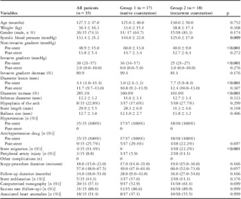
Native coarctations were grouped as Group 1 and recoarctations seen after balloon angioplasty or surgery were grouped as Group 2. The two groups were compared in terms of procedural success, pre-procedural and post-procedural echocardiography and catheterisation data, hypertension rates, and complication rates during follow-up (Table 1).
Procedure and patient characteristics.

Values are expressed as mean ± standard deviation or median (25th–75th percentiles)
Catheterisation technique
All the procedures were performed under general anaesthesia and some were performed with intubation. In all patients, the cover was placed; thereafter, 100 U/kg heparin was administered and the heparin doses were repeated to yield an activated clotting time of more than 200 seconds during the procedure. For stent implantation, the femoral arterial route was used in all patients.
Coarctation was generally visualised by administering an injection of contrast in the lateral and left anterior-oblique or anterior-posterior positions in the proximal of the lesion using a pigtail catheter. The ascending aorta, isthmus, coarctation, and descending aorta diameters, length of the coarcted segment, and distance to the brachiocephalic artery were recorded. The presence of a duct was evaluated. The pressure gradient across the lesion area was measured by performing a pull-back. The length of the stent to be administered was selected based on the length of the aorta involved. The diameter of the balloon to be used was selected to be not larger than the diameter of the aorta at the level of the diaphragm and at the most 2 mm larger than the isthmus. Bioenterics intragastric balloon was used in all patients. In order to prevent technical complications such as balloon migration and rupture, a balloon as long as the stent or one slightly longer than the stent was used. The procedure was conducted and terminated as previously described.Reference Holzer, Qureshi and Ghasemi 12 The position and presence of vascular complications were evaluated by injecting a contrast in the proximal of the coarctation after the procedure. The diameter of the enlarged lesion area was measured. Careful pull-back re-measurements were performed to determine the post-stenting gradient. If the position of the stent was appropriate and the decrease in the diameter and gradient was adequate, the procedure was terminated.
The success criteria of the procedure included the lack of catheterisation-related death, a systolic gradient lower than 10 mmHg on the stent, and the absence of residual stenosis, which is dilatation of the narrowest segment by more than 50%. Complications existing during the intervention or those that required observation/treatment before discharge were considered acute.
Follow-up
The patients were followed up for 24 hours in the ward after the procedure for eventual blood pressure changes and acute complications. Electrocardiographic monitoring was performed. Acetyl salicylic acid was not initiated and heparin infusion was not continued after the catheter room. All patients received propranolol therapy for 1 month to prevent rebound hypertension immediately after the procedure. The patients who were still hypertensive in their follow-up visits continued to receive treatment at appropriate doses. Patients were seen on the 15th day, 1st month, 3rd month, and 6th month for echocardiography, electrocardiography, blood pressure monitoring in the four extremities, and other examinations. Thereafter, their follow-up visits were performed at 6-month intervals. Patients who completed a 2-year follow-up and gave written consent were evaluated using multi-slice computerised tomography. The multi-slice computerised tomography examinations were performed using a 16-row multi-slice computerised tomography scanner (LightSpeed 16; GE Healthcare, Milwaukee, Wisconsin, United States of America). Patients who showed systemic hypertension with an upper to lower limb systolic blood pressure difference of more than 20 mmHg and a pressure gradient on echocardiography of more than 20 mmHg during follow-up were evaluated by repeated angiocardiography.
Results
The pre-procedural, post-procedural, and follow-up demographics as well as procedural characteristics of all 35 patients, 26 of whom were boys, are summarised in Table 1. Of the patients, five (14.3%) weighed less than 20 kg, 11 (31.4%) weighed between 20 and 30 kg, and 19 (54.3%) weighed over 30 kg (11–70 kg). These 35 patients underwent a total of 38 stent, 12 bare and 26 covered, implantation therapies – three patients underwent a second stent implantation in the same session because of migration of the stent. Among the patients, 18 (51.4%) developed recoarctation – 14 patients who had undergone angioplasty; three patients, both angioplasty and surgery; and one patient, both angioplasty and stent implantation. Although these two patient groups showed statistically significant differences in terms of pre-procedural blood pressure values, non-invasive and invasive gradients, coarctation diameters, and coarctation site diameter to the descending aorta diameter ratios, they had similar data in terms of other properties. There was no statistically significant difference in terms of post-procedural data (Table 1).
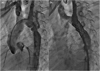
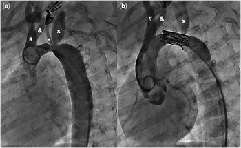
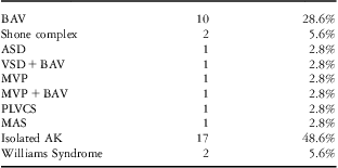
The coarctation was subatretic-discrete in six patients with native coarctation. In addition, it was long-segmented in seven patients and was tortuous in one patient (Fig 1). The coarctations were localised in the distal isthmus in 24 (68.6%) patients, in the proximal isthmus in seven patients, in the distal transverse arch in two patients, in the proximal transverse arch in one patient (Fig 2), and in the thoracic aorta in one patient. The coarctations associated with different anomalies are shown in Table 2.
An 8-year-old boy with an aneurysmatic tortuous coarctation of the aorta on the distal arch and proximal isthmus. The left anterior oblique aortogram ( a ). The aortogram image in the same position after bare stent implantation ( b ).

A 3-year-old girl weighing 11 kg with complex coarctation of the proximal aortic arch. The left anterior oblique aortogram views before ( a ) and after the stenting (28 mm bare stent) procedure. #The brachiocephalic artery; &The left common carotid artery; +The vertebral artery; xThe left subclavian artery.

Coarctation and associated anomalies.

ASD = atrial septal defect; BAV = bicuspid aortic valve; MAS = middle aortic syndrome; MVP = mitral valve prolapses; PLVCS = persistent left vena cava superior; VSD = ventricular septal defect
Before the procedure, all patients had been receiving antihypertensive medication – metoprolol or propranolol. During the post-procedural follow-up, the drug was discontinued in 26 patients (p < 0.001) and nine patients (25.7%) continued to use antihypertensive medications – six patient used propranolol and three patients used metoprolol; however, all patients remained normotensive (p < 0.001). Of these patients, seven developed recoarctation and two had a native coarctation.
The procedure was successful in all patients during the acute phase. The pre-procedural and post-procedural non-invasive and invasive gradients, coarctation diameters, and coarctation site diameter to the descending aorta diameter ratio changes were statistically significant in both groups (p < 0.001). Before the procedure, 11 (31.4%) patients showed minimal and moderate aortic regurgitation on echocardiography. After the procedure, minimal aortic regurgitation was resolved in three patients and aortic regurgitation regressed from moderate to minimal in three patients. Stent implantation was found to be significantly efficient in the resolution or reduction of aortic valve failure (p = 0.031) even when other reasons were not taken into account. The bicuspid aortic valve was present in all patients with sustained aortic regurgitation.
The 25 covered stents (68.4%) that were used were generally preferred for subatretic coarctations – six patients; long-segment coarctations – three patients; tortuous coarctations – one patient; patent ductus arteriosus – one patient; as well as in patients with an aortic aneurysm – four patients. Other patients in whom covered stents were preferred included those who developed recoarctation after balloon or surgical intervention.
In one patient with subatretic coarctation, the catheter could not be passed from the distal of the narrowing to its proximal. In this patient, pre-dilatation was performed using 3 and 6 mm balloons through a second axillary arterial route. Moreover, in all patients, the femoral arterial route was used. In seven patients who did not show an efficient enlargement of the stent, a second stent dilatation was performed by preferring a one size larger balloon diameter during the same session.
There was no statistically significant difference between the five patients who weighed less than 20 kg and the other 30 patients who weighed over 20 kg in terms of demographic and procedural characteristics, procedure success and complication rates, and follow-up duration.
Complications
No procedure-related deaths were reported. Of the patients, four (11.4%) exhibited stent migration during balloon dilatation. In these four patients, the stent migrated to the aneurysmatic dilatation zone in the distal of the lesion. The first patient, who had middle aortic syndrome, had coarctations in two different regions of the thoracic aorta and had received a bare stent for the proximal coarctation at another centre. In this patient, a 45 mm covered stent shifted to the distal of the coarctation after being fully expanded. A 39 mm bare stent was placed between the two stents and was successfully fixed. The second, third, and fourth patients had discrete recurrent coarctations in the distal of the isthmus. The stent was deployed in the aneurysmatic enlargement of the abdominal aorta because it could not be retrieved and was thus fixed at that location. Thereafter, another bare stent was placed for appropriate localisation and was fixed using a bioenterics intragastric balloon balloon. Although these four patients developed recoarctation, the patients with native coarctations did not show stent-related technical complications.
A total of three patients exhibited peripheral arterial injuries. In the first patient, a 3-year-old child who weighed 11 kg and had a native coarctation localised in the proximal transverse arch, the distal part of the 10 Fr long sheath could not be pulled out. The sheath was removed without any complications during the surgery. During follow-up of these three patients, no circulatory complications were observed in the associated extremity. Peripheral arterial pulselessness was reported in the second and third patients who were recanalised by anticoagulant therapy. During the procedure, none of the patients showed dissection, aneurysm rupture, or stent fracture.
Follow-up
The mean follow-up duration was 34 ± 16 months. The follow-up duration was over 60 months – long-term – in three patients (8.6%), 18–60 months – intermediate term – in 26 patients (74.2%), and less than 18 months – short-term – in six patients (17.1%). The echocardiographic gradient measurements obtained 24 hours after the procedure (17.8 ± 11) and the last echocardiographic gradients (13.8 ± 5.4) were not statistically significantly different (p = 0.09). Of the patients, nine who underwent stent implantation continued to use antihypertensive medications after the procedure: 5/15 (33%) in Group 1 and 4/18 (22%) in Group 2 (p = 0.697). The follow-up duration was similar between the nine patients who continued to receive antihypertensive therapy (mean: 29 ± 23 months) and the other patients (p = 0.468). On average, 21 (6–42) months after the procedure, six patients underwent cardiac catheterisation because of concerns for restenosis. In these patients, the pre-procedural mean non-invasive gradient was 29 mmHg and the pre-procedural mean invasive gradient was 15 mmHg. Only one patient had an invasive gradient greater than 20 mmHg. Of these six patients, four underwent balloon dilatation. This was accompanied by hypoplasia of the aortic arch in all four patients. Of the patients, two who underwent balloon dilatation had native coarctations and the other two had recurrent coarctations.
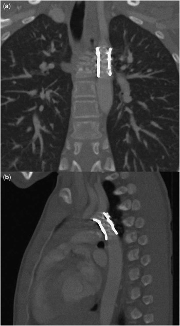
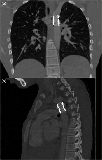
A total of 20 patients (57.1%) who completed the 2-year follow-up and gave written consent underwent multi-slice computerised tomography. The multi-slice computerised tomography was not performed because of a concern for restenosis or stent-related technical complications, but to evaluate the efficacy of additional imaging tools in follow-up. On multi-slice computerised tomography, none of the patients showed stent migration or apparent fractures, important restenosis, formation of an aneurysm, or intimal ruptures (Figs 3 and 4).
Thoracic multi-detector computed technology images of a 13-year-old boy with recoarctation after stent implantation. Coronal view ( a ) and sagittal view ( b ). Restenosis, migration, and apparent fractures and aneurysms cannot be seen.

Thoracic multi-detector computed technology images of a 17-year-old boy with native coarctation of the aorta after stenting. Coronal view ( a ) and sagittal view ( b ). Restenosis, migration, and apparent fractures and aneurysms are not seen.

Discussion
During the past two decades, stent implantation has become a widely accepted therapeutic modality for the treatment of native and recurrent coarctations in children and adults.Reference Singer, Rowen and Dorsey 1 – Reference Krasemann, Bano, Rosenthal and Qureshi 13 In these studies presenting the short-term and intermediate-term results of children, satisfactory results were obtained for efficient correction of the narrowing in both native and recurrent coarctations. In contrast, studies involving long-term results also reported favourable results that regard the safety and efficacy of the procedure.Reference Shah, Hijazi, Sandhu, Joseph and Cao 7 , Reference Pedra, Fontes and Esteves 8 , Reference Holzer, Qureshi and Ghasemi 12 , Reference Krasemann, Bano, Rosenthal and Qureshi 13 Our accepted success criteria were: the lack of procedure-related death, the reduction of the invasive gradient to below 10 mmHg, and an enlargement of the lesion area by more than 50%. The success rate according to these criteria was determined to be 100%. Stent implantation was seen to be a useful technique to reduce invasive/non-invasive gradients, eliminate the narrowing of the aorta, reduce the incidence of hypertension and the need for antihypertensive therapy, improve aortic valve failure, and increase the effort capacity. However, as four of six patients who had undergone recatheterisation underwent stent dilatation an average of 21 months after the procedure, during follow-up the long-term cumulative success rate remained 88.6% (31/35). The patients were taken into the catheterisation laboratory because of blood pressure differences between the upper and lower extremities and because of a pressure gradient more than 20 mmHg across the lesion area on echocardiography. In the catheterisation laboratory, only one of the six patients had a measured invasive gradient of more than 20 mmHg. In some studies, patients in whom an important gradient was detected by echocardiography despite no invasive gradient received dopamine in the catheterisation laboratory. Thus, it was demonstrated that the gradient may be decreased and could not be detected under deep sedation and may appear after pharmacological stimulation.Reference Holzer, Qureshi and Ghasemi 12 , Reference Krasemann, Bano, Rosenthal and Qureshi 13 The other three patients who had an anatomical narrowing in the stent lumen underwent stent redilatation. None of these three patients with an invasive gradient of less than 20 mmHg were stimulated with dopamine during the catheterisation procedure.
Finally, in the largest multi-centre study performed by Holzer et al, it was reported that 12% of patients in whom a stent was implanted an intervention was expected; only 4% underwent redilatation.Reference Holzer, Qureshi and Ghasemi 12 However, in several studies, during the intermediate-term and long-term follow-ups after stent implantation, the incidence of re-intervention ranged between 7%Reference Chessa, Carrozza and Butera 14 , Reference Sua′rez de Lezo, Pan and Romero 15 and 33%.Reference Qureshi, McElhinney, Lock, Landzberg, Lang and Marshall 16 – Reference Thanopoulos, Eleftherakis, Tzanos, Skoularigis and Triposkiadis 18 The incidence of re-intervention was reported to vary between 6% and 20% after surgical repair.Reference Hager, Kanz, Kaemmerer, Schreiber and Hess 19 , Reference Wright, Nowak, Goldberg, Ohye, Bove and Rocchini 20 In our study, we performed re-catheterisation in 16.6% and re-intervention in 11.4% of patients.
Despite all these satisfactory haemodynamic and anatomical results, stent implantation involves some rare risks. It was reported that the rupture of the aortic wall, which is a catastrophic complication of the procedure, was more commonly observed in older patients with decreased aortic wall compliance and in children with a concomitant vascular pathology. In a large multi-centre study, the incidence of such a rupture was reported to be 4/521.Reference Forbes, Garekar and Amin 3 In our study, covered stents, which protect against eventual wall rupture and dissection development, were successfully used in children with aneurysmatic, long-segment, tortuous, and subatretic coarctations. None of the patients developed ruptures or dissections. No procedure-related deaths were observed during follow-up.
Although procedure-related technical complications have decreased over the past 10 years because of the introduction of new balloons and stents, even if rarely, complications may still be observed. In our study, as a technical complication, stent migration was observed at a high rate of 4/35 (11.4%). However, all of these four patients had recurrent coarctation with an aneurysmatic aortic segment in the distal of the lesion. Further, they had complex coarctations – two patients with aortic arch hypoplasia and one patient with multiple narrowing in the abdominal aorta. This complication did not lead to procedural failure or important morbidity. In these four patients, the procedures were terminated by using appropriate manipulations without the need for a surgical intervention. In the multi-centre study conducted by Forbes et al, the incidence of procedure-related technical complications was reported to be 10.7%, and it was demonstrated that the majority of these complications (5%) were stent migrations. In the same study, in half of the 28 patients in whom stent migration was observed the most common reason was shown to be the presence of a large aortic segment after the lesion. Other reasons were cited as the preference for small balloons and balloon rupture.Reference Forbes, Garekar and Amin 3 However, in some centres, right ventricle rapid pacing via a catheter or the use of several drugs is recommended just before inflation of the balloon in order to prevent sudden falls in the blood pressure and stroke volume.Reference Forbes, Garekar and Amin 3 , Reference Nienaber, Kische and Rehders 21 – Reference Kannan and Srinivasan 23 In our study, we did not use the right ventricle rapid pacing technique during the inflation of the balloon. Further, none of the patients showed other technical complications such as balloon rupture, occlusion of the brachiocephalic artery with the stent, or stent fracture. Only one of three patients in whom a femoral arterial injury was observed required surgery. This patient, who underwent femoral arterial repair, did not experience any circulation problems in subsequent follow-ups. Only one of three patients weighed less than 20 kg; the other two patients weighed over 40 kg. Similarly, in the literature, peripheral vascular complications reported with an incidence rate of 2–5% were mostly reported in patients with a low weight and in whom a large cover was used.Reference Forbes, Garekar and Amin 3 , Reference Tanous, Collins, Dehghani, Benson and Horlick 24 , Reference Carr 25
After the procedure, all patients received antihypertensive therapy to reflex hypertension for 1 month. The antihypertensive medication was discontinued in normotensive patients during the first visit. In the second visit after the procedure, that is, the third month, the systolic blood pressure of the upper extremity was found to be more than 95% in seven patients (20%) and 99% in two patients (5.6%). In these nine patients, antihypertensive therapy was continued. Interestingly, during follow-up, only three of these nine patients showed a value less than 20 mmHg across the lesion area by echocardiography. This indicates that, in patients with coarctation, despite the elimination of the anatomical narrowing with a stent, hypertension could be permanent because of decreased aortic elasticity and impaired aortic compliance.
In the literature, it was reported that stent implantation can be applied for the treatment of coarctation in infants and even in premature babies and that successful results can be obtained.Reference Dryżek, Ostrowska, Góreczny, Moszura, Politowska and Sysa 26 – Reference Rao 30 However, the patients who were reported in these reports were critical patients with therapy-resistant cardiac failure and thus surgical therapy was risky. The stents that were used for the procedure included low-profile stents such as Palmaz 8 coronary stents that do not extend up to adult aortic diameters. Therefore, stent implantations for coarctations performed using these stents were palliative procedures that required a second intervention and were performed to gain time. As cheatham platinum stents can be extended to adult aortic diameters, they may be used as a curative therapeutic modality in the treatment of coarctations. However, the age and weight range for a safe application of these stents is known to be above 6 years and over 25 kg. In our centre, stenting was performed in seven (20%) patients who weighed less than 25 kg. The patients with a lesser weight who underwent cheatham platinum stent implantation for the treatment of coarctation showed similar results in terms of demographics, clinical characteristics, procedural properties, and follow-up findings when compared with other patients who weighed over 20 kg. In addition, the incidence of complications among these patients was also not higher when compared with other patients. We believe that, by increasing the experience, the age and weight limits recommended by the manufacturer may be forced beyond the limits in the hands of experienced interventionists.
In many centres, follow-ups of patients who underwent stent implantation have remained limited to chest X-rays, blood pressure measurements, electrocardiography, and echocardiography. In these centres, advanced imaging methods such as computerised tomography, magnetic resonance imaging, or cardiac catheterisation are performed in patients because of the concern for complications such as recoarctation, formation of an aneurysm, intimal proliferation, and stent migration. In the report of a multi-centre study conducted on 578 adults and children by Forbes et al, 160 patients underwent imaging methods such as computerised tomography, magnetic resonance imaging, and catheterisation.Reference Forbes, Moore and Pedra 17 The imaging results were normal in 103 patients (71.5%). However, 41 patients (28.5%) had pathological results, including neointimal hyperplasia, formation of an aneurysm, dissection/intimal rupture, and stent fracture; further, nine patients (22%) needed another stent implantation. Therefore, it was stated that limiting follow-ups to echocardiographic examination may result in patients with coarctation who need repeated intervention remaining undiagnosed. A total of 20 patients who had their regular follow-ups at our centre and gave written consent underwent multi-slice computerised tomography. None of the patients who underwent multi-slice computerised tomography showed complications such as restenosis, intimal proliferation, formation of an aneurysm, and apparent stent fracture/migration. We believe that this resulted from the fact that our follow-up results mostly contained short-term and intermediate-term data and that the patients at risk of complications such as stent restenosis had undergone cardiac catheterisation before undergoing multi-slice computerised tomography. We think that the pathological results exhibited by 28.5% of the imaging methods, except echocardiography, in the study performed by Forbes et al might have resulted from the administration of the majority of these imaging tools to patients with extraordinary results in whom some complications were expected.
Conclusion
Stent implantation for coarctation of the aorta is a method yielding satisfactory results in the reduction of both invasive and non-invasive gradients and efficient enlargement of the lesion area. Technical complications such as stent migration are the main adverse effects of this procedure and are associated with the anatomical properties of the lesion. However, because of the nature of the coarctation, the need for antihypertensive drugs may persist in some patients. Cheatham platinum stents may also be applied to selected patients whose anthropometric measures are below the age and weights recommended by the manufacturer. In patients with both native and recurrent coarctations, repeated intervention may be needed, even in short-term and intermediate-term follow-ups. Multi-slice computerised tomography results alone do not seem to be a marker in the determination of patients who need a re-intervention. Multi-disciplinary evaluation using the available imaging methods seems to be the best follow-up model in patients at risk.
Acknowledgements
This is an original research and has not been previously published or submitted to another journal. This project was performed at Erciyes University Medical Faculty. There was no funding for the work and there were no affiliations of the authors. This research received no specific grant from any funding agency in the public, commercial, or not-for-profit sectors.