Introduction
Discrete subaortic stenosis is characterised by the formation of a fibrous membrane obstructing the left ventricular outflow tract. Reference Massé, Bové and Khairy1,Reference Etnel, Hakim and Papageorgiou2 Subaortic stenosis usually occurs with other congenital lesions such as ventricular septal defect, coarctation of the aorta, interrupted aortic arch, double-outlet right ventricle, or atrioventricular septal defect. Reference Theocharis, Wray and Anderson3 The isolated lesion accounts for 8–20% of overall left ventricular outflow tract abnormalities. Reference Theocharis, Wray and Anderson3
Subaortic stenosis is characterised by rapid progression and is associated with both a high-velocity jet and a high-pressure gradient across the left ventricular outflow tract. There are many morphologies of the membrane that cause discrete subaortic stenosis. The most common type is the fibro-muscular ring; other types include incomplete shelf and ridge-like structures. Reference Massé, Bové and Khairy1,Reference Etnel, Hakim and Papageorgiou2
Subaortic stenosis may cause aortic insufficiency through turbulent blood flow, which results in scarring and prolapse of the valve, or by direct extension of subaortic tissue onto the valve. If untreated, severe subaortic stenosis carries high morbidity and mortality. Reference Donald, Yong and d’Udekem4 Surgery for subaortic stenosis provides safe and effective alleviation of the obstruction in the majority of patients. Reference Donald, Yong and d’Udekem4 However, subaortic stenosis can be a recurring problem after surgical resection. Recurrence occurs in up to 55% of cases, and reoperation is required in 30% of patients. Reference Theocharis, Wray and Anderson3 The mechanisms of recurrence remain unclear. Reference Theocharis, Wray and Anderson3
Many anatomical and echocardiographic features have been associated with the recurrence of subaortic stenosis after primary resection. Reference Theocharis, Wray and Anderson3 Echocardiographic features that strongly predict postoperative recurrence requiring reintervention include unfavourable left ventricle geometry and interrupted aortic arch. Reference Theocharis, Wray and Anderson3 Other factors associated with a higher rate of reintervention are younger age at surgery, lesions closer to the aortic valve, and higher early postoperative left ventricular outflow tract gradients. Reference Theocharis, Wray and Anderson3 There are insufficient data on the characteristic morphological features of the subaortic membrane and their relationship to the recurrence rate and aortic valve competency following membrane resection.
Our study aims to determine the prevalence of subaortic stenosis requiring reoperation and to identify morphological predictors of recurrence of subaortic stenosis after initial surgical resection, as well as the effects of initial repair on aortic valve competency.
Methodology
We reviewed the paediatric cardiology database from 2002 to 2019 to identify patients with significant subaortic stenosis requiring surgical correction. Demographic and clinical data were retrieved from patients’ medical records. Patients were included in the analysis if they fulfilled the following criteria:
-
1. Paediatric age group (0–14 years).
-
2. Isolated subaortic stenosis with normal intracardiac anatomy and normally related great vessels.
-
3. A minor associated anomaly that would not affect the diagnosis of subaortic stenosis (small closing ventricular septal defect, atrial septal defect, patent ductus arteriosus, and coarctation of the aorta without ventricular septal defect).
Patients with Shone complex, hypertrophic cardiomyopathy, or those diagnosed with subaortic stenosis only after primary intracardiac surgical correction were excluded. Furthermore, patients with congenital aortic valve stenosis as primary diagnosis were also excluded except those with bicuspid aortic valve.
The study was approved by the institutional review board (RC19/407/R).
Definitions
-
• Successful subaortic stenosis resection: post-resection mean gradient <15 mmHg
-
• Recurrence subaortic stenosis: defined as peak instantaneous left ventricular outflow tract Doppler gradient >40 mmHg at any time after the first postoperative month. Reference Donald, Yong and d’Udekem4
-
• Progression of aortic regurgitation: progression of aortic insufficiency was defined as worsening of preoperative aortic regurgitation, new aortic regurgitation developing postoperatively, or aortic valve repair or replacement occurring postoperatively. Reference Donald, Yong and d’Udekem4
Indications for surgical subaortic stenosis resection included a mean left ventricular outflow tract gradient >30 mmHg or moderate to severe aortic insufficiency. Reference Waqar, Khan and Riaz5 The decision to proceed with surgery, whether primary or reoperation, was made in a multidisciplinary paediatric cardiology team meeting and was based on clinical and echocardiographic criteria.
Echocardiographic analysis
The initial preoperative, postoperative, and most recent follow-up echocardiograms, or those performed before reoperation, were reviewed. Still frames at systole were used for analysis by three independent paediatric cardiologists. Two-dimensional echocardiographic images were used to calculate the following:
-
1. Left ventricular ejection fraction.
-
2. Distance of the lesion from the hinge point of the right coronary cusp of the aortic valve in systole from the parasternal long-axis view (mm).
-
3. Length of the membrane or fibro-muscular ridge (mm), measured from the parasternal long-axis view as the distance from the septal insertion to the free edge of the obstructive lesion (Figure 1).
-
4. Aorto-septal angle, measured using the method described by Fowles et al., Reference Tani, Minich and Pagotto6 by superimposing an online protractor on the angle formed by the long axis of the ascending aorta and the plane of the ventricular septum. [The AS angle was measured by using an online protractor at this website: https://www.ginifab.com/feeds/angle measurement/]. (Figure 1).
-
5. Distance between the aortic and mitral valves in systole in the parasternal long-axis view, measured from the hinge point of the non-coronary aortic cusp to the hinge point of the anterior mitral valve leaflet (mm).
X: Aorto-septal angle measurement between the long axis of the ascending aorta and the plane of the ventricular septum. The angle labelled as X represents the aorto-septal angle. Y: Membrane ridge length: measured from the parasternal long-axis view as the distance from the septal insertion to the free edge of the obstructive lesion.

The z-score of the aortic valve annulus diameter and left ventricular outflow tract peak and mean gradients were measured. Aortic regurgitation was graded as none = 0, trivial = 1, mild = 2, moderate = 3, or severe = 4. Aortic valve morphology was classified as tricuspid, bicuspid, or unicuspid. Reference Tani, Minich and Pagotto6 Associated lesions, if any, were recorded.
Surgical technique
Following median sternotomy and institution of cardiopulmonary bypass via aortic and right atrial cannulation, the aorta was cross-clamped and antegrade cardioplegia was administered. A transverse aortotomy provided exposure of the left ventricular outflow tract, where the obstructive subaortic membrane was identified and completely excised using blunt dissection. The aortotomy was closed, the heart was de-aired, and the patient was weaned from cardiopulmonary bypass following reperfusion. Intraoperative transoesophageal echocardiography confirmed complete relief of the left ventricular outflow tract obstruction without significant residual lesions. After decannulation and haemostasis, drains and temporary pacing wires were placed, and the chest was closed in the standard fashion.
Data collection
The study was conducted as a retrospective cohort study at a single centre to delineate risk factors for significant subaortic stenosis after primary resection. The principal investigator reviewed all available digital medical charts and records (such as Best Care and Accelerating workloads in data centres and clouds [Xelera R4.1L14.1.1.1133-2013]) to identify all patients who were diagnosed with subaortic stenosis and underwent primary resection in our institution. Patients were selected to meet the inclusion and exclusion criteria. Demographic data, type of surgery, and echocardiographic data were analysed.
Study participants were divided into 2 groups based on recurrence of subaortic stenosis after primary resection: a control group who had initial membrane resection with no need for reoperation and a reoperation group. Means and proportions were calculated for the overall cohort and for each group.
To determine factors associated with recurrence, the two groups were compared using the chi-square or Fisher’s exact test for categorical variables and the Student’s t-test or Mann–Whitney U test for continuous variables, as appropriate. Subsequently, to identify independent risk factors associated with increased or decreased incidence of recurrence, a multivariate logistic regression model was used, with recurrence of subaortic stenosis after primary resection as the dependent variable and all potential risk factors as independent variables. The level of significance was set at α = 0.05. Statistical analysis was conducted using subaortic stenosis 9.4 (SAS Institute Inc., Cary, NC, USA).
Result
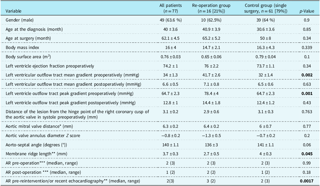
After initial screening of 80 patients diagnosed with subaortic stenosis, three patients with associated aortic valve stenosis were excluded. We included 77 paediatric patients with isolated subaortic stenosis or subaortic stenosis with a minor associated anomaly who underwent successful primary subaortic stenosis resection in the study. Table 1 summarises the demographic and echocardiographic data for both groups. A peak left ventricular outflow tract gradient >50 mmHg or a mean gradient >30 mmHg before surgery remained the main threshold for surgery. Figure 2 summarises the associated cardiac lesions and surgical interventions performed in both groups.
Surgical interventions for 77 patients with subaortic membrane stenosis who underwent repair during 25 years of follow-up.

Demographic and echocardiographic data of all patients and patients in control and re-operation groups

Table 1. Long description
A table comparing demographic and echocardiographic data of patients undergoing surgery for subaortic stenosis. The table has 19 rows and 5 columns. Column headers are Variable, All patients (n = 77), Re-operation group (single surgery, n = 16), Control group (single surgery, n = 61), and p-Value. Row 1: Gender (male), 49 (63.6 percent), 10 (62.5 percent), 39 (64 percent), 0.9. Row 2: Age at the diagnosis (month), 40 plus or minus 3.6, 40.9 plus or minus 3.9, 30.6 plus or minus 3.6, 0.85. Row 3: Age at surgery (month), 62.1 plus or minus 4.5, 65.2 plus or minus 5.2, 50 plus or minus 8, 0.34. Row 4: Body mass index, 16 plus or minus 4, 14.7 plus or minus 2.1, 16.3 plus or minus 4.3, 0.339. Row 5: Body surface area (square meters), 0.76 plus or minus 0.03, 0.65 plus or minus 0.06, 0.79 plus or minus 0.04, 0.1. Row 6: Left ventricle ejection fraction preoperatively (percent), 74.2 plus or minus 1, 76 plus or minus 2.2, 73.7 plus or minus 1.1, 0.34. Row 7: Left ventricular outflow tract gradient preoperatively (millimeters of mercury), 34 plus or minus 1.3, 41.7 plus or minus 2.6, 32 plus or minus 1.4, 0.002. Row 8: Left ventricular outflow tract mean gradient postoperatively (millimeters of mercury), 6.6 plus or minus 0.5, 7.1 plus or minus 0.8, 6.5 plus or minus 0.6, 0.63. Row 9: Left ventricle outflow tract peak gradient preoperatively (millimeters of mercury), 64.7 plus or minus 2.3, 78.4 plus or minus 4, 64.7 plus or minus 2.3, 0.001. Row 10: Left ventricle outflow tract peak gradient postoperatively (millimeters of mercury), 12.8 plus or minus 1, 14.4 plus or minus 1.8, 12.4 plus or minus 1.2, 0.43. Row 11: Distance of the lesion from the hinge point of the right coronary cusp of the aortic valve in systole preoperatively (millimeters), 3.1 plus or minus 0.2, 2.9 plus or minus 0.6, 3.1 plus or minus 0.3, 0.763. Row 12: Aortic mitral valve distance (millimeters), 6.3 plus or minus 0.2, 6.4 plus or minus 0.2, 6 plus or minus 0.7, 0.77. Row 13: Aortic valve annulus Z score, -0.8 plus or minus 0.2, -1.3 plus or minus 0.5, -0.7 plus or minus 0.2, 0.2. Row 14: Aorto-septal angle (degrees), 140 plus or minus 1.1, 136 plus or minus 3, 141 plus or minus 1.1, 0.06. Row 15: Membrane ridge length (millimeters), 3.7 plus or minus 0.3, 2.7 plus or minus 0.5, 4 plus or minus 0.3, 0.045. Row 16: A R pre-operation (median, range), 2 (3), 2 (3), 2 (3), 0.99. Row 17: A R post-operation (median, range), 1 (2), 2 (2), 1 (2), 0.18. Row 18: A R pre-reintervention or recent echocardiography (median, range), 2 (3), 3 (2), 2 (3), 0.0017.
Demographic and echocardiographic data of all patients and in the control and re-operation groups. All continuous data are expressed as mean ± standard error of the mean (SEM). The bold correlate with statistically significant values.
* Distance between aortic and mitral valves in systole in the parasternal long-axis view (mm).
** The length of the membrane/fibro-muscular ridge from the septal insertion to the free edge of the obstructive lesion in mm measured from the parasternal long-axis view preoperatively (mm).
*** AR: aortic regurgitation. 0 = no aortic regurgitation, 1 = trivial aortic regurgitation, 2 = mild aortic regurgitation, 3 = moderate, 4 = severe.
Of the 77 patients, 16 (21%) required reintervention for recurrent subaortic stenosis, whereas 61 underwent a single surgical intervention. There were no significant differences in most demographic characteristics between the groups. Among patients who required reoperation, 50% and 75% of them had their reoperation done within 5 and 10 years of initial resection, respectively. Figure 3 shows the Kaplan–Meier curve for freedom from reintervention.
Kaplan–Meier curve for freedom from re-intervention in 77 children operated on for subaortic membrane stenosis.

Predictors of re-intervention
Several preoperative echocardiographic findings were associated with recurrent subaortic stenosis and reintervention. The reintervention group had significantly higher preoperative left ventricular outflow tract peak and mean gradients than the control group (p = 0.001 and p = 0.002, respectively). In contrast, postoperative peak and mean left ventricular outflow tract gradients were not significantly different between the groups.
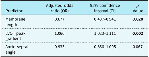
Shorter membrane/fibro-muscular ridge length, measured from the septal insertion to the free edge of the obstructive lesion, was significantly associated with reintervention on univariate analysis (p = 0.045). In the multivariable logistic regression model (Table 2), membrane length remained independently associated with reintervention (adjusted odds ratio 0.677 per 1 mm increase, 95% confidence interval 0.487–0.941; p = 0.020), corresponding to approximately a 32% reduction in the odds of reintervention for each 1 mm increase in membrane length.
Adjusted multivariate logistic regression of recurrent subaortic membranous stenosis predictors

Table 2. Long description
A table with three rows and four columns comparing predictors of recurrent subaortic membranous stenosis. The columns are labeled Predictor, Adjusted odds ratio (OR), 95% confidence interval (CI), and p Value. The row labels are Membrane length, LVOT peak gradient, and Aorto-septal angle. Membrane length has an adjusted odds ratio of 0.677, a 95% confidence interval of 0.487 to 0.941, and a p value of 0.020. LVOT peak gradient has an adjusted odds ratio of 1.066, a 95% confidence interval of 1.023 to 1.111, and a p value of 0.002. Aorto-septal angle has an adjusted odds ratio of 0.933, a 95% confidence interval of 0.866 to 1.005, and a p value of 0.067.
The bold correlate with statistically significant values.
The aorto-septal angle was smaller in patients requiring reintervention and approached significance on univariate analysis (p = 0.06). After adjustment for membrane length and preoperative left ventricular outflow tract peak gradient, the association remained a trend but did not reach conventional statistical significance (adjusted odds ratio 0.933 per 1-degree increase, 95% confidence interval 0.866–1.005; p = 0.067).
Higher preoperative left ventricular outflow tract peak gradient was an independent predictor of reintervention (adjusted odds ratio 1.066 per 1-mmHg increase, 95% confidence interval 1.023–1.111; p = 0.002). Thus, each 1-mmHg increase in preoperative peak gradient was associated with approximately a 6.6% increase in the odds of reintervention.
The distance between the aortic and mitral valves, distance of the lesion from the aortic valve, preoperative ejection fraction, and aortic valve annulus diameter Z score were not significantly associated with reintervention.
Regarding aortic regurgitation, there was no significant difference between groups before the primary operation or immediately after repair. However, during follow-up, aortic regurgitation was significantly worse in the reintervention group (p = 0.0017).
Discussion
We studied a cohort of patients with subaortic stenosis who underwent successful primary resection. Postoperative follow-up demonstrated that shorter membrane length was associated with a higher risk of membrane recurrence and the need for reoperation, a finding that has not been well described in previous studies. In multivariable analysis, membrane length remained independently associated with reintervention (adjusted odds ratio 0.677, 95% confidence interval 0.487–0.941; p = 0.020), indicating that each 1-mm increase in membrane length was associated with approximately a 32% reduction in the odds of reintervention. This phenomenon may be explained by the fact that smaller membranes/ridges may be more difficult to remove completely, especially given the observation that no myomectomy was performed at the initial operation, thereby potentially increasing recurrence in patients with shorter membranes. Recent studies have shown that septal myomectomy at initial surgery can minimise the risk of reoperation. Reference Binsalamah, AlHabshan and AlFagih7–Reference Chan, Gibbs and Wright10
As demonstrated in previous studies, echocardiographic indices of unfavourable left ventricular outflow tract geometry strongly predict recurrence of subaortic stenosis requiring reoperation. Reference Theocharis, Wray and Anderson3,Reference Barkhordarian, Madani and Zaretsky11,Reference Kleinert and Geva12 In our study, higher preoperative left ventricular outflow tract obstruction was strongly associated with reintervention. The preoperative peak left ventricular outflow tract gradient was significantly higher in patients requiring reintervention and remained an independent predictor in the multivariable model (adjusted odds ratio 1.066 per 1-mmHg increase, 95% confidence interval 1.023–1.111; p = 0.002). Thus, each 1 mmHg increase in the preoperative peak gradient was associated with approximately a 6.6% increase in the odds of reintervention. The preoperative mean left ventricular outflow tract gradient was also significantly higher in the reintervention group on univariate analysis, supporting the relationship between greater baseline obstruction and subsequent recurrence. Reference Theocharis, Wray and Anderson3,Reference Anderson, Pundi and Baker13,Reference Dodge-Khatami, Pretre and Seifert14 In contrast, the postoperative peak and mean gradients were not significantly different between groups, suggesting that the severity of preoperative obstruction may be more informative for recurrence risk than the early residual gradient in this cohort.
A steeper aorto-septal angle, reflecting less favourable left ventricular outflow tract geometry, was observed in patients requiring reintervention and showed a trend towards an independent association with recurrence. However, after adjustment for membrane length and preoperative peak left ventricular outflow tract gradient, this association did not reach conventional statistical significance (adjusted odds ratio 0.933 per 1-degree increase, 95% confidence interval 0.866–1.005; p = 0.067). Therefore, although the direction of the association is consistent with previous reports linking unfavourable left ventricular outflow tract geometry to recurrent subaortic stenosis, Reference Theocharis, Wray and Anderson3,Reference Barkhordarian, Madani and Zaretsky11,Reference Kleinert and Geva12 the aorto-septal angle should be interpreted as a potential risk marker rather than an independent predictor in the present cohort. The limited number of reintervention events may have reduced the statistical power to demonstrate an independent effect.
Younger age has been identified in previous studies as a risk factor for reoperation. Reference Theocharis, Wray and Anderson3,Reference Donald, Yong and d’Udekem4,Reference Anderson, Pundi and Baker13 In our cohort, age at the time of primary resection was not significantly associated with reintervention. Similarly, in contrast to previous studies, Reference Theocharis, Wray and Anderson3,Reference Nawaytou, Lee and Eleftheriou15 which reported the proximity of the membrane to the aortic valve as a risk factor for recurrent subaortic stenosis, the distance between the subaortic membrane and the aortic valve was not significantly associated with recurrent left ventricular outflow tract obstruction. During follow-up, however, aortic regurgitation was significantly worse in patients requiring reintervention than in the control group (p = 0.0017). Because this measurement was obtained at recent follow-up or before reintervention, it was not included as a baseline predictor in the multivariable model; nevertheless, its progression may represent an important marker of recurrent obstruction and supports continued echocardiographic surveillance of aortic valve function after subaortic stenosis resection.
Limitations
The retrospective design and single-centre experience may limit the generalisability of the results. In addition, the relatively small cohort and the limited number of reintervention events reduced the number of variables that could be included reliably in the multivariable model and may have limited the power to detect some associations, particularly the effect of aorto-septal angle.
Conclusion
Subaortic stenosis is not definitively curable, as recurrence can occur even after initial resection. In this study, shorter subaortic membrane length and higher preoperative left ventricular outflow tract peak gradient were independent predictors of reintervention. A smaller aorto-septal angle showed a clinically relevant trend but did not reach statistical significance after adjustment. Recognition of these features may improve risk stratification, guide postoperative surveillance, and identify patients who may benefit from closer long-term echocardiographic follow-up.