Left ventricular pseudoaneurysm is a clinically rare condition. The most common causes are myocardial infarction, cardiac surgery, trauma, and endocarditis.Reference Frances, Romero and Grady1–Reference Graupner, Vilacosta and SanRoman3 Pseudoaneurysm is characterised by rapid growth, a narrow neck, and a large external sac.Reference Frances, Romero and Grady1, Reference Brown, Gropler and Harris2 This report describes a 9.5-year-old boy with endocarditis who had a large left ventricular pseudoaneurysm causing inferior caval vein syndrome and congestive cardiac failure. He was treated by a transcatheter approach using an Amplatzer Vascular Plug 4 (AGA Medical, Golden Valley, Minnesota, United States of America) for the pseudoaneurysm and using a bare Cheatham–Platinum stent (NuMED, Hopkinton, New York, United States of America) for inferior caval vein syndrome.
Case report
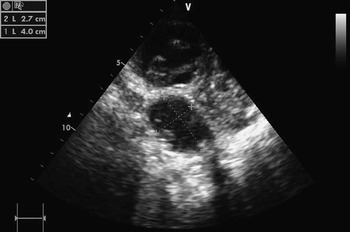
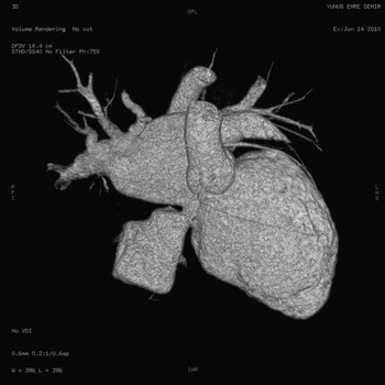
A 9.5-year-old-boy was previously operated on for the left femoral osteomyelitis. Staphylococcus aureus was cultured from the blood. Echocardiographic examination showed large mobile vegetation at the native mitral valve, and moderate pericardial effusion. He had undergone a mitral repair operation in the acute phase of infective endocarditis on day 8 of the treatment; however, his mitral valvular regurgitation worsened and cardiac decompensation developed during the post-operative days. The left ventricular end-diastolic diameter was 53 millimetres, mitral regurgitation was severe, and pulmonary artery pressure was nearly 60 millimetres of mercury at the echocardiographic control during the first month after surgery. After 3 months, a perivalvular extension of infection complication was detected; a large aneurysmal sac was seen at the posterior left ventricle. Its dimensions were 27 × 40 millimetres at the echocardiographic view (Fig 1). Dense thrombotic material was seen inside. A three-dimension volume-rendering tomography showed an approximately 33-millimetre-sized outpouching lesion arising from the atrioventricular groove, just beneath the mitral valve (Fig 2). Functional class was evaluated as class IV according to the New York Heart Association. The second unsuccessful surgical approach was performed as an external suture of the pseudoaneurysmal sac instead of resection. The pseudoaneurysm, organised pericardial fibrinous material, and some possible surgical patches were compressing into the inferior caval vein. The physical examination documented distended and swollen superficial veins, ascites, abdominal distension, and lower extremity oedema. Inferior caval vein syndrome was diagnosed clinically and by abdominal ultrasonography.
Transthoracic echocardiography with modified long-axis view showing the large pseudoaneurysmal sac with thrombus.

Three-dimension volume-rendering computed tomography showing the large pseudoaneurysm at the left atrioventricular groove.

He had a septic appearance, entubated, and was also suffering renal and hepatic insufficiency. We decided to attempt transcatheter closure of the defects. We entered the pseudoaneurysm and tried a standard device delivery catheter; however, we could not provide adequate length, angle, and loop for stable device deployment with the Nit-Occlud coil system (PFM Medical, Cologne, Germany), Amplatzer Vascular Plug (AGA Medical), and Cook detachable coil (Cook Medical Inc., Bloomington, Indiana, United States of America).
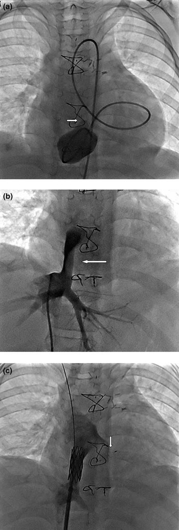
After 15 days, a second angiocardiography was performed. A hydrophilic guidewire was entered into the pseudoaneurysm through the neck. Selective hand angiography of the pseudoaneurysm revealed the neck as measuring 3.8 millimetres in diameter (Fig 3a). An 8-millimetre Amplatzer Vascular Plug 4 was placed in the neck of the pseudoaneurysm. The distal disk was deployed in the just distal part of the neck, and the proximal part was deployed in the ventricular aspect of the pseudoaneurysm. A left ventricular angiography confirmed a stable device position.
(a) Selective hand angiography of the left ventricle pseudoaneurysm showing a large cavity arising from the left ventricle; the cavity appeared to connect to the left ventricle by an orifice with a narrow neck (arrow). (b) Venogram showing significant stenosis in the inferior caval vein due to compression of the sac (arrow). (c) Venogram after a bare Cheatham–Platinum stent placement in the inferior caval vein with a good forward flow. The arrow indicates the position of the Amplatzer Vascular Plug 4.

The bare Cheatham–Platinum stent of length 34 millimetres was placed in the compressing inferior caval vein (Fig 3b). The final diameter of the compressing region was increased to 13 millimetres from 3.5 millimetres (Fig 3c). The pressure gradient at the compressed region changed from 25 millimetres of mercury to 3 millimetres of mercury. After the stenting, the clinical features of the inferior caval vein symptoms decreased dramatically. After 6 months, the echocardiographic control showed a well-seated device and a clot within the aneurysmal cavity without any residual shunt. At follow-up, he remained well in class 1 clinical evaluation, and echocardiography showed a normally functioning mitral valve leaflet with a very eccentric moderate jet of mitral regurgitation.
Discussion
The left ventricle pseudoaneurysm, even when small, can rupture.Reference Frances, Romero and Grady1, Reference Brown, Gropler and Harris2 A suitable approach to treat the left ventricle pseudoaneurysm is to consider performing surgery in patients.Reference Frances, Romero and Grady1, Reference Brown, Gropler and Harris2 Cases of successful percutaneous closure mostly in adults have been reported,Reference Clift, Thorne and de Giovanni4–Reference Breinholt, Rodefeld and Hoyer8 and this treatment may be considered in patients who are poor candidates for surgery. We considered a transcatheter approach to close the neck of the pseudoaneurysm because of his clinical conditions. We first tried out other different devices; however, we could implant the Amplatzer Vascular Plug 4 on the basis of low profile, retrievability, and ability to reposition the device. The lower profile of the device can extend to reach difficult areas. In addition, it can be deployed through a diagnostic catheter following angiography without exchanging a guiding catheter. It also has a floppy distal section of the delivery wire to enable the device to travel through tortuous anatomy.
Only two experiences with percutaneous device closure have been reported in the literature in the childhood period.Reference Clift, Thorne and de Giovanni4, Reference Elshershari, Gossett and Hijazi6 Elshershari et alReference Elshershari, Gossett and Hijazi6 described the successful occlusion of a pseudoaneurysm at the proximal suture line of the pulmonary autograph in a 13-year-old boy. They pointed out the potential for perforation risk with a catheter or wire manipulation. Our case had unique characteristics; he suffered from subacute phase endocarditis disease and had not only the presence of a large pseudoaneurysm, but also symptoms of compressing the inferior caval vein. Only closing the pseudoaneurysm with the device might increase the mass effect symptoms of the inferior caval vein. Stenting of the inferior caval vein is needed to relieve the compression.
Acknowledgements
We are grateful to Feyza G. Yılmaz, MD, for her role in the care of the patient image acquisition. There is no conflict of interest to declare.