Introduction
Whiteflies are classified in the family Aleyrodidae (Sternorryncha: Hemiptera) (Campbell et al., Reference Campbell, Steffen-Campbell and Gill1994). Of the nearly 1200 whitefly species described (Mound & Halsey, Reference Mound and Halsey1978; Martin et al., Reference Martin, Mifsud and Rapisarda2000), Bemisia tabaci (Gennadius) is the most widely distributed, and it is of greatest importance as a crop pest in the tropics, subtropics and the Mediterranean Basin. This polyphagous whitefly complex colonizes more than 500 annual and perennial (Eudicot) hosts, causing direct damage due to feeding and indirect damage by transmitting a great number of plant viruses (Bedford et al., Reference Bedford, Briddon, Brown, Rosell and Markham1994; Jones, Reference Jones2003).
High genetic polymorphism has been demonstrated in B. tabaci populations worldwide, based on several types of molecular analyses. This observation suggests that it is most aptly considered a species complex. The complex comprises multiple genetic haplotypes and a number of well-characterized, behaviorally distinct variants, referred to as biotypes (Brown et al., Reference Brown, Frohlich and Rosell1995a; Perring, Reference Perring2001). Despite significant intraspecific genetic variation, B. tabaci biotypes and haplotypes are morphologically indistinguishable, lacking clear-cut morphological characters in either the pupae or the adults. Genetic polymorphisms were first demonstrated in B. tabaci, based on unique esterase banding patterns. The method, when applied to a suite of B. tabaci from different locations and hosts worldwide, yielded numerous haplotypes, each arbitrarily assigned an alphabetical designation from A to T (Costa & Brown, Reference Costa and Brown1991; Costa et al., Reference Costa, Brown, Sivasupramaniam and Bird1993; Brown et al., Reference Brown, Coats, Bedford, Markham, Bird and Frohlich1995b). This nomenclature is still in use by the scientific community to refer to particular biotypes and for historical reasons. Although most of the esterase-based designations coincide with well-studied biological types, certain esterase morphotypes were found to share relatively low % nucleotide sequence identities, based on the mtCOI sequence, e.g. B biotype relatives. Contemporary identification of B. tabaci variants now relies almost exclusively on molecular diagnostics, with the most common being polymerase chain reaction (PCR) amplification and DNA sequencing of the mtCOI or 16S rRNA gene, followed by PCR-restriction fragment length polymorphism (PCR-RFLP) of the mtCOI or mt16S rRNA gene, or RFLP alone, PCR and sequencing of the nuclear ITS-1 region, random amplified polymorphic DNA analysis (RAPD-PCR) and, most recently, for population studies, microsatellite DNA analysis (Brown et al., Reference Brown, Coats, Bedford, Markham, Bird and Frohlich1995b; Frohlich et al., Reference Frohlich, Torres-Jerez, Bedford, Markham and Brown1999; Brown et al., Reference Brown, Perring, Cooper, Bedford and Markham2000; Cervera et al., Reference Cervera, Cabezas, Simón, Martínez-Zapater, Beitia and Cenis2000; De Barro et al., Reference De Barro, Driver, Trueman and Curran2000; Viscarret et al., Reference Viscarret, Torres-Jerez, Agostini de Manero, López, Botto and Brown2003; Berry et al., Reference Berry, Rey, Rogan, Fondong, Fauquet and Brown2004; De la Rúa et al., Reference De la Rúa, Simón, Cifuentes, Martínez-Mora and Cenis2006; Qiu et al., Reference Qiu, Coats, Ren, Idris, Caixia and Brown2007). Studying the geographical dispersal and population dynamics of the insect's different biotypes is of great importance as they exhibit differences on host range, dispersal behaviour, virus transmission competency and susceptibility to various insecticides. Presently, B and Q biotypes are the two most widespread and important variants of B. tabaci in Europe and in countries surrounding the Mediterranean basin (Bosco et al., Reference Bosco, Loria, Sartor and Cenis2006; De la Rua et al., Reference De la Rúa, Simón, Cifuentes, Martínez-Mora and Cenis2006).
In Cyprus, the B biotype is widely distributed and is believed to be the predominant B. tabaci type in the island associated with whitefly-transmitted virus epidemics in solanaceous and cucurbit crops, such as Tomato yellow leaf curl virus (TYLCV) (Ioannou, Reference Ioannou1985), Tomato chlorosis virus (ToCV) (Papayiannis et al., Reference Papayiannis, Ioannou, Dovas, Maliogka and Katis2006), Cucurbit yellow stunting disorder virus (CYSDV) and Cucumber vein yellowing virus (CVYV) (Papayiannis et al., Reference Papayiannis, Ioannou, Boubourakas, Dovas, Katis and Falk2005). This biotype is also responsible for causing physiological disorders on plants and inducing the toxin-regulated silverleaf symptom on squash plants (Costa & Brown, Reference Costa and Brown1991; Schuster et al., Reference Schuster, Kring and Price1991), as well as tomato irregular ripening (Yokomi et al., Reference Yokomi, Hoelmer and Osborne1990). Recently, the Q biotype was identified for the first time in Cyprus, based on the unique esterase pattern of the biotype produced using native polyacrylamide gel electrophoresis (PAGE).
In recent years, advanced molecular diagnostic tools have been widely used for the rapid detection of DNA targets. Implementation of 5′-nuclease fluorogenic assay, also known as TaqMan® real-time PCR, has been demonstrated for the identification of human, animal and plant pathogens. It utilizes a fluorogenic probe that anneals to the DNA sequence target during amplification, emitting a readable fluorescent signal which can be monitored through advanced equipment. This technology provides sensitive and accurate results and alleviates the need of post-PCR manipulations, such as gel electrophoresis. During this study, a real-time (TaqMan®) PCR assay was developed and evaluated for the rapid, simultaneous identification of B. tabaci biotypes B and Q, which was used along with other DNA based techniques in a two-year survey to evaluate the incidence of B. tabaci biotypes in Cyprus.
Materials and methods
Collection of plant material and whiteflies
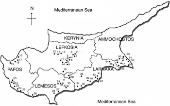
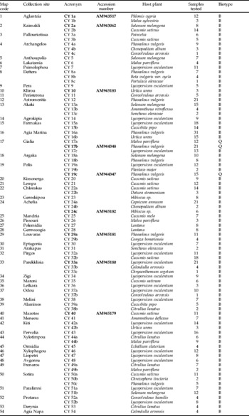
The samples used in this study were adult B. tabaci collected during 2005–2007 from open field and greenhouse cultivated crops, as well as from weeds and ornamental plants cultivated on Cyprus. The survey was carried out in the main vegetable-growing areas, house gardens and plant nurseries of Lefkosia, Larnaca, Lemesos, Paphos and Ammochostos districts. The numbers assigned to the B. tabaci populations in table 1 correspond to the collection sites shown in fig. 1. A total of 775 adult whiteflies identified as B. tabaci were collected from 54 areas located within the five districts and on 35 distinct cultivated and wild plant species (table 1). The insect samples were collected using an electrical hand-held aspirator and processed immediately or preserved in 96% ethanol at –30°C until DNA extraction and analysis were performed.
Map of the collection sites for B. tabaci samples from the island of Cyprus. Samples were collected from more than one host in some locations. Locality codes are given in table 1.

Map code, geographic locale, acronym, GenBank accession number (for selected populations), host plant, number of samples and results of biotyping for the Bemisia tabaci populations collected from Cyprus.

Nucleic acid extractions
Three methods were used for DNA extraction for comparative purposes. The first was based on the ChargeSwitch® gDNA Micro Tissue commercial kit (Invitrogen Corporation, CA), which uses a novel switchable magnetic-based surface technology depending on the pH of the surrounding buffer. This method was used for DNA isolation from all collected samples. The second procedure was according to Cenis et al. (Reference Cenis, Perez and Fereres1993) and was applied to 100 B. tabaci individuals. For the third approach, 100 B. tabaci adults were ground using the TissueLyser in 50 μl of ice-cold lysis buffer (100 mm NaCl, 10 mm Tris-HCl, pH:8.0), containing 0.4 mg ml−1 proteinase K and incubated at 55°C for 30 min and at 94°C for 10 min, followed be a 5 min centrifugation step at 10,000 g to pellet debris. The supernatant was used for DNA template for the PCR assays.
B. tabaci biotype detection using PCR amplification, digestion and DNA sequencing of the mitochondrial cytochrome oxidase I (mtCOI) gene
Two primers, C1-J-2195 and L2-N-3014, were used for partial amplification and analysis of mtCOI gene sequences (816 bp) (table 2) (Frohlich et al., Reference Frohlich, Torres-Jerez, Bedford, Markham and Brown1999). PCR assays were performed in a final volume of 25 μl containing 10 mm Tris-HCl (pH:8.8), 50 mm KCl, 1.5 mm MgCl2, 0.25 of each deoxyribonucleoside triphosphates (dNTPs), 1 Unit of Platinum® TaqPCR DNA Polymerase (Invitrogen Corporation, CA) and a final concentration of 0.5 μm for each primer (C1-J-2195/L2-N-3014) using 4 μl of template DNA extracted with the commercial kit. The amplification was carried out in a PTC 200 Thermal Cycler (MJ Research, USA) and the cycling profile was as follows: 94°C for 4 min followed by 35 cycles segmented in 94°C for 30 s, 50°C for 60 s and 72°C for 60 s, followed by a final extension step of 72°C for 10 min.
Primers and probes used in the experiments.

PCR products were then subjected to RFLP analysis using Taq I and Mse I, separated by electrophoresis on a 7% acrylamide/bisacrylamide (29:1) gel and visualized by staining with ethidium bromide. The amplified mtCOI PCR fragments (~750 bp) from 15 samples were purified using PureLink™ (Invitrogen Corporation, CA) gel extraction system and both strands were sequenced using the ABI PRISM BigDye Terminators v3.0 Cycling Sequencing Kit in an ABI automated sequencer (Applied Biosystems, Foster City, CA). Sequencing data were analyzed and deposited in the NCBI GenBank database as the Accession Numbers of AM943517, AM943062, AM944347, AM944348 and AM945179-AM945183. Multiple sequence alignment was carried out using ClustalX (Thompson et al., Reference Thompson, Gibson, Plewniak, Jeanmougin and Higgins1997), and phylogenetic trees were reconstructed using the neighbour-joining algorithm implemented in the Mega 3.1 software package (Kumar et al., Reference Kumar, Tamura and Nei2004).
Real-time PCR assay design and establishment
A multiple sequence alignment of Bemisia tabaci mtCOI sequences available from the GenBank database, including the Cypriot sequenced isolates, was made using the ClustalX program (AF418671, AJ517768, AJ550173, AY686065, DQ365872, AY747688, AY766369, DQ365877, AF342775, AF342776, AJ517769, AY766370, DQ365860, DQ365863) (Thompson et al., Reference Thompson, Gibson, Plewniak, Jeanmougin and Higgins1997). Variable regions were identified among biotypes B and Q and probes and primers were designed for a TaqMan® assay using the PRIMER EXPRESS® software (Applied Biosystems) to amplify an 87 bp and a 73 bp fragment for B and Q, respectively. The probes were designed to have the T m values approximately 10°C higher (70°C) than that of the primers (60°C) with its length not exceeding 30 nt and its % GC content higher than 40%. The sequences of the primers and probes used are given in table 2.
The 5′ terminal reporter dye used was FAM (6-carboxyfluorescein) or Texas Red, and the 3′ quencher dye used was Blackhole Quencher. All primers and probes were supplied by MWG AG Biotech, Anzinger, Germany. The real-time PCR assays were performed in a final volume of 25 μl containing 12.5 μl of 2X IQ™ Supermix (BIO-RAD Laboratories, CA), 0.3 μm of each primer, 0.1 μm TaqMan® probe and 2 μl template DNA. Cycling parameters were the following: 3 min at 94°C followed by 40 cycles of 15 s at 94°C and 60 s at 60°C.
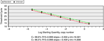
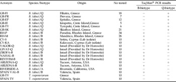
The assay was developed and evaluated using the iCycler IQ™ 5 Multicolor Real-time PCR Detection System (Bio-Rad Laboratories). Cycle thresholds (the cycle at which a significant increase of fluorescence occurs, Ct) for a ten-fold dilution series of mtCOI gene plasmid DNA were plotted to yield a standard curve for the assays (fig. 2). A total of 20 B. tabaci reference isolates (B and Q) from different areas were also tested using this assay (table 3).
Standard curves showing the multiplex detection efficiency of B. tabaci biotype B and Q using TaqMan® PCR ( , FAM;
 , TexasRed).

Reference B. tabaci from different geographic regions tested using the TaqMan® assay.

Results
Biotype discrimination using RFLP analysis of mtCOI gene
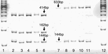
Two types of patterns were obtained after digestion of the mtCOI PCR products using Taq I restriction endonuclease: (i) a restriction pattern of three fragments (414, 162 and 144 bp) corresponding to the B biotype and (ii) a restriction pattern of two fragments (634 and 144 bp) which is typical for the Q biotypes (fig. 3). The first digestion profile was observed for most tested samples which were collected within the districts of Ammochostos, Larnaka, Lefkosia, Lemesos and Paphos. The second profile in which Taq I endonuclease produced only one restriction site was observed only in populations from two areas of Paphos district, which are located on the northwest part of the island. Results were corroborated based on simulated digestion of mtCOI sequences from sequences available at NCBI and with reference isolates, which were collected from Greece and USA. Several restriction sites were present in COI amplicons using Mse I digestion enzyme, and two different patterns were obtained for the samples tested. Both patterns produced three visible fragments of 203, 184 and 150 bp corresponding to the B biotype and 184, 138 and 107 bp corresponding to the Q biotype.
Restriction fragment length polymorphism (RFLP) analysis of the amplified mtCOI gene of B. tabaci individual samples from Cyprus, following digestion with Taq I restriction enzyme. Lanes: 1, 12: DNA size marker 100 bp ladder; 2–6: B. tabaci biotype B samples, 7–11: B. tabaci biotype Q samples.

Mitochondrial COI sequence analysis
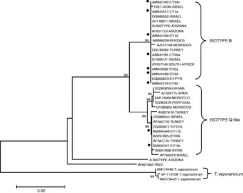
Phylogenetic analysis of the mtCOI fragment sequences for nine B. tabaci individuals collected in Cyprus (table 1) were compared with reference sequences available from NCBI Genbank database for the B, Q, A, Ms and T biotypes, as well as Trialeurodes vaporariorum (Westwood), previously reported surrounding the Mediterranean countries, indicated that all B. tabaci individuals from Cyprus clustered with haplotypes of the major North African-Mediterranean-Middle Eastern clade (Brown, Reference Brown2000) (fig. 4). This analysis revealed two subclusters, one comprising the B biotype and another with sequences reported for the Q biotype and related haplotypes. All sequences obtained from Cyprus population grouped one of these two B. tabaci sister clades (fig. 4).
Phylogenetic analysis of nine B. tabaci mtCOI sequences from Cyprus and selected B. tabaci mtCOI reference sequences available in the NCBI Genbank database.

TaqMan® PCR assay
The TaqMan® primers and probes were initially subjected to a primer optimisation matrix on extracted DNA from reference samples of B and Q biotypes. The concentration of primers and probes that gave the highest reporter fluorescence and the lowest threshold cycle was 300 nm for primers and 100 nm for probes on both tests (data not shown). The two biotype specific tests were validated by analyzing known laboratory populations and confirmed the identity of the B and Q biotypes, respectively. The results showed that the two assays were specific and did not cross react with each other. Primer pair BTB-F/BTB-R and probe BTB-TAQ amplified and produced increased fluorescence only for ‘known’ biotype B samples. Similarly, primers BTQ-F/BTQ-R and probe BTQ-TAQ reacted only with Q biotypes.
Further tests were conducted in order to test the possibility of multiplexing the two reactions by using a Texas Red labelled probe. Although the Ct values obtained for simplex assays were lower than those obtained from multiplex tests, both formats gave exactly the same results in terms of detection for the two biotypes.
Comparison of sensitivity and DNA template preparation used in real-time and conventional PCR assays
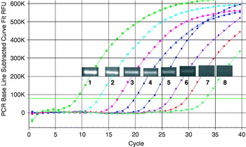
A comparison was made between real-time and conventional PCR assays in order to test the sensitivity of the mtCOI amplification. TaqMan PCR was far more sensitive with a dilution end-point of 10−6, whereas normal PCR produced visible band only up to 10−3 (fig. 5). The relative cost, duration, use of organic solvents, final volume and amplification efficiency for the TaqMan and conventional PCR assay of three extraction methods were compared (table 4). All of the methods examined here allowed the detection of the target sequence using Real-time and conventional PCR for both biotypes. Among them, magnetic bead-based extraction technique showed the best results producing a lower mean Ct value in TaqMan® PCR and a brighter amplification band in the conventional PCR assay. Although it was the most expensive extraction method, the approach allowed the DNA storage for longer period at both 4°C and −20°C (data not shown). The Proteinase K method also proved to be a very reliable and rapid extraction method for biotyping large numbers of samples using the molecular methods described. The low extraction volume and the use of volatile compounds such as chloroform were the main disadvantages of that procedure (Cenis et al., Reference Cenis, Perez and Fereres1993).
Sensitivity of the TaqMan® assay for B. tabaci biotype B detection compared with conventional PCR, determined by end point dilution. (1) 10 ng plasmid DNA, (2) 1 ng plasmid DNA, (3) 10−1 ng plasmid DNA, (4) 10−2 ng plasmid DNA, (5) 10−3 ng plasmid DNA (6) 10−4 ng plasmid DNA (7) 10−5 plasmid DNA (8) 10−6 ng plasmid DNA.

Results for the comparison of whitefly template preparation procedures.

Discussion
The B. tabaci complex is an excellent example of a cryptic species for which genetic variation is evident in the absence of morphological differences. Biological and genetic variants exhibit no corresponding morphological characters that permit their discrimination. In this light, a number of biochemical and molecular markers have been employed to detect polymorphisms within this species and determine the identity of biotypes or reveal the presence of unclassified haplotypes (Brown et al., Reference Brown, Coats, Bedford, Markham, Bird and Frohlich1995b; Vos et al., Reference Vos, Hogers, Bleeker, Reijians, Lee, Hornes, Frijters, Pot, Peleman, Kuiper and Zabeau1995; Brown, Reference Brown2000; Cervera et al., Reference Cervera, Cabezas, Simón, Martínez-Zapater, Beitia and Cenis2000; Viscarret et al., Reference Viscarret, Torres-Jerez, Agostini de Manero, López, Botto and Brown2003; Berry et al., Reference Berry, Rey, Rogan, Fondong, Fauquet and Brown2004; Khasdan et al., Reference Khasdan, Levin, Rosner, Morin, Kontsedalov, Maslenin and Horowitz2005; Bosco et al., Reference Bosco, Loria, Sartor and Cenis2006; Tsangarakou et al., Reference Tsangarakou, Tsigenopoulos, Gorman, Lagnel and Bedford2006; Jones et al., Reference Jones, Gorman, Denholm and Williamson2008). Among the several coding and non-coding regions examined as molecular markers, the mtCOI sequence reveals the highest variability for the species complex, and subsequent analysis of this gene has been highly informative and capable of differentiating B. tabaci at the level of biotypes. In this report, the mtCOI sequences of B. tabaci were analyzed, and a real-time (TaqMan®) PCR diagnostic assay was developed for the rapid discrimination of B. tabaci B and Q biotypes and was used to identify the variants collected from Cyprus.
The TaqMan® PCR assays employ a sequence specific dually labeled oligonucleotide probe, which is able to anneal to the target DNA between the PCR primers. During the extension step, the probe is cleaved off the target, the reporter is separated from the quencher, and a fluorogenic tag is emitted and measured in each cycle. The resulting fluorescence signal is proportional to the amount of amplified product in the sample and can be observed in real-time without the need for post-PCR manipulations such as gel electrophoresis. As noted before, some great advantages of real-time PCR assays using TaqMan® chemistry include high accuracy, sensitivity and specificity, as well as the use of generic cycling conditions (Boonham et al., Reference Boonham, González Pérez, Mendez, Lilia Peralta, Blockley, Walsh, Barker and Mumford2004). The major disadvantage of this method is that it requires expensive equipment and reagents. The results presented show that this duplex real-time PCR assay allows the specific and sensitive detection of biotypes B and Q within the B. tabaci complex. The new assay has been successfully tested against a number of whitefly samples from Cyprus, Greece, Israel, Spain and the USA. In addition, by database searching with the sequences of the primers and probes, it is possible to predict that the assay will be able to detect available sequences of the B and the Q biotype, respectively reported from different isolates in many regions where this whitefly has emerged.
RFLP analysis of mtCOI amplicons has successfully allowed the differentiation of almost all biotypes reported in the Mediterranean (Bosco et al., Reference Bosco, Loria, Sartor and Cenis2006). Digestion of the mtCOI gene was used to study the polymorphism in whitefly isolates collected in the island and results were compared with those obtained from the real-time PCR assays. Although RFLP analysis of the mtCOI gene has proven to be time consuming and expensive, restriction profiles obtained with two different endonucleases previously reported and used for B. tabaci biotype differentiation provided an additional accurate diagnostic tool that gave results consistent to the TaqMan® assay. Restriction patterns from Cypriot populations, using Taq I and MseI digestion endonucleases, made it possible to identify B and Q biotypes and exclude the existence of three other biotypes (M, S, T), which have been reported in Mediterranean countries (Sseruwagi et al., Reference Sseruwagi, Legg, Maruthi, Colvin, Rey and Brown2005; Bosco et al., Reference Bosco, Loria, Sartor and Cenis2006). The identities of the biotypes of B. tabaci examined from Cyprus were consistently corroborated, based on mtCOI sequence analysis.
Phylogenetic studies of nine B. tabaci collections from different localities in the island infesting cultivated and weed plants have been shown to belong in the major North African-Mediterranean-Middle Eastern clade, together with isolates reported in Greece, Spain, Turkey, Morocco, Israel and France (Brown, Reference Brown2000; Berry et al., Reference Berry, Rey, Rogan, Fondong, Fauquet and Brown2004). Comparison of the sequences for the Cyprus B biotype populations indicated that they share high nucleotide identity and cluster together with other similar haplotypes noted as B biotype. Although the Q biotype was not found frequently in this study, it is not known if it has been established in Cyprus. Even so, the Q-like samples from Cyprus grouped with other Q biotype sequences and its close relatives, reported from neighbouring countries such as Syria, Israel and Turkey, suggesting that it may have been introduced or migrated from these areas. This is not surprising given that the proximity of Cyprus to these countries is less than 50 km from Turkey and the Middle East (Lebanon, Syria and Israel). Thus, taken together with the high-density whitefly migrations often observed arriving to Cyprus on ocean winds, the B. tabaci biotype composition in Cyprus may potentially be altered within the next few years.
The genetic structure of biotypes B and Q in the Mediterranean is expected to be influenced by a combination of factors, including host species, habitat (greenhouse crops vs. field) and local insecticide regimes. In Israel, for example, B and Q biotypes occurred sympatrically in certain parts of the country (Horowitz et al., Reference Horowitz, Denholm, Gorman, Cenis, Kontsedalov and Ishaaya2003), whereas in southeastern Spain the B biotype-mediated displacement by Q was attributed to the application of neonicotoinoids (Pascual & Callejas, Reference Pascual and Callejas2004). Whether insecticide selection pressure or other ecological factors drive the current distribution and ratio of the B and Q biotypes in Cyprus is not understood. Further sampling from indigenous plants in isolated areas, compared to intensive agricultural areas, will lead to an understanding of the population dynamics of these two biotypes in the Mediterranean region.
Acknowledgements
The authors would like to thank Mrs A. Stylianou and A. Hadjinicoli for their help with DNA extractions and Drs N. Vouzounis and Mr. G. Economides for their assistance in weed identification. The authors would also like to thank Drs R. Horowitz and A. Paraskevopoulos for supplying whitefly samples. This work was funded by Cyprus Research Promotion Foundation.