





Non-technical Summary
In this paper we re-examine previously described fossilized body impressions of mayfly larvae from the Wapiti Formation (deposited ca. 72.5 million years ago) in Alberta, Canada. While most insect fossils occur in ancient lake deposits or inside amber, these mayfly fossils most likely formed in a river environment with a current strong enough to scour the riverbeds and carry sand particles. In order to understand the sequence of events that led to their fossilization, we examined the orientation of the mayfly fossils relative to current-generated features preserved on the sandstone. All 16 of the fossils analyzed in this study were found with the abdomen facing downwards and the head facing into the current. This suggests that the fossilized impressions were actually produced by live mayfly larvae. Finally, we propose a new trace fossil name for traces that represent shallow self-burial of an insect larva: Kalyptichnus belli.
Introduction
More than a decade ago, Bell et al. (Reference Bell, Fanti, Acorn and Sissons2013) reported on unusual three-dimensional body impressions of mayfly larvae from the fluvial Wapiti Formation in Alberta, Canada. These fossil larvae were found on the underside of a sandstone slab that was recovered from interbedded sand and silt deposits displaying erosional sedimentary features (Bell et al., Reference Bell, Fanti, Acorn and Sissons2013). Body fossils of mayflies and other aquatic insects are most commonly found in lacustrine deposits or as inclusions in amber (e.g., Wootton, Reference Wootton1988; Smith, Reference Smith2012; Greenwalt et al., Reference Greenwalt, Rose, Siljestrom, Goreva, Constenius and Wingerath2014; Labandeira, Reference Labandeira2014; Sroka et al., Reference Sroka, Godunko and Prokop2023; Storari et al., Reference Storari, Salles, Fonseca, Saraiva and Rodrigues2025). In contrast, preservation of soft-bodied insects within a high-energy fluvial setting is unusual and raises important taphonomic questions, given that such depositional conditions are more typically associated with erosion and physical disruption. In this short contribution, we aim to propose a taphonomic model that explains how insect larvae came to be exceptionally preserved within a high-energy fluvial environment.
Geological setting
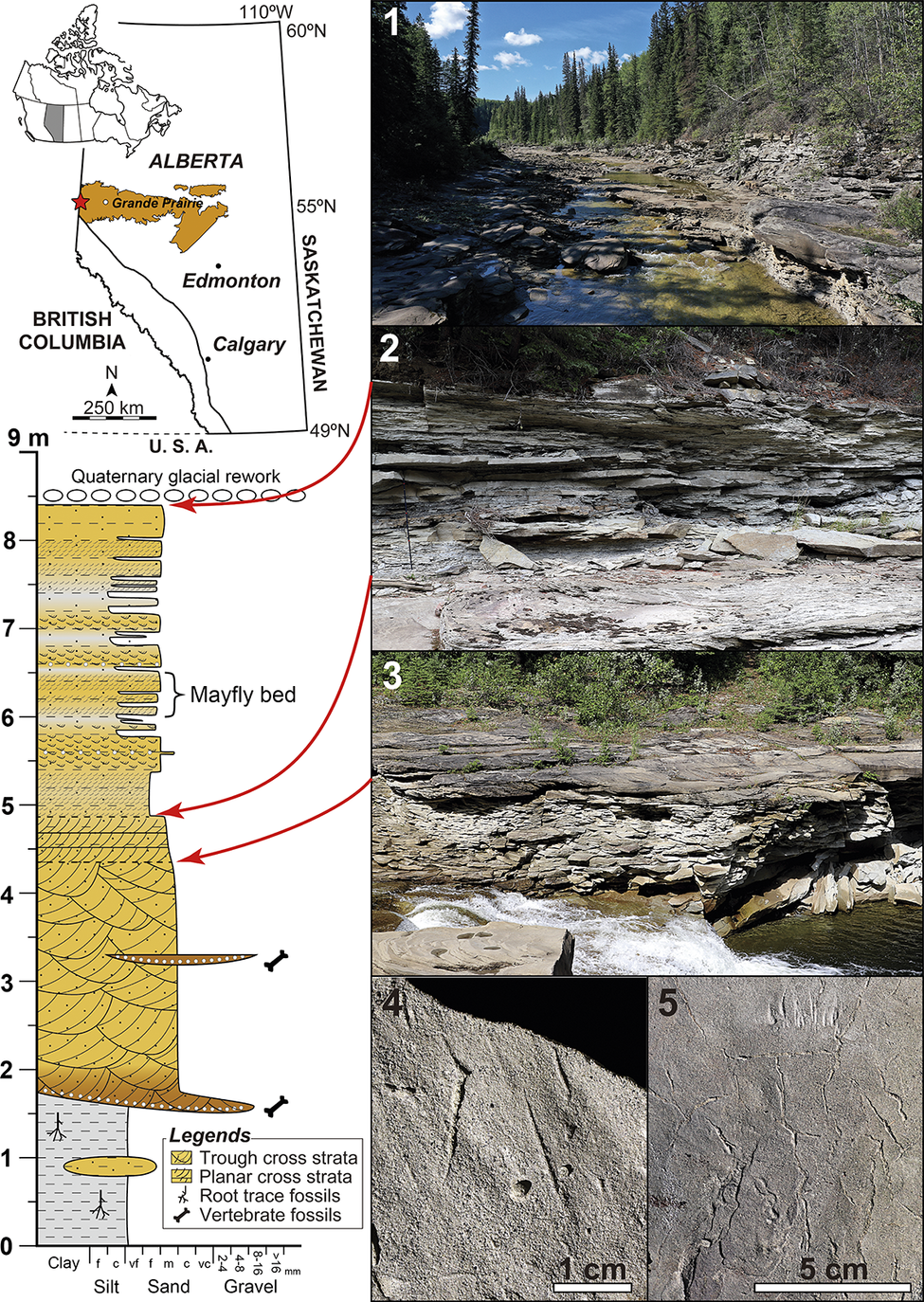
The Upper Cretaceous Wapiti Formation consists of fluvial and alluvial-plain strata deposited in west-central Alberta and east-central British Columbia, Canada (Fig. 1). The Wapiti Formation is well known for its exceptionally well-preserved vertebrate body and trace fossils, as well as occasional invertebrate body fossils and numerous, mostly undescribed plant fossils and invertebrate trace fossils (e.g., Fanti and Miyashita, Reference Fanti and Miyashita2009; Koppelhus and Fanti, Reference Koppelhus, Fanti, Murray, LeBlanc and Holmes2019; Cockx et al., Reference Cockx, McKellar, Tappert, Vavrek and Muehlenbachs2020; Enriquez et al., Reference Enriquez, Campione, White, Fanti, Sissons, Sullivan, Vavrek and Bell2022; Fanti et al., Reference Fanti, Bell, Vavrek, Larson, Koppelhus, Sissons, Langone, Campione and Sullivan2022; Hamilton et al., Reference Hamilton, Paparella, Bell, Campione, Fanti, Larson, Sissons, Vavrek, Balsai and Sullivan2023). The fossils discussed herein were collected from an outcrop near Red Willow Falls along the Redwillow River (Fig. 1.1), ~80 km WSW of Grande Prairie, Alberta, Canada. Aside from the materials reassessed here, fossils described from Red Willow Falls include a “mummified” hadrosaurid dinosaur with soft tissue preservation (Bell et al., Reference Bell, Fanti, Currie and Arbour2014; Sharpe et al., Reference Sharpe, Wang, Dudgeon, Powers, Whitebone, Coppock, Dyer and Sullivan2025), tracks of small vertebrates (Fanti et al., Reference Fanti, Bell and Sissons2013), and vertebrate swim traces (Kimitsuki et al., Reference Kimitsuki, Rodriguez, Sullivan, Zonneveld, Sissons, Bell, Campione, Fanti and Gingras2024).
The study area at Red Willow Falls. Geological map at top left modified from Prior et al. (Reference Prior, Hathway, Glombick, Pana, Banks, Hay, Schneider, Grobe, Elgr and Weiss2013). (1) General view of the outcrop along the Redwillow River; (2) Trace fossil bearing, interbedded mud and sand unit; the fossils described here were found at the base of a sandstone layer; (3) the basal sandstone unit, including trough cross-bedded strata underlying finer-grained, planar tabular cross bedded ones; (4) tool marks preserved in positive hyporelief on the same surface as the trace fossils described in this study; (5) incipient desiccation cracks and parting lineations preserved in the same stratigraphic interval as the trace fossils, but on a different bedding surface.

Figure 1. Long description
The image is divided into a left column containing maps and a stratigraphic log, and a right column with five numbered photographs.
Left Column:
- Top: A map of Canada with Alberta highlighted, followed by a detailed map of Alberta showing the study area marked with a red star near Grande Prairie, along with the locations of Edmonton, and Calgary. Coordinates range from 49 degrees North to 60 degrees North and 110 degrees West. A scale bar indicates 250 km.
- Bottom: A stratigraphic column from 0 to 9 meters. The base (0 to 1.5 meters) consists of grey silt with root trace fossils. From 1.5 to 5.5 meters, a thick yellow unit shows trough cross strata that fines upwards into planar cross strata and vertebrate fossils. A bracket from 6 to 6.5 meters identifies the Mayfly bed. The top is capped by Quaternary glacial rework. Red arrows link specific stratigraphic levels to the photographs on the right.
Right Column:
- Panel 1: A wide-angle photo of the Redwillow River flowing through the outcrops of the Wapiti Formation.
- Panel 2: A close-up of interbedded mud and sand units in the outcrop wall.
- Panel 3: A view of the basal sandstone unit showing distinct trough cross-bedded layers.
- Panel 4: A high-detail photo of tool marks in positive hyporelief on a rock surface, including a 1 cm scale bar.
- Panel 5: A photo showing incipient desiccation cracks and parting lineations on a bedding surface, including a 5 cm scale bar.
Legend:
- Trough cross strata: yellow with curved lines.
- Planar cross strata: yellow with diagonal straight lines.
- Root trace fossils: vertical branching symbols.
- Vertebrate fossils: bone-shaped icons.
The Wapiti Formation is informally subdivided into five lithostratigraphic units (units 1 to 5; sensu Fanti and Catuneanu, Reference Fanti and Catuneanu2009). The sediments at Red Willow Falls belong to Unit 4 of the Wapiti Formation (Bell et al., Reference Bell, Fanti, Acorn and Sissons2013, Reference Bell, Fanti, Currie and Arbour2014; Fanti et al., Reference Fanti, Bell and Sissons2013; Kimitsuki et al., Reference Kimitsuki, Rodriguez, Sullivan, Zonneveld, Sissons, Bell, Campione, Fanti and Gingras2024). According to Fanti and Catuneanu (Reference Fanti and Catuneanu2009, Reference Fanti and Catuneanu2010), Unit 4 of the Wapiti Formation records a time of medium to high sediment-accommodation space. The fossil-bearing strata at Red Willow Falls, which are characterized by a predominantly fining upwards succession, were interpreted by previous researchers as crevasse splay deposits formed in a meandering channel belt (Bell et al., Reference Bell, Fanti, Acorn and Sissons2013; Kimitsuki et al., Reference Kimitsuki, Rodriguez, Sullivan, Zonneveld, Sissons, Bell, Campione, Fanti and Gingras2024). An Ar/Ar date obtained from volcanic ash collected ~2 m above the “mummified” hadrosaurid from the same locality yielded an age of 72.58 ± 0.09 Ma (Bell et al., Reference Bell, Fanti, Currie and Arbour2014). This age falls within the late Campanian, a time that witnessed the regressive phase of the marine Bearpaw Formation farther southeast (Eberth and Braman, Reference Eberth and Braman2012; Fanti et al., Reference Fanti, Bell, Vavrek, Larson, Koppelhus, Sissons, Langone, Campione and Sullivan2022).
Materials and methods
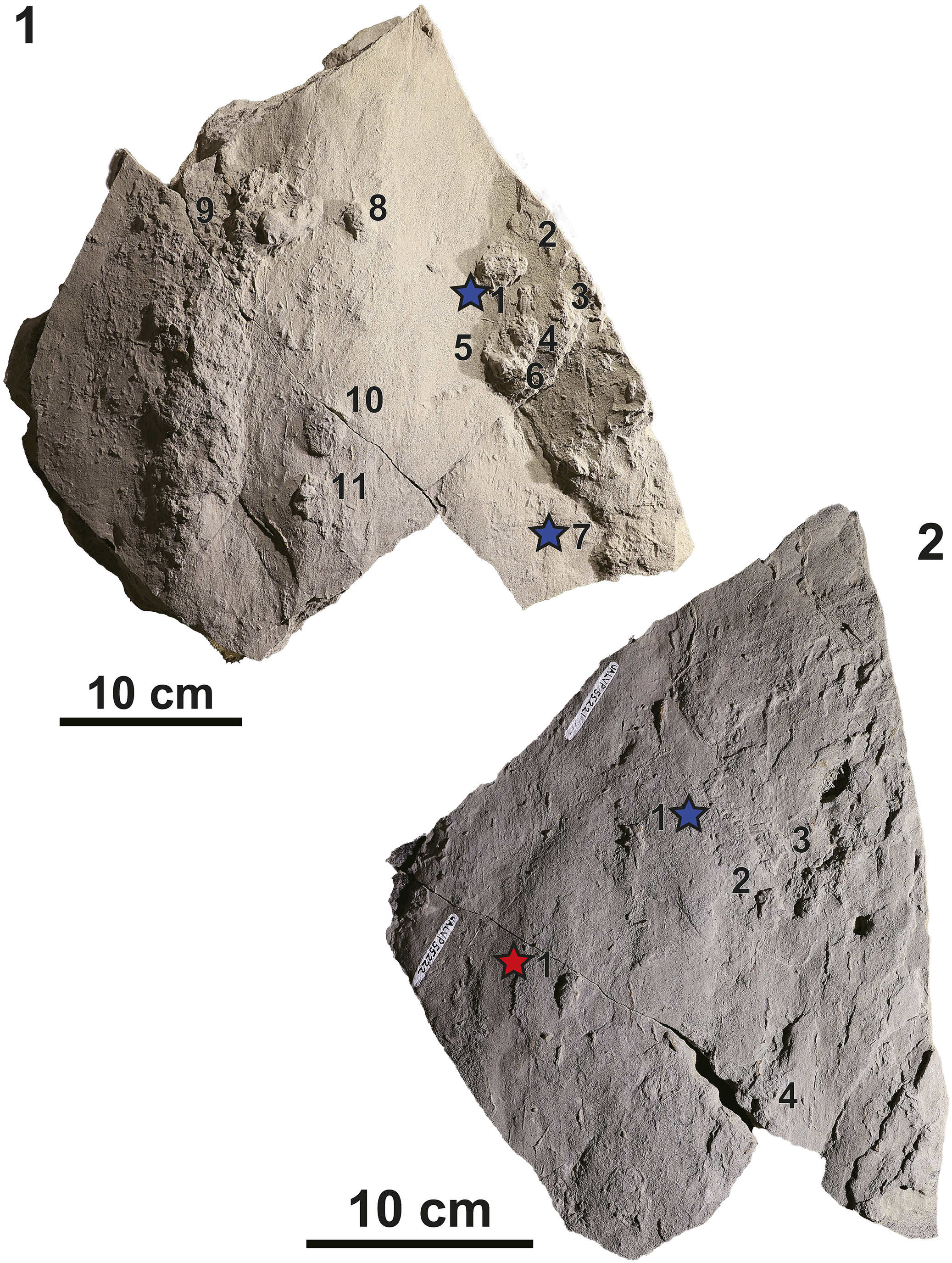
The fossil specimens described herein were collected at Red Willow Falls in 2011. The site was revisited in the summer of 2024 and 2025 to conduct detailed observations of the sedimentological characteristics of the fossil-bearing units. The fossils preserved on slab UALVP 53483 (originally collected and described by Bell et al., Reference Bell, Fanti, Acorn and Sissons2013; UALVP = University of Alberta Laboratory of Vertebrate Palaeontology) were re-examined, and an undescribed two-part sandstone slab with sample numbers UALVP 55221 and UALVP 55222 was also included in the study (Fig. 2). Each sandstone slab was photographed, and the morphology and orientation of each fossil were noted along with the orientations of tool marks on the slab surface. The fossils on each sandstone slab were individually numbered for simplicity (e.g., UALVP 53483-1, UALVP 55221-1; Fig. 2). In addition, one specimen (UALVP 53483-11) was sectioned and polished to observe its relationship with sedimentary features of the sandstone.
The sandstone slabs studied herein. (1) Slab UALVP 53483; and (2) slabs UALVP 55221 + UALVP 55222. Both images are oriented so that the current flow direction is from the top to the bottom of the figure. The mayfly fossils are numbered, and the holotype of Kalyptichnus belli n. igen. n. isp. is indicated with a red star, whereas the paratypes are indicated by blue stars.

Figure 2. Long description
Two panels showing fossiliferous sandstone slabs.
Panel 1, top left, shows slab UALVP 53483. It features eleven numbered mayfly fossils. Numbers 1 through 6 are clustered on the right side, with a blue star indicating the paratype located next to number 1. Number 7 is at the bottom right, also marked with a blue star. Numbers 8 and 9 are near the top, while 10 and 11 are located in the lower-middle section. A 10 cm scale bar is positioned at the bottom left.
Panel 2, bottom right, shows slabs UALVP 55221 and UALVP 55222 joined together. Four mayfly fossils are numbered. A red star indicating the holotype of Kalyptichnus belli is located at the bottom left next to number 1. A blue star paratype is located in the upper center next to another number 1. Numbers 2, 3, and 4 are distributed toward the right and bottom edges. A 10 cm scale bar is at the bottom center.
Orientations were measured for 11 fossils and 48 tool marks on UALVP 53483, and for five fossils and 58 tool marks on UALVP 55221 + UALVP 55222. Determining flow direction solely from the tool marks proved difficult, due to their small size. These marks were therefore recorded as bidirectional features—aligned with the current but not indicative of the direction of sediment transport. Flow direction was later constrained using cross-bedding visible on the edges of the sandstone slabs. Because the original compass orientation of the slabs was not recorded, the orientations of the fossils and tool marks were measured from a single photograph using Adobe Photoshop. The tool mark orientations were averaged to estimate the mean flow direction in the image, which was then used to produce a rose plot of the tool mark and fossil orientations with 180° representing the downstream direction.
Repository and institutional abbreviation
Types, figured, and other specimens examined in this study are reposited at the University of Alberta Laboratory of Vertebrate Palaeontology (UALVP).
Results
Sedimentological description
The outcrop at Red Willow Falls comprises a basal fine- to medium-grained sandstone with decimeter-scale trough crossbedding, erosively overlying a thick mudstone bed. Both the bedding scale and grain size decrease upward through the succession (Fig. 1.2, 1.3). According to Bell et al. (Reference Bell, Fanti, Acorn and Sissons2013), the fossil-bearing sandstone slabs were collected from a distinct interval (~3 m thick) of interbedded sandstone and mudstone, with the mudstone–sandstone intervals ranging from a few centimeters to ~35 cm.
We were able to relocate this heterolithic unit near the top of the measured section (Fig. 1.2). The fossils described herein were observed as positive hyporelief structures at the base of a sandstone bed. The sandstone beds in the measured section vary in thickness from 1 to 25 cm, and are laterally extensive, giving them a sheet-like appearance. They exhibit planar or trough cross laminae with bedding thicknesses of up to 1 cm. Their bases are associated with erosional features such as tool marks (Fig. 1.4) and parting lineations (Fig. 1.5), as well as abundant trace fossils of vertebrates and invertebrates. The mudstone layers vary from 2 to 10 cm in thickness, and are dark gray, organic-rich, with planar laminae, and rare desiccation cracks (Fig. 1.5). In addition, laterally restricted (usually 2–5 m wide), erosionally based, buff-colored mudstone lenses occur throughout the interbedded sand–mud deposit.
Other trace fossils reported from the same interval as the ephemeropteran specimens have included examples of Planolites, Skolithos, and Taenidium, as well as traces attributed to nematodes, bivalves, a small tetrapod, mammals, and an enigmatic swimming vertebrate (Bell et al., Reference Bell, Fanti, Acorn and Sissons2013; Fanti et al., Reference Fanti, Bell and Sissons2013; Kimitsuki et al., Reference Kimitsuki, Rodriguez, Sullivan, Zonneveld, Sissons, Bell, Campione, Fanti and Gingras2024). However, many additional trace fossils from this interval remain undescribed. The package of interbedded sandstone and mudstone continues laterally for ~400 m along the Redwillow River. The package passes to the south into a more homolithic trough cross bedded sandstone deposit, which is truncated to the north by broad and dish-shaped trough cross bedded sandstone associated with inclined heterolithic strata.
Fossil description
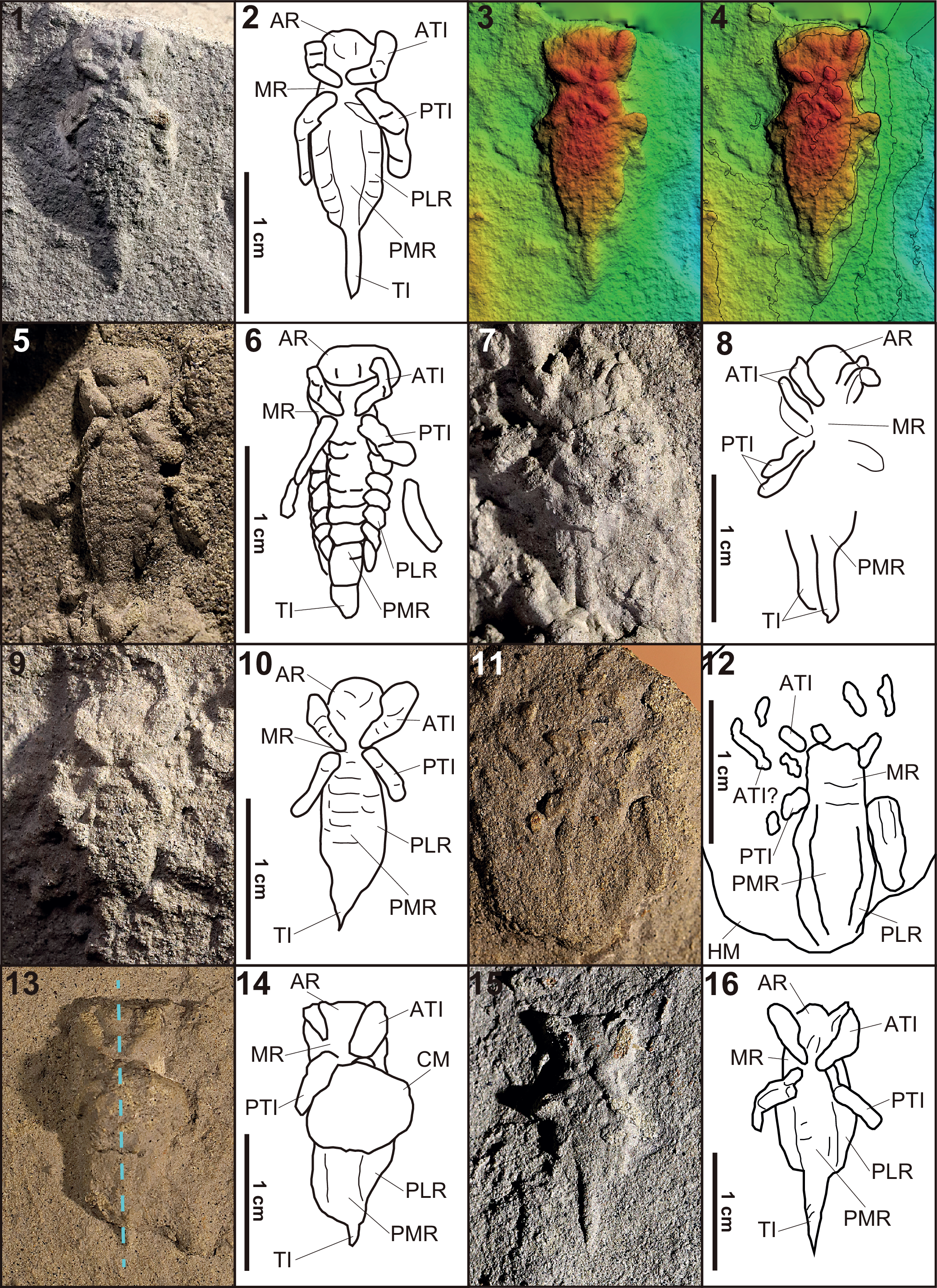
The morphological characteristics of the larval fossils were described in detail by Bell et al. (Reference Bell, Fanti, Acorn and Sissons2013). Preserved in positive hyporelief, the fossils protrude downwards out of the bedding surface of the sandstone slabs. The best-preserved specimen (UALVP 53483-1; Fig. 3.5, 3.6) consists of an elliptical head imprint, a short thorax with two pairs of legs in an X-shaped configuration, an abdominal section with at least eight body segments, and an elongate cercal base. The abdominal section has seven pairs of lateral elliptical impressions that each extend from between two adjoining abdominal segments. However, many of the fossils do not preserve the body segments as well as UALVP 53483-1, showing only the outline of the insect body (Fig. 3.1–3.4, 3.9–3.16). Notably, all of the fossils were preserved with their ventral sides facing down, in life orientation. Two specimens (UALVP 53483-8 and UALVP 53483-11) were found with a mound superimposed on the central part of the ventral surface of the body (Fig. 3.13, 3.14). As well, UALVP 53483-10 was preserved on the surface of an oval-shaped mound that is ~25 mm long, ~20 mm wide, and ~3 mm high (Fig. 3.11, 3.12). This fossil somewhat incompletely preserves three pairs of legs, the anterior two pairs directed anterolaterally, and the posteriormost pair extending in the opposite direction. In addition, UALVP 53483-4 comprises two overlapping partial impressions, resulting in two sets of appendages, and two terminal imprints, which are slightly mutually offset (Fig. 3.7, 3.8).
Type specimens and morphological variants of Kalyptichnus belli n. igen. n. isp. (1–4) Holotype specimen (UALVP 55222-1); (1) photo of holotype; (2) interpretive line drawing of holotype; (3) digital elevation model (DEM) of holotype; (4) DEM of holotype with elevation contours at intervals of 0.5 mm. (5, 6) Paratype specimen (UALVP 53483-1); (5) photo showing exceptional preservation of the tracemaker’s morphology; (6) interpretive line drawing. (7, 8) Photo and interpretive line drawing of a morphological variant showing overlapping traces (UALVP 53483-4). (9, 10) Photo and interpretive line drawing of paratype specimen (UALVP 53483-7). (11, 12) Photo and interpretive line drawing of a morphological variant (UALVP 53483-10) with the trace fossil situated on a shallow mound. (13, 14) Photo and interpretive line drawing of a morphological variant (UALVP 53483-11) with a mound superimposed on the center of the trace fossil; the blue dashed line shows where the specimen was sectioned as shown in Figure 5. (15, 16) Photo and interpretive line drawing of a paratype (UALVP 55221-1). Abbreviations: AR = Anterior region; ATI = Anterior transverse imprint; CM = Central mound; HM = Horizontal mound; MR = Middle region; PTI = Posterior transverse imprint; PLR = Posterior lateral region; PMR = Posterior middle region; TI = Terminal imprint.

Figure 3. Long description
The grid contains 16 panels numbered 1 through 16.
Panels 1 to 4 show the holotype specimen UALVP 5 52 22-1. Panel 1 is a photo of the fossil in rock. Panel 2 is a line drawing labeling the A R, AT I, M R, PT I, PL R, PM R, and TI. Panel 3 is a DEM with a color gradient from green at the base to red at the highest elevation. Panel 4 adds contour lines at 0.5 millimeter intervals to the DE M.
Panels 5 and 6 show paratype UALVP 5 34 83-1. The photo in panel 5 shows a highly textured surface, and the drawing in panel 6 highlights segmented structures in the PM R.
Panels 7 and 8 show specimen UALVP 5 34 83-4. The drawing in panel 8 illustrates overlapping traces with multiple TI and PTI features.
Panels 9 and 10 show paratype UALVP 5 34 83-7. The drawing in panel 10 shows a wide AR and distinct transverse imprints.
Panels 11 and 12 show specimen UALVP 5 34 83-10. The drawing in panel 12 shows the trace situated on a shallow horizontal mound labeled H M.
Panels 13 and 14 show specimen UALVP 5 34 83-11. Panel 13 includes a vertical blue dashed line indicating a cross-section. Panel 14 labels a large central mound as CM superimposed on the trace.
Panels 15 and 16 show paratype UALVP 5 52 21-1. The drawing in panel 16 shows a narrow, elongated T I.
Each drawing includes a 1 centimeter scale bar. The anatomical regions are consistently labeled: AR for anterior region, ATI for anterior transverse imprint, MR for middle region, PTI for posterior transverse imprint, PLR for posterior lateral region, PMR for posterior middle region, and TI for terminal imprint.
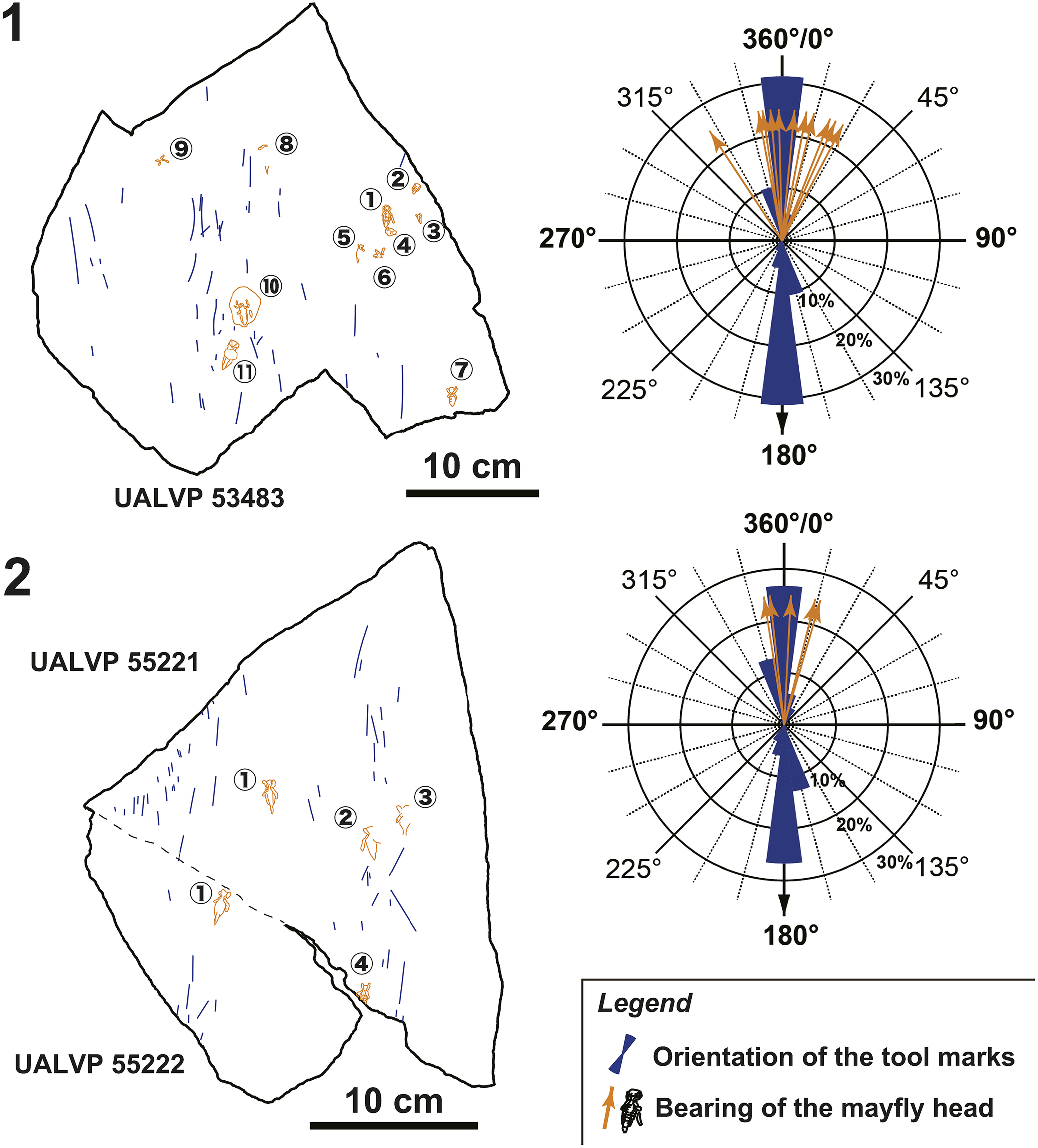
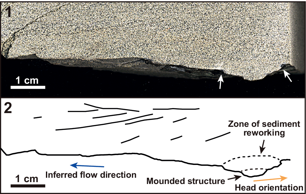
Measurements of the orientations of the fossils indicate that, on each slab, they are roughly parallel to each other. Moreover, they are approximately aligned with the average current orientation inferred from tool marks. Except for one specimen, the fossils have bearings within 30° of the mean current orientation (n = 16, standard deviation = 16.0°; Fig. 4). A sagittal cross section through UALVP 53483-11 confirms the geometric relationship between the fossil and the current orientation inferred from the pattern of crossbedding, indicating that the head of the insect was directed into the current (Fig. 5). The cross section reveals a zone of sediment reworking characterized by clasts that are lighter-colored and finer-grained than those of the host sediment. The zone of reworking continues stratigraphically upwards to about 5 mm from the base of the body impression (Fig. 5).
Interpretive line drawings and rose plots of the current features and the mayfly fossils on the sandstone slabs studied herein. The drawings are oriented so that the current flow direction is from the top to the bottom of the figure. The mayfly fossils are numbered as in Figure 2. (1) Interpretive line drawing of UALVP 53483 and a rose plot showing the orientations of the fossils and tool marks. (2) Interpretive line drawing of UALVP 55221 and UALVP 55222, and a rose plot showing the orientations of the fossils and the tool marks. The dashed line indicates the boundary between UALVP 55221 and UALV 55222. For both line drawings, the outlines of the mayfly fossils are in orange, and the tool marks are in blue. Both rose plots show the tool marks as bidirectional features, whereas the arrows indicate the orientations of the anterior ends of the mayfly fossils.

Figure 4. Long description
The figure consists of two horizontal sections, labeled 1 and 2.
Section 1 (Top):
* Left: Line drawing of slab UALVP 53483. It features orange outlines of mayfly fossils numbered 1 through 11 and blue linear tool marks. A 10 cm scale bar is at the bottom right of the drawing.
* Right: A rose plot showing tool mark orientations as bidirectional dark blue bars concentrated along the 0-180 degree axis. Orange arrows indicate the bearing of mayfly heads, primarily clustered between 315 and 45 degrees.
Section 2 (Bottom):
* Left: Line drawing of slabs UALVP 55221 (top portion) and UALVP 55222 (bottom portion), separated by a dashed line. Mayfly fossils are numbered 1 through 3 on the top slab and 1 and 4 on the bottom slab. Blue tool marks are scattered across both. A 10 cm scale bar is at the bottom right.
* Right: A rose plot showing tool mark orientations in dark blue, heavily weighted toward the 0-180 degree axis. Orange arrows for mayfly head bearings are clustered near the 0/360-degree mark.
Legend (Bottom-Right):
* A dark blue bidirectional bar represents the orientation of the tool marks.
* An orange arrow with a mayfly icon represents the bearing of the mayfly head.
(1) Parasagittal section through the fossil shown in Figure 3.13 (UALVP 53483-11). The anterior and posterior ends of the fossil are indicated by the white arrows, and the head is towards the right of the image. (2) Line drawing of the section through UALVP 53484-11. Note the mounded structure at the center of the fossil.

Figure 5. Long description
Panel 1 is a photo of a rock section with a fine, speckled tan and grey granular texture. At the bottom edge, a dark, bedding plane is visible. Two white arrows at the bottom right point upward to specific dark depressions in the fossil. A 1 cm scale bar is located at the bottom left.
Panel 2 is a line drawing of the same section.
* The top half contains several thin, horizontal and slightly angled black lines representing sediment layers.
* A thick, irregular horizontal line across the middle represents the bedding plane with the fossil.
* On the left, a blue arrow points left with the label Inferred flow direction.
* On the right, a depression in the fossil line is labeled Mounded structure.
* Above this depression, a dashed oval area is labeled Zone of sediment reworking.
* Below the mounded structure, an orange arrow points right with the label Head orientation.
* A 1 cm scale bar is at the bottom left.
Interpretation
Sedimentological interpretations
As briefly mentioned in a previous section, Bell et al. (Reference Bell, Fanti, Acorn and Sissons2013) and Kimitsuki et al. (Reference Kimitsuki, Rodriguez, Sullivan, Zonneveld, Sissons, Bell, Campione, Fanti and Gingras2024) interpreted the fossil-bearing interval at the Red Willow Falls site as overbank and crevasse splay deposits laid down in a meandering channel belt. Indeed, the overall fining-upwards sequence and presence of thick mud beds are common features of meandering river sedimentation (e.g., Jackson, Reference Jackson and Miall1977; Halfar et al., Reference Halfar, Riegel and Walther1998; Viseras et al., Reference Viseras, Henares, Yeste, Garcia-Garcia, Ghinassi, Colombera, Mountney, Reesink and Bateman2018). Vertically aggrading crevasse splays commonly form as a result of repeated deposition of normally graded sand-sheets through flooding events (e.g., Burns et al., Reference Burns, Mountney, Hodgson and Colombera2017; Gulliford et al., Reference Gulliford, Flint and Hodgson2017).
On the other hand, the general lack of clear indicators of pedogenic alteration, such as rooting, mottling, soil horizons, and soil structures (e.g., Retallack, Reference Retallack1988; Tabor et al., Reference Tabor, Myers, Michel, Zeigler and Parker2017), suggests that the depositional environment did not experience significant subaerial exposure. The absence of pedogenic alteration is commonly found in submerged channel levees (e.g., Fielding, Reference Fielding1984; Farrell, Reference Farrell1987) or crevasse deltas, where the splay elements prograde into a standing body of water (e.g., Farrell, Reference Farrell1987, Reference Farrell2001; Smith and Pérez-Arlucea, Reference Smith and Pérez-Arlucea1994; Plint et al., Reference Plint, Helm and Lockley2023). Notably, some sedimentologists treat crevasse deltas as synonymous with crevasse splays that form under subaerial conditions (e.g., Colombera and Mountney, Reference Colombera and Mountney2021) because both are geomorphologically similar and result from the same mode of sediment transportation to the floodplain (i.e., levee breach during flooding). However, we consider the distinction between crevasse deltas and crevasse splays to be crucial because paleohydrology has a strong influence on the distribution of plants and animals, and therefore on the distribution of their traces in continental environments (e.g., Hasiotis, Reference Hasiotis and Hasiotis2002, Reference Hasiotis and Miller2007; Gibert and Sáez, Reference Gibert and Sáez2009; Genise, Reference Genise2017).
The Cumberland Marshes in Saskatchewan, Canada, provide a modern analogue of the depositional setting that may have existed in the Red Willow Falls area in the Late Cretaceous. The Cumberland Marshes were drastically altered when the “Old Channel” of the Saskatchewan River was clogged by an ice jam in 1873, and the river’s discharge was forced into a smaller channel system running through a wetland north of the Old Channel (Smith et al., Reference Smith, Cross, Dufficy and Clough1989). Overwhelmed by the sudden increase in discharge, the northern channels experienced extensive levee breaching, which delivered sediment-laden water into the adjacent wetlands. Initially, sediment and water were transported through sheetfloods, which became progressively channelized as the splays evolved (Smith et al., Reference Smith, Cross, Dufficy and Clough1989; Pérez-Arlucea and Smith, Reference Pérez-Arlucea and Smith1999).
According to Smith and Pérez-Arlucea (Reference Smith and Pérez-Arlucea1994) and Farrell (Reference Farrell2001), heterolithic deposits with minor rooting are found on the levees of both trunk and crevasse channels, and in the “prodelta” (sensu Smith and Pérez-Arlucea, Reference Smith and Pérez-Arlucea1994) or “distal mouth bar complex” (sensu Farrell, Reference Farrell2001) of the crevasse splay/delta. The heterolithic strata represent a repeating fining-upwards sequence (described as rhythmites), with each bed measuring a few millimeters to 30 cm thick and having a sharp erosional base overlain by ripple-laminated intervals and graded laminates (Farrell, Reference Farrell2001). Heterolithic rhythmites form during flooding events, in which coarse sediments are deposited first, and grain size then decreases under waning flow conditions (Farrell, Reference Farrell1987; Smith et al., Reference Smith, Cross, Dufficy and Clough1989).
Despite having similar sedimentological attributes, submerged levee deposits and crevasse deltas can be differentiated based on geomorphology. The former are ribbon-like and occur parallel to the channel with which they’re associated, whereas the latter are sediment lobes extending lateral to the trunk channel (e.g., Fielding, Reference Fielding1984; Pérez-Arlucea and Smith, Reference Pérez-Arlucea and Smith1999; Farrell, Reference Farrell2001). Unfortunately, determining the depositional setting for the Red Willow Falls strata through geomorphology is challenging because the exposure is restricted to a narrow area (< 50 m wide) that has been eroded by the present-day Redwillow River. On the other hand, we interpret the homolithic trough cross bedded sand to the south of the interbedded sandstone and mudstone to be crevasse channel fill (cf., Farrell, Reference Farrell2001), while the interspersed mud lenses throughout the outcrop section most likely represent mud plugs of abandoned distributary channels (cf., Tye and Coleman, Reference Tye and Coleman1989). These are a sedimentological constituent of crevasse splays and deltas (e.g., Smith et al., Reference Smith, Cross, Dufficy and Clough1989; Farrell, Reference Farrell2001); thus, we favor the interpretation that the Red Willow Falls strata formed as crevasse deltas rather than as levee deposits.
Fossil interpretations
Our reanalysis has identified the following notable characteristics of the specimens described here: (1) the fossils protrude about 1.5–2 mm from the base of the sandstone; (2) all of the fossils are in life orientation, with their ventral sides facing down; (3) all of the fossils have their anterior ends pointing into the current, mostly within 30° of the mean current-flow direction; and (4) the limb impressions exhibit features expected from live insect larvae and do not indicate disarticulation, dislocation, or abnormal positioning of the limbs (Figs. 2, 3). We are unaware of abiotic processes that can orient 16 insect bodies in the same direction and body position (i.e., ventral side down), and subsequently force them into the underlying substrate. In a recent experiment, Storari et al. (Reference Storari, Salles, Fonseca, Saraiva and Rodrigues2025) studied the postmortem processes that operate on modern mayfly and dragonfly larvae in a tank setup. According to them, only 47% of the deceased mayflies were found with the dorsal side up, whereas 36% of the dragonfly larvae were in dorsal-side-up position.
On the other hand, according to Vallon et al. (Reference Vallon, Schweigert, Bromley, Röper and Ebert2015), elongate objects (they gave fish carcasses and logs as examples) that are subjected to a current have a tendency to become oriented parallel to the current direction, with whichever end is closer to the center of gravity pointing upstream. However, when the objects that have settled into this rheotactically stable position are disturbed, they will regain stability by flipping over (Vallon et al., Reference Vallon, Schweigert, Bromley, Röper and Ebert2015). Therefore, while it is physically possible for a set of insect carcasses to become oriented in the same direction under a unidirectional flow regime, they would be unlikely to all adopt the same body position in this situation, with each individual’s ventral side facing down. It is also unlikely that overlapping pairs of larval carcasses would have been preserved in three dimensions as seen in UALVP 53483-4 (Fig. 3.7). Instead, a more parsimonious interpretation is that the specimens are not body fossils as suggested by Bell et al. (Reference Bell, Fanti, Acorn and Sissons2013) but rather represent traces of insects that shallowly buried themselves in the substrate, while orienting their heads into the current flow. The two overlapping body impressions in UALVP 53483-4 can be explained as traces successively produced by a single live insect repositioning itself within the substrate.
Notably, Knecht et al. (Reference Knecht, Benner, Dunlop and Renczkowski2024) established four criteria for identifying body impressions made by living organisms, and the specimen described here meets three of them. The three criteria observed are that the impressions are exclusively of the ventral surface, the limbs represented in the impressions are those expected to make contact with the substrate, and the limb impressions do not indicate postures that exceed the range of motion likely present in the living animal. The fourth criterion, which these fossils do not meet, is that body impressions may be accompanied by trackways approaching and/or departing from them. This fourth criterion would of course not be met if the living organism flew or swam to the location in which the body impression occurs or walked to the location on a surface that was unsuitable for preserving trackways and/or subsequently eroded away. Although the taphonomic study by Storari et al. (Reference Storari, Salles, Fonseca, Saraiva and Rodrigues2025) revealed that undisturbed larval bodies may long remain in articulation, the current flow experienced by the larvae described herein would likely have sufficed to cause major amputations or disarticulations if the larvae had been deceased. The legs are among the first parts of the body to disarticulate under mechanical disturbance, along with antennae, gills, and caudal filaments in the case of mayfly larvae, and labia in the case of dragonfly larvae (Storari et al., Reference Storari, Salles, Fonseca, Saraiva and Rodrigues2025).
As an alternative to interpreting the body impressions as resulting from active self-burial, one might argue that insect larvae clinging to the sediment surface at a specific orientation against a rapid current could have been pressed into the substrate through the weight of the sediment that piled up on top of them. However, insects are less dense than the surrounding sand/porewater medium, and the relatively broad ventral surfaces of the larvae described here would have prevented their bodies from sinking into the underlying mud. The lack of load structures (e.g., Owen, Reference Owen2003) also indicates that the substrate was not soft enough to be deformed under the weight of the larvae and/or that of the sediment itself. Thus, active self-burial by the larvae is the only mechanism available to explain why these fossils are found under the bedding surface.
Shallow self-burial behaviors are observed in modern dragonfly and mayfly larvae, both of which have similar body plans to the fossils described here. For example, some stream-dwelling dragonfly naiads, particularly those in the family Gomphidae, are known to burrow shallowly in the substrate to ambush prey and hide from predators (e.g., Chamberlain, Reference Chamberlain and Frey1975; Tennessen, Reference Tennessen2019). Huggins and DuBois (Reference Huggins and DuBois1982) observed the behaviors of two burrowing dragonfly naiads (Progomphus obscurus (Rambur, Reference Rambur1842) and Gomphus externus Hagen in Selys, Reference Selys1858) and found that the larvae always directed their heads upstream to stabilize themselves before burrowing. These larvae burrow in sand- to silt-sized sediments, although P. obscurus prefers coarser-grained substrates than G. externus. They also found that the burrowing depths were shallow, with P. obscurus larvae “never observed to burrow deeper than 2 cm”, and G. externus “normally burrowing only 3–5 mm deep” (Huggins and DuBois, Reference Huggins and DuBois1982, p. 5–6). Notably, Huggins and DuBois (Reference Huggins and DuBois1982) observed that the larvae of P. obscurus would move around within the substrate after burial, leaving an irregular trail. Such behavior was not observed in G. externus larvae.
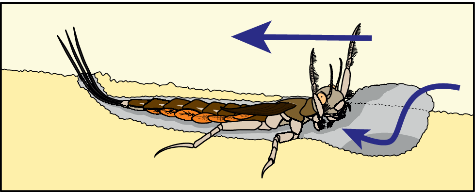
On the other hand, Soluk and Craig (Reference Soluk and Craig1988) documented suspension-feeding mayfly naiads (Ametropus neavei McDonnough, Reference McDonnough1928) that bury themselves in the sediment in a ventral-down position and extend their forelegs to capture food particles (mainly detritus) or remove unwanted sediment particles from around their mouths (Fig. 6). Burial depth is extremely shallow, and the larvae keep their eyes projecting above the sediment surface (Clifford and Barton, Reference Clifford and Barton1979). The larvae also orient themselves facing into the current to trap suspended food particles near their mouthparts and to allow water to flow over their abdominal gills. Ametropus neavei occur natively in the bed of the Athabasca River in Alberta, typically on very fine-grained sand (average grain size of ~100 μm) substrate on the downstream side of sandbars and on mud–sand substrates in deeper waters (Clifford and Barton, Reference Clifford and Barton1979). These mayfly nymphs are documented to be “excellent swimmers”, although they spend most of their time stationary in the substrate under lab conditions (Clifford and Barton, Reference Clifford and Barton1979, p. 856–857). A few other mayfly species have been reported to bury themselves in the sediment (Riek, Reference Riek1973; Courtney, Reference Courtney and Giberson2018), although details about their behavior are not available. It is also noteworthy that some “burrowing” mayfly naiads sometimes produce poorly defined burrows with unstable walls (Bae and McCafferty, Reference Bae, McCafferty, Corkum and Ciborowski1995), which may collapse over the larvae through current action.
Schematic drawing of modern suspension-feeding mayfly larvae, Ametropus neavei. Soluk and Craig (Reference Soluk and Craig1988) observed captive larvae of this species burrowing in the substrate with their heads oriented into the current (shown by blue arrows), as they captured suspended particles in pits excavated near their heads. A similar burrowing strategy may have been used by the producers of the body impressions from the Wapiti Formation. Image redrawn and modified from Soluk and Craig (Reference Soluk and Craig1988, p. 641, fig. 2) with permission number 6307301195953.

Although we interpret the specimens described here as trace rather than body fossils, we agree with Bell et al. (Reference Bell, Fanti, Acorn and Sissons2013) that they are attributable to mayfly naiads rather than to dragonfly naiads, which have a broadly similar overall body plan. This is based on the fact that the impressions do not show the prominent labium found in dragonfly naiads (e.g., Kukalová-Peck, Reference Kukalová-Peck2009; Schädel et al., Reference Schädel, Müller and Haug2020; Storari et al., Reference Storari, Salles, Fonseca, Saraiva and Rodrigues2025) but do show lobe-like structures in the posterior lateral region resembling the tracheal gills of mayfly naiads (e.g., Sroka et al., Reference Sroka, Godunko and Prokop2023) (Fig. 3.5, 3.6). Furthermore, the presence of only two pairs of leg impressions in most of the fossils described here is consistent with the tendency of some mayfly naiads, but not dragonfly naiads, to hold the forelegs clear of the substrate and use them for feeding purposes (Fig. 6).
While we are less certain about the correct identification of the tracemakers at lower taxonomic levels, the suspension-feeding behavior of modern Ametropus naiads hints that the tracemakers could have been referable to Ametropodidae rather than to cf. Heptageniidae as suggested by Bell et al. (Reference Bell, Fanti, Acorn and Sissons2013). Extant Ametropodidae have highly reduced forelimbs and seven pairs of ovate tracheal gills, and the impressions described herein indicate similar features in the tracemakers (e.g., Allen and Edmunds, Reference Allen and Edmunds1976; Wang et al., Reference Wang, Zhou and Zhou2013; Memishishi et al., Reference Memishishi, Bikashvili, Japoshvili and Mumladze2023). However, the impressions do not capture the morphology of the tracemakers completely enough for them to be definitively identifiable as ametropodids.
Based on our sedimentological interpretations and our observations of the fossils found in association with tractional sedimentary structures (i.e., tool marks), we propose the following taphonomic scenario: (1) the depositional event that preserved the fossils took place in a flooded overbank setting adjacent to a breach in the levee of a river’s trunk channel; (2) outflowing water from the trunk channel created hydrologic conditions optimal for suspension-feeding mayfly larvae (likely belonging to Ametropodidae or a similar group), prompting them to bury themselves in the muddy substrate with their bodies oriented against the current (cf., Soluk and Craig, Reference Soluk and Craig1988); (3) during seasonal flooding and increased fluvial discharge, sand-sized sediment was transported to the study area; (4) rapid deposition of sand preserved the impressions of the mayfly larvae as part of a crevasse delta deposit, possibly on the lee side of a migrating mouth bar complex (cf., Clifford and Barton, Reference Clifford and Barton1979).
Discussion
Sedimentological significance
Biogenic sedimentary structures can be useful indicators in sedimentological studies, yielding insights beyond those available from their abiogenic counterparts. Crevasse splay and crevasse delta deposits (such as those described here) represent event-driven sedimentation in which the levee of a riverbank is breached during flooding, causing erosion in areas proximal to the source channel, surface scouring farther out, and subsequent rapid sedimentation on the floodplain (Farrell, Reference Farrell2001; Burns et al., Reference Burns, Mountney, Hodgson and Colombera2017). Benner and Knecht (Reference Benner and Knecht2025) recently discussed the utility of full body impressions in recognizing true substrates, although in general, the ichnological signatures of fluvial event beds remain poorly documented. Similarly, trace fossils preserved in marine event beds (such as tempestites and turbidites) have been useful in stratigraphy and in understanding the responses of benthic organisms to such events (Frey and Goldring, Reference Frey and Goldring1992; Pemberton and MacEachern, Reference Pemberton, MacEachern, Brett and Baird1997; MacEachern et al., Reference MacEachern, Pemberton, Bann and Gingras2007). Further investigation, focusing on tiering of body impressions in relation to other associated trace fossils, may reveal similarities and differences from marine event beds, and significantly enhance our understanding of fluvial event beds.
Recently, King et al. (Reference King, Timmer, Furlong, Gingras, Hayes, Nadon, Tweet and Santucci2025) demonstrated the use of Lockeia (resting traces of freshwater bivalves) as indicators of paleocurrent direction in fluvial channels. This is based on the tendency of unionid bivalves to orient themselves in a manner that maximized the efficiency of water exchange through their siphons. Comparable rheotactic behaviors are also demonstrated to have determined the orientation of the arthropod cubichnion Rusophycus (and ‘Isopodichnus’) in both marine and non-marine settings (e.g. Pollard, Reference Pollard1985; Pickerill, Reference Pickerill1995; Hartung et al., Reference Hartung, Augustin, Kampouridis and Chure2021). The preferential orientation of the larval mayfly body impressions presumably has a similar utility as a paleocurrent indicator in environments with unidirectional flow. Although paleocurrent assessment can also be achieved through foreset dip measurements, it is challenging to accurately measure three-dimensional bedforms, especially in outcrops (Dott, Reference Dott1973; Dasgupta, Reference Dasgupta2002; King et al., Reference King, Timmer, Furlong, Gingras, Hayes, Nadon, Tweet and Santucci2025). Moreover, dip measurements may not be possible for upper flow regime planar beds, which are commonly preserved during rapid flow events in fluvial environments (Fielding, Reference Fielding2006).
Ichnotaxonomic implications
As discussed above, available evidence indicates that the fossils from Red Willow Falls described in this study record aspects of ancient mayfly larval behavior. The question that follows is whether these specimens should be formally recognized as trace fossils by referring them to an ichnotaxon. In the International Code of Zoological Nomenclature (Reference Ride, Cogger, Dupuis, Kraus, Minelli, Thompson and Tubbs1999, Article 1.2, p. 2–3), (body) fossils are characterized as “substitutions (replacements, impressions, molds and casts) for the actual remains of animals”, and trace fossils as “fossilized work of organisms”. Ichnologists have come up with a stricter definition for trace fossils, namely “a morphologically recurrent structure resulting from the life activity of an individual organism (or homotypic organisms) modifying the substrate” (Bertling et al., Reference Bertling, Braddy, Bromley, Demathieu and Genise2006, p. 266), and lists of traces and non-traces can be found in Bertling et al. (Reference Bertling, Braddy, Bromley, Demathieu and Genise2006, Reference Bertling, Buatois, Knaust, Laing and Mángano2022).
Fine details of the bodies of the Red Willow Falls fossil mayfly larvae are preserved in some cases (e.g., UALVP 53483-1; Fig. 3.5), which suggests that some (if not all) of the larvae failed to escape the deposition of sand and were killed at their burial site (Kesidis et al., Reference Kesidis, Slater, Jensen and Budd2019 similarly interpreted tubular impressions preserving fine details as annelid body fossils, rather than burrows). The lack of any distinct escape structure (fugichnion) on the visible sagittal cross section of one of the larvae (Fig. 5) weakly supports this idea. Intuitively, this might suggest that the fossils should be treated as casts of the mayfly larvae, which would qualify as body fossils, rather than as trace fossils. However, in our opinion, the presence of the ‘actual remains of animals’ is not sufficient to categorically disqualify body impressions produced through the work of animals as trace fossils. Although each preserved body impression is essentially a partial cast of a mayfly naiad’s body, the cast would not exist if not for the animal’s behavior in life, and the extent of the cast is determined by the specifics of that behavior (keeping the head and front legs clear of the substrate while feeding). Thus, the fossil could be seen as a cast actively created by the naiad (i.e., a modification of the substrate), rather than passively created post-mortem by sedimentological processes, and therefore as a trace fossil representing the work of an animal. This interpretation is also consistent with that of cubichnial trace fossils that closely replicates the external morphology of their producers, as in Asteriacites and Lockeia (e.g., Seilacher, Reference Seilacher2007).
Moreover, if an animal perished inside an excavation that it produced, then the preserved remains of the body and the excavation should be treated separately, as a body fossil and a trace fossil respectively. A good analog is provided by cases where body fossils of tracemakers are preserved in their own burrows. This is not uncommon when the burrows are borings in firm/hard substrates (e.g., Donovan and Hensley, Reference Donovan and Hensley2006; Kumar et al., Reference Kumar, Singh and Rana2011) but can also occur when the burrows penetrate soft substrates (e.g., Sellwood, Reference Sellwood1971; Bromley and Asgaard, Reference Bromley and Asgaard1975; Hasiotis and Mitchell, Reference Hasiotis and Mitchell1993; Vallon et al., Reference Vallon, Schweigert, Bromley, Röper and Ebert2015; Paz et al., Reference Paz, Mángano, Buatois, Campetella, Sproat, Pérez-Pueyo, Piñuela and García-Ramos2023). In these examples, the burrows and body fossils normally receive independent taxonomic identifications. Turning to Red Willow Falls, we have ample evidence that the mayfly body impressions described herein were produced by active self-burial on the part of the larvae.
While shallow excavations and resting traces produced by marine decapods have been recognized and named Solusichnium by O’Brien et al. (Reference O’Brien, Braddy and Radley2009), to our knowledge, fossil traces representing shallow self-burial behavior by insect larvae have never previously been reported. Given the novelty of ichnofossils of this type, and their potential utility in paleoecological and sedimentological interpretation, the shallow burial trace fossils described here merit assignment to a new ichnotaxon.
Conclusions
By integrating detailed sedimentological analyses with re-evaluation of body impressions of mayfly larvae from the Wapiti Formation, we were able to demonstrate that these fossils can tell us much more than what was originally inferred by Bell et al. (Reference Bell, Fanti, Acorn and Sissons2013). Specifically, we were able to deduce that the fossils record the behavior of live mayfly larvae that had shallowly buried themselves in the substrate, with their heads directed upstream. The event likely took place in a flooded channel overbank setting affected by seasonal deposition of sand. These shallow burial casts of insect larvae represent a novel type of trace fossil, which we have named Kalyptichnus belli n. igen. n. isp. (see section below). Our study emphasizes the necessity of considering sedimentological context when interpreting fossils and highlights that re-examination of invertebrate body casts can shed fresh light on ancient animal behaviors.
Systematic ichnology
Ichnogenus Kalyptichnus new ichnogenus
Type ichnospecies
Kalyptichnus belli n. isp. by monotypy.
Diagnosis
A small, bilaterally symmetrical, trace fossil preserved in positive hyporelief and oriented subparallel to the bedding plane. The trace fossil shallowly projects out of the bottom of the bedding surface with sheer, locally overhanging edges. It consists of a small subcircular to oval anterior region, a broad middle region with two pairs of elongate transverse imprints projecting laterally at oblique angles to the fossil’s long axis, and a posterior region that gradually tapers towards the end. An elongate terminal impression is commonly present.
Etymology
Derived from the Greek words “Kalyptos (Καλυπτός)” meaning “covered”, “concealed” or “hidden”, and “Ichnos (Ίχνος)” meaning “trace”.
Remarks
Kalyptichnus n. igen. is a distinct ichnogenus for shallow excavations of arthropods, representing three-dimensional structures that closely reflect the form of much of the tracemaker’s body. Excavations of this kind can be differentiated from many arthropod (e.g., insect or chelicerate) resting traces, such as Alacranichnus, Cardinichnus, Chliceratichnus, Hedriumichnus, Inmontibusichnus, Narragansettichnus, and Rotterodichnium, which are primarily surface traces of tracemakers that did not fully dig themselves into the substrate (e.g., Braddy and Briggs, Reference Braddy and Briggs2002; Lucas et al., Reference Lucas, Lerner and Voigt2013; Benner et al., Reference Benner, Knecht and Engel2015; Dalman and Lucas, Reference Dalman and Lucas2015; Getty et al., Reference Getty, Sproule, Stimson and Lyons2017; Knecht et al., Reference Knecht, Benner, Dunlop and Renczkowski2024). These trace fossils are expressed as shallow, poorly defined impressions of a portion of the ventral surface of the tracemakers, often limited to parts of the abdomen and the appendages. Furthermore, these ichnogenera typically differ from Kalyptichnus n. igen. in lacking a well-defined circular anterior region. Tonganoxichnus is another shallow arthropod resting trace fossil that sometimes features exceptional preservation of a ventral body impression of the tracemakers (Mángano et al., Reference Mángano, Buatois, Maples and Lanier1997, Reference Mángano, Labandeira, Kvale and Buatois2001). However, Tonganoxichnus is diagnosed based on the presence of anteriormost appendage imprints that are parallel to the main axis and common abdominal appendage imprints (Mángano et al., Reference Mángano, Buatois, Maples and Lanier1997; Benner et al., Reference Benner, Knecht and Engel2015), both of which are absent in Kalyptichnus n. igen. Conversely, Tonganoxichnus often lacks a well-defined subcircular anterior region, and the three pairs of anteromedian appendage imprints (which is positionally equivalent to the two pairs of transverse imprints in the type ichnospecies Kalyptichnus belli n. igen. n. isp.) are short and ellipsoidal. On the other hand, Tripartichnus (particularly the type ichnospecies T. triassicus Vallon and Röper, Reference Vallon and Röper2006) has a similar gross overall appearance to Kalyptichnus n. igen., but Tripartichnus is diagnosed based on its three-part morphological division into three parts, with the anterior part broadest, the middle part medium in width, and the posterior part narrow (Vallon and Röper, Reference Vallon and Röper2006). By contrast, Kalyptichnus has a relatively small anterior region that is typically narrower than, or at most similar in width to, the posterior region. Tripartichnus also lacks distinct transverse imprints as in Kalyptichnus n. igen.
Some examples of arthropod cubichnia that constitute shallow excavations include Rusophycus, Selenichnites, and Surculichnus, all of which differ distinctly in outline from Kalyptichnus n. igen. (overall coffee-bean shape in Rusophycus; broad, crescentic anterior region in Selenichnites and Surculichnus) (e.g., Gand et al., Reference Gand, Garric, Schneider, Walter, Lapeyrie, Martin and Thiéry2008; Knecht et al., Reference Knecht, Benner, Rogers and Ridge2009; Buhler and Grey, Reference Buhler and Grey2017). Kalyptichnus n. igen. is similar to Solusichnium, a shallow, suboval excavation that represents the impression of a marine decapod tracemaker (O’Brien et al., Reference O’Brien, Braddy and Radley2009). However, Kalyptichnus n. igen. differs from Solusichnium in not being suboval in outline and in having a divergent overall body plan: the anterior imprint of Solusichnium is circular to arrow-shaped with two pairs of linear (antennal) imprints, whereas that of Kalyptichnus is consistently rounded and lacks linear imprints; Kalyptichnus n. igen. lacks the narrow medial imprint seen in Solusichnium; and Solusichnium has only one pair of posteriorly directed lateral imprints with bifurcating ends, whereas Kalyptichnus has two pairs of non-bifurcating transverse imprints that form an x-shaped configuration. In addition, Solusichnium has a posterior region preserved in convex epirelief reflecting the use of the uropod to push clear of the substrate, which is absent in our specimen.
According to Minter et al. (Reference Minter, Braddy and Davis2007) and Bertling et al. (Reference Bertling, Buatois, Knaust, Laing and Mángano2022), an ichnotaxobase should reflect the morphology of the trace fossil, which in turn is a function of tracemaker anatomy, tracemaker behavior, the nature of the substrate, and arrangement of subunits. The differences between Kalyptichnus n. igen. and Solusichnium mainly arise from the contrasting body plans of the tracemakers (hexapods and decapods, respectively) and the inclusion of an escape structure produced by action of the uropod in Solusichnium. In addition, although both ichnogenera represent shallow excavations, Solusichnium is primarily an escape trace related to a storm event with rapidly shifting sediments, whereas Kalyptichnus n. igen. is a dwelling trace related to less dynamic bottom conditions. We deem these differences significant enough to justify separating the two types of trace fossil at the ichnogenus level. The diagnosis for Kalyptichnus n. igen. is broadly formulated to accommodate similar trace fossils that may be produced by other insect larvae.
Kalyptichnus belli new ichnospecies
Holotype
UALVP 55222-1.
Paratypes
UALVP 53483-1, UALVP 53483-7, UALVP 55221-1.
Materials
Sixteen trace fossils preserved on sandstone slabs UALVP 53483, UALVP 55221, and UALVP 55222.
Diagnosis
Kalyptichnus n. igen. with two pairs of transverse imprints in the middle region. The anterior pair of transverse imprints are directed anterolaterally and the posterior pair posterolaterally, forming an X shape. The posterior region can be subdividedinto a single posterior-middle region and paired posterior-lateral regions, all of which may exhibit segmentation in exceptionally preserved examples.
Occurrence
The type specimens occur in the Upper Cretaceous (upper Campanian) Wapiti Formation of northwestern Alberta, Canada.
Description
The trace fossils are preserved in positive hyporelief, and protrude out of the bedding surface to depths of 1.5–2.0 mm. The trace fossils measure ~14 mm in length, not including the terminal imprint, and 4–6 mm in width excluding the lateral imprints. The anterior region is generally wider than long, with a length of 2–3 mm and a width of ~3 mm. The lengths of the transverse imprints are highly variable, ranging from 2.5–8.5 mm. The anterior transverse imprints diverge from the midline at 35–80° (usually ~45°), whereas the posterior transverse imprints diverge at between 140–160°. The posterior region is antero-posteriorly elongated, with a length of 7–9 mm and a width of 4–5 mm. The posterior-lateral regions are ~1 mm in width. Where present, the terminal imprint can be as long as 7 mm.
Etymology
Named after Dr. Phil R. Bell, now of the University of New England in Armidale, NSW, Australia, in recognition of his having led the original description of the fossils redescribed herein.
Remarks
Kalyptichnus belli n. igen. n. isp. is a distinctive trace fossil that preserves three body segments and two pairs of appendages, interpreted to be a shallow burial trace created by an insect larva. Although insects have three pairs of walking legs, only two pairs are interpreted as having been used to dig into the substrate during formation of K. belli n. igen. n. isp. traces, whereas the anteriormost pair was raised dorsally. Such self-burial behavior is observed in modern mayfly naiads belonging to Ametropodidae (Clifford and Barton, Reference Clifford and Barton1979; Soluk and Craig, Reference Soluk and Craig1988). Where multiple individuals are preserved in association with current indicators, all of them are similarly oriented, with their heads pointing in the inferred upstream direction.
Acknowledgments
This research was funded by NSERC Discovery Grants to M.K.G., J.-P.Z. and C.S., and by an endowment associated with the Philip J. Currie Professorship in Vertebrate Palaeontology at the University of Alberta. Northwestern Polytechnic generously provided accommodation during recent geological fieldwork at Red Willow Falls. We would also like to thank the field crew members of the Boreal Alberta Dinosaur Project for their assistance in the field. Finally, we would like to thank the associate editor G. Mángano, and the reviewers R. Knecht and L. Vallon for their professional insights on arthropod body impressions, which helped improve this manuscript.
Competing interests
The authors declare none.

 Open access

