Although major advances have been made in the field of congenital cardiac surgery, mortality remains high. According to some, the 30-day mortality for all patients with congenitally malformed hearts is 5%, rising to 11% in neonates.Reference Lacour-Gayet, Clarke and Jacobs1 Inappropriate myocardial protection is still considered the main cause of mortality and morbidity.Reference Allen2 For myocardial protection in children, cardioplegic solutions are universally popular. Although the application of cardioplegia is an effective strategy, it does not eradicate ischaemia reperfusion injury, especially in patients who are cyanotic prior to correction.Reference Allen2 Protection of the myocardium during the cardiac operation has two functions, first to reduce ischaemic injury by means of cardioplegia and hypothermia during cardiac arrest, and second to decrease reperfusion injury by using modified reperfusion. The phenomenon of postconditioning, achieved by means of multiple brief periods of ischaemic reperfusion just after the prolonged ischaemic insult, was initially described by Zhao et al.Reference Zhao, Corvera and Alkos3, Reference Zhao and Vinten-Johansen4 using dogs, and has proved to provide strong endogenous myocardial protection by reducing reperfusion injury. The phenomenon of postconditioning also has been shown to exist in humans in the setting of percutaneous cardiac intervention.Reference Staat, Rioufol and Piot5, Reference Laskey6 These results are very encouraging. It remains to be determined, however, whether postconditioning has an additive effect to cardioplegia in the setting of cardiac surgery, one of the few controlled models of human myocardial ischaemia. As far as we are aware, the effect of postconditioning on myocardial protection has not been investigated in the field of cardiac surgery. We conducted, therefore, a controlled trial of patients undergoing cardiac surgery. We hypothesized that postconditioning would provide protection against myocardial ischaemic reperfusion in children undergoing cardiopulmonary bypass for repair of their congenitally malformed hearts.
Methods
The ethics committees of the hospital approved the study, and written informed consent was obtained from the parents of the patients. We enrolled 40 patients scheduled for elective repair of congenitally malformed hearts from July to December, 2006, at the Xiang Ya Hospital, Central South University, China. The patients, referred to us for elective correction of ventricular septal defects and tetralogy of Fallot without other major anomalies, were randomized into a group to undergo postconditioning, and a control group, by use of a randomized number table. The surgeon performing the operation was aware of the grouping just prior to aortic cross de-clamping. The anaesthesiologist was unaware of the grouping until postconditioning was performed after aortic de-clamping. The staff caring for postoperative patients, those collecting the data, and the laboratory staff were all blinded to the groupings.
Study criterions
Criterions for inclusion were being scheduled for elective closure of ventricular septal defects, or correction of tetralogy of Fallot, on cardiopulmonary bypass with cardiac arrest, and with an age between 1 and 14 years. We excluded patients undergoing concomitant aortic or valvar surgery, those with concomitant infections such as infective endocarditis, those having emergency procedures, those with tetralogy with pulmonary atresia or absent pulmonary valve, those showing haemodynamic instability requiring inotropic support within 24 hours prior to surgery, and those with an aortic cross clamping time of less than 30 minutes. No patient in either group received preoperative medications, such as furosemide, or inhibitors of angiotensin converting enzyme. Preoperative hepatic and renal function in both groups was normal.
Anaesthesia and surgical techniques
During the surgical procedure, anaesthesia was induced with midazolam and vecuronium bromide intravenously, followed by the use of intravenous fentanyl and intermittent inhalation of isoflurane to maintain anaesthesia. The operations were performed using standard hypothermic cardiopulmonary bypass, with bicaval venous cannulation and venting via the atrial septum, and with a disposable hollow fibre oxygenator and aprotinin administered to the priming volume. The myocardium was protected using intermittent perfusion of cold blood cardioplegia. The cardioplegic solution was mixed through the deliver system of the heart-lung machine with autologous blood obtained from the extracorporeal circuit in a ratio of 1 to 4 parts of crystalloid cardioplegia to autologous blood while the patient was on bypass prior to its application. The cold blood cardioplegia was infused into the aortic root, using an initial dose of 20 ml/kg body weight at a flow rate of 200–250 ml/min. Thereafter, the cardioplegia was reinfused every 20 to 30 minutes, at a dose of 10 ml/kg. All operations were performed by same surgeon, using identical methods in both groups for closure of the ventricular septal defect or repair of tetralogy. A right ventricular incision was used for correction of tetralogy in both groups, using a right atrial or pulmonary arterial incision for those needing closure of ventricular septal defects. After surgery, all patients were admitted to the intensive care unit. Patients from both groups received the same routine postoperative care as determined by the caring physicians. Postoperatively, dopamine was used as first-line inotrope therapy for patients needing such support. If needed, epinephrine or milrinone was added for appropriate haemodynamic support.
Postconditioning protocol
Postconditioning was started at 30 seconds after aortic cross de-lamping, and the aorta was re-clamped for 30 seconds, rendering ischaemia, and meanwhile aortic root suction was established during aortic re-clamping. Thereafter, the aortic clamp was released for 30 seconds for full myocardial reperfusion. This cycle was repeated twice after cardioplegic arrest. In the control group, the same protocol was performed as in postconditioning patients except that the aorta was not repeatedly re-clamped after the cardioplegic arrest.
Blood sampling and biochemical analysis
Blood samples were obtained preoperatively, and 4, 20, and 48 hours after aortic declamping for determination of creatine kinase-MB and troponin I. The levels of creatine kinase were determined immediately after sampling using an immunoturbidimetric assay. The value was expressed as U/L, the normal value in our hospital being less than 24 U/L. For measurement of troponin, the plasma was transferred to a sterile polypropylene tube and stored at −20 until assayed using a commercially available enzyme-linked immunosorbent kit. The value was expressed as ng/ml, the normal value in our hospital being less then 0.15 ng/ml.
Transcardiac white blood count
Blood samples were drawn simultaneously from the aorta and coronary sinus just before aortic cross clamping, and at 5 minutes after aortic declamping, to obtain the routine count of white blood cells, correcting for haemodilution. We calculated the difference of the count between the blood from aorta and from the coronary sinus.
Transcardiac lactate
The same blood samples as taken in for the blood count were also analyzed for blood gases and lactate. Positive values of lactate were defined as release, and negative values were defined as uptake.
Haemodynamic measurements and follow-up
We recorded mean arterial pressure, heart rate, central venous pressure, use of inotropes, ventilatory support; and stay in intensive care and hospital during the studies. We calculated the inotropic score for the first 24 hours postoperatively. All patients were followed up for at least 30 days after the operation, making echocardiograms before discharge and one month after the operation to detect any residual lesions, and to evaluate cardiac function.
Statistical analysis
If not otherwise indicated, values were expressed as a mean plus and minus standard deviation. Statistical analysis was performed with the SPSS13.0 software (SPSS Inc, Chicago, IL). The differences were assessed by unpaired Student’s t-test for parametric data, and the Mann-Whitney test for nonparametric data. Two-factor repeated-measures analysis of variance was used to evaluate differences over time between the groups for troponin and haemodynamic parameters, using post-hoc tests to compare these parameters at each time point between the groups. Categorical data was analyzed using the two-tailed Fisher exact test or chi-square test as appropriate. A p-value less than 0.05 was considered significant.
Results
Clinical outcomes
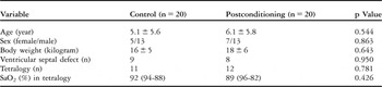
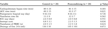
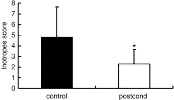
There was no hospital mortality or major complication in either group. The preoperative clinical data is shown in Table 1. No significant differences were noted with regard to age, gender, or weight in either group. The operative and postoperative data are shown in Table 2. Patients from the two groups were similar with regard to the types of procedure, bypass time, periods of aortic clamping, ventilatory duration, and length of stay in the intensive care unit. Analysis of variance revealed that there were no significant differences within groups or between groups for heart rate, mean arterial pressure, and central venous pressure (Table 3). The inotrope score used during the first 24 hours postoperatively in those undergoing postconditioning was significantly less than in the control group (p = 0.036, Fig. 1). Follow-up results revealed no significant residual lesions in any patient one month after discharge.
Demographics and preoperative characteristics.

SaO2 = saturation of oxygen by pulse oximeter.
Intraoperative and postoperative characteristics.

Cardiopulmonary bypass = cardiopulmonary bypass; ACC = aortic cross clamping; ICU = intensive care unit; PRBC = packed red blood cell.
Haemodynamics dataa.

HR = heart rate; MAP = mean artery pressure; CVP = central venous pressure; CPB = cardiopulmonary bypass;
a p > 0.05, postconditioning group versus control group.
Effect of postconditioning on the inotropic score. There was significant reduction in the requirement of inotropic in postconditioning group compared with in control for the first 24 hours postoperatively (*p = 0.036).

Creatine kinase activity
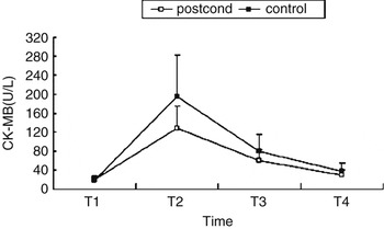
Preoperative concentrations of creatine kinase in the plasma were within normal limits in all patients, and no differences were found between the groups. A significant postoperative increase in activity was observed in both groups. There was a significant difference over time between the groups (group effect, p = 0.03), and significant differences between the groups were found 4 hours after aortic declamping, with values of 195 ± 98, and range from 50 to 490 U/L for the controls, and 128 ± 48, with range from 46 to 320 U/L for those undergoing postconditioning (Fig. 2, p = 0.016).
Effect of postconditioning on creatine kinase-MB. A significantly decrease is noted in levels 4 hours after reperfusion in those undergoing postconditioning compared with control group (*p = 0.016). T1 = before cardiopulmonary bypass; T2 = 4 hours after aortic declamping; T3 = 20 hours after aortic declamping; T4 = 48 hours after aortic declamping.

Levels of troponin
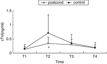
The levels collected at several sampling times are presented in Figure 3. The preoperative levels were similar in both groups. Repeated measures of analysis of variance revealed significant increases at 4, 20, and 48 hours after aortic declamping in both groups (time effect, p = 0.017). The levels were significantly lower in those undergoing postconditioning than in the controls (group effect, p = 0.043). Significant differences between the groups were found at 4 hours after aortic de-clamping, with values of 0.74 ± 0.65, and range from 0.18 to 2.6 ng/ml for the controls, and 0.35 ± 0.21, with range from 0.11 to 1.47 ng/ml for those undergoing postconditioning (p = 0.05).
Effect of postconditioning on Troponin I. A significantly decrease is noted 4 hours after reperfusion in those undergoing postconditioning compared with controls (*p = 0.05). T1 = before cardiopulmonary bypass; T2 = 4 hours after aortic declamping; T3 = 20 hours after aortic declamping; T4 = 48 hours after aortic declamping.

Transcardiac white blood count
The difference in white blood cell count between the aorta and the coronary sinus was similar before aortic clamping in both groups (3.2 ± 0.8% vs. 3.8 ± 1.0%, p = 0.46). The difference was not significant in either group 5 minutes after aortic declamping (10.8 ± 6.3% for those undergoing postconditioning compared to 14.0 ± 8.7% for control, p = 0.128).
Transcardiac lactate and other metabolic parameters
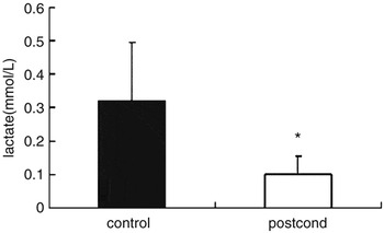
Before cardioplegic arrest, the values for transcardiac lactate were all negative, without significant difference between groups. At 5 minutes after aortic declamping, the values were positive in 80% of the control patients, and in 70% of those undergoing postconditioning, albeit that the net release in those undergoing postconditioning was significantly less than in the control group (0.10 ± 0.27 mmol/l vs. 0.37 ± 0.43 mmol/l, Fig. 4, p = 0.048). There was no significant difference with regard to myocardial uptake of oxygen 5 minutes after reperfusion between the groups, at 4.49 ± 1.5 vol% for controls as compared to 4.57 ± 1.8 vol% for those undergoing postconditioning (p = 0.42).
Lactate release 5 minutes after aortic declamping in both groups. A significantly decrease is seen after reperfusion in those undergoing postconditioning compared with the controls (*p = 0.048).

Discussion
In this study, we have provided further data that blood cardioplegic arrest remains a suboptimal technique in paediatric cardiac surgery. Although we used cold blood cardioplegia to provide cardiac protection in both groups, the significant elevation of biomarkers, known as sensitive and specific markers for myocardial injury, demonstrated that myocardial damage still occurred during the operation. The myocardial injury is manifested clinically by low cardiac output, hypotension, and the need for inotropes, or even death. Although myocardial damage is relatively mild or reversible in the majority of patients, subclinical injury still exists, and is manifested by the elevation of markers for myocardial injury.Reference Mildh, Pettilä, Sairanen and Rautiainen7 This finding is consistent with previous studies from Imura et al.Reference Imura, Caputo, Parry, Pawade, Angelini and Suleiman8
Our major finding is that postconditioning applied after cardioplegic arrest reduced the postoperative peak release by one-third for creatine kinase, and by half for troponin. Release of lactate during the early period of reperfusion was also decreased in those undergoing postconditioning compared with controls. These findings suggest that postconditioning not only reduces myocardial necrosis, but also improves myocardial metabolism. Our results provide further evidence that postconditioning can be initiated in the setting of cardiac surgery with cardioplegia as effectively as that without cardioplegia in percutaneous coronary intervention.Reference Staat, Rioufol and Piot5, Reference Laskey6 To our knowledge, ours is the first controlled and randomized clinical trial evaluating the effect of postconditioning on myocardial protection in the setting of cardiac surgery. The phenomenon of postcondtioning has been demonstrated previously in animal models,Reference Zhao, Corvera and Alkos3, Reference Zhao and Vinten-Johansen4, Reference Tsang, Hausenloy, Mocanu and Yellon9 but there are few studies that examined whether the phenomenon can occur in humans.Reference Staat, Rioufol and Piot5, Reference Laskey6 These encouraging results obtained during percutaneous intervention are attractive to the cardiac surgeon, because cardioplegic arrest of the heart during cardiac surgery is one of the few controlled models of myocardial ischaemia in which postconditioning as a simple procedure can be invoked early during reperfusion by using intermittent aortic cross clamping.
Another finding of our study is that two cycles of intermittent brief episodes of ischaemia induced by repeated aortic clamping after cardioplegic arrest is effective in children undergoing repair of their congenitally malformed hearts. This result is in contrast to the majority of previous experimental reports, which needed 3 or more cycles of postconditioning, and from humans undergoing percutaneous cardiac intervention when 4 cycles were used. There are few reports about the effect of two cycles of postconditioning on myocardial protection.Reference Galagudza, Kurapeev, Minasiana, Valen and Vaage10, Reference Serviddio, Di Venosa and Federici11 In most animal models, at least three or more cycles of brief ischaemia have been applied to trigger postconditioning.Reference Kin, Zhao, Sun and Vinten-Johansen12, Reference Yang, Proctor, Cui, Krieg, Downey and Cohen13 We speculate that the differences could be due to the multiple factors, including those that are pathology-dependent, age-dependent, species-dependent, and cardioplegia-dependent. These issues need further investigation.
There are several limitations to our present study. Firstly, the small size of our groups is not clinically sufficient to evaluate the effects of on the reduction of morbidity and mortality. Determination of such effects will need larger samples, and involve a multicentric study. Secondly, we did not investigate the optimal cycles and intervals for postcondtioning. These also are worthy of further study. Thirdly, our study did not extensively explore the mechanism of postconditioning, but used the transcardiac difference in white blood cells as an index for sequestration of neutrophils in the myocardium during reperfusion. Although much experimental evidence has now accumulated on mechanisms of postconditioning for cardiac protection,Reference Argaud, Gateau-Roesch, Raisky, Loufouat, Robert and Ovize14, Reference Hausenloy and Yellon15 clinically relevant evidence of mechanism is currently lacking. Finally, use of intermittent aortic cross-clamping to provide postconditioning in patients undergoing coronary arterial bypass grafting courts the risk of stroke. The applicability of postconditioning induced by intermittent aortic cross-clamping, therefore, may be limited to certain clinical situations. The strategy of postconditioning, nonetheless, must be attractive to cardiac surgeons because of its simplicity, its cost-effectiveness, and the easy with which it can be performed in the setting of cardiac surgery.Reference Ramzy, Rao and Weisel16 Our study, nonetheless, has shown for the first time that ischaemic postconditioning protects the myocardium of children undergoing cardioplegic arrest. The data supports the need for a larger clinical trial in children undergoing cardiac surgery.