Introduction
In nearly all mammals, the oocyte is surrounded by compact multilayers of cumulus cells, which form cumulus–oocyte complexes (COCs). During oocyte maturation, the COCs expand dramatically; this phenomenon is termed cumulus expansion. The structure of the expanded COCs promotes its dissociation from the follicle wall. Moreover, it supports the expulsion of the oocyte through the ruptured follicle wall during ovulation. Additionally, it has been reported that the expansion of the COCs is essential for fertilization and the developmental potential of early embryos (Chen et al., Reference Chen, Russell and Larsen1993; Hess et al., Reference Hess, Chen and Larsen1999). Hence, the degree of cumulus expansion is often used as a major indicator for the selection of mature oocytes in in vitro fertilization protocols (Ball et al., Reference Ball, Leibfried, Lenz, Ax, Bavister and First1983; Chen et al., Reference Chen, Russell and Larsen1993; Yang et al., Reference Yang, Kadam and Koide1993; Qian et al., Reference Qian, Shi, Ding, Sha and Fan2003; Somfai et al., Reference Somfai, Kikuchi, Onishi, Iwamoto, Fuchimoto, Papp, Sato and Nagai2004).
Cumulus expansion is the result of the synthesis and accumulation of hyaluronan around the cumulus cells (Chen et al., Reference Chen, Wert, Hendrix, Russell, Cannon and Larsen1990; Salustri et al., Reference Salustri, Yanagishita and Hascall1990; Zhuo et al., Reference Zhuo, Yoneda, Zhao, Yingsung, Yoshida, Kitagawa, Kawamura, Suzuki and Kimata2001). Hyaluronan is synthesized by hyaluronan synthase (HAS), which has been reported to exhibit three isoforms (HAS1, HAS2 and HAS3) (Spicer & McDonald, Reference Spicer and McDonald1998). Studies on mice, pigs and cattle have demonstrated that the HAS2 mRNA is strongly expressed in cumulus cells after a surge of gonadotropin and that it controls cumulus expansion (Fulop et al., Reference Fulop, Salustri and Hascall1997; Kimura et al., Reference Kimura, Konno, Miyoshi, Matsumoto and Sato2002; Schoenfelder & Einspanier, Reference Schoenfelder and Einspanier2003). The volumetric expansion of COCs is organized not only by the synthesis of hyaluronan but also by its accumulation within the COCs. Hyaluronan secreted from the cumulus cells remains in the extracellular matrix of COCs and is stabilized by hyaluronan-binding proteins, which include serum-derived factors of the inter-α-trypsin inhibitor (ITI) family and tumor necrosis factor-induced protein-6 (TNFIP6). Therefore, a close relationship is present between the degree of cumulus expansion and hyaluronan accumulation.
In our previous studies, we demonstrated that the hyaluronan receptor CD44 is expressed in cumulus cells during oocyte maturation in pigs (Kimura et al., Reference Kimura, Konno, Miyoshi, Matsumoto and Sato2002; Yokoo et al., Reference Yokoo, Miyahayashi, Naganuma, Kimura, Sasada and Sato2002a, Reference Yokoo, Tientha, Kimura, Niwa, Sato and Rodriguez-Martinezb). Moreover, we reported that the rate of oocyte maturation in vitro was inhibited by blocking the antibodies to CD44, which attenuated the hyaluronan-binding ability of CD44 (Yokoo et al., Reference Yokoo, Shimizu, Kimura, Tunjung, Matsumoto, Abe, Sasada, Rodriguez-Martinez and Sato2007). Therefore, on the basis of our previous studies on CD44, it may be inferred that hyaluronan accumulation within the COCs plays an important role in the oocyte maturation process.
In this study, we examined the influence of hyaluronan accumulation during cumulus expansion on the rate of porcine oocyte maturation in vitro by using 6-diazo-5-oxo-l-norleucine (DON), an inhibitor of hyaluronan synthesis and we demonstrated the correlation between the hyaluronan accumulated within the COCs and oocyte maturation.
Materials and Methods
Isolation and culturing of porcine COCs
Gilt ovaries were obtained from a slaughterhouse and transported to the laboratory within 30 min in a container maintained at 37 °C. Ovarian follicles that were 2–5 mm in diameter were aspirated with a 5 ml syringe using a 20G needle and only the COCs with uniform and compact cumulus cells were collected in modified tissue culture medium (TCM)-199 (mTCM-199) supplemented with 10% (v/v) porcine follicular fluid and 10 IU/ml equine chorionic gonadotropin (eCG) (Serotropin; Teikoku Zouki Pharmaceutical Co.). In mTCM-199 (Gibco BRL Life Technologies Inc.) containing Earle's salt and l-glutamine, we added 2.2 mg/ml sodium bicarbonate (Nacalai Tesque Inc.), 0.1 mg/ml sodium pyruvate (Sigma), 10 mg/ml bovine serum albumin (BSA) (Sigma), 100 IU/ml penicillin (Meiji Seika Co.) and 100 μg/ml streptomycin (Meiji Seika). The collected COCs were washed three times with mTCM-199 and 50 COCs were cultured in 500 μl drops of the same medium supplemented with 0–1.0 mM DON (Sigma) and covered with paraffin oil (Nacalai Tesque) for 48 h at 37 °C in an atmosphere containing 5% CO2.
Morphologic assessment of cumulus expansion
COCs in each treatment group were evaluated to determine the degree of cumulus expansion at 48 h of culturing. Cumulus expansion was assessed on the basis of the area occupied by the cumulus cell aggregate of the COCs (Daen et al., Reference Daen, Sato, Naito and Toyoda1994). The length and width of the COCs were measured with an eyepiece micrometer. The length was defined as the distance between the two most distant points and the width was defined as the distance between the two closest points. The area occupied by each COC was calculated using the following formula: area = length × width × 0.7854.
Assessment of nuclear maturation
Oocytes were evaluated at 48 h after maturation of the culture to assess the maturation rate. COCs were denuded by pipetting in 0.1% hyaluronidase (Sigma). The denuded oocytes were fixed for 48 h with acetoethanol (acetic acid : ethanol, 1 : 3 v/v), stained with 1% aceto-orcein and examined under a phase-contrast microscope for evaluating their chromatin configurations.
Immunofluorescence analysis
The hyaluronan retention in COCs was examined following a method previously published by Zhuo et al. (Zhuo et al., Reference Zhuo, Yoneda, Zhao, Yingsung, Yoshida, Kitagawa, Kawamura, Suzuki and Kimata2001). In brief, the COCs were fixed with 4 % (w/v) paraformaldehyde–phosphate-buffered saline (PBS) (pH 7.4) at room temperature. After 30 min, they were rinsed three times with PBS and permeabilized with 0.5 % (v/v) Triton X-100–PBS for 30 min at room temperature. The fixed COCs were incubated with biotinylated hyaluronan-binding protein (Seikagaku) in 5 % BSA in PBS for 1 h at 4 °C, rinsed with PBS and incubated with a fluorescein-conjugated streptavidin (Sigma) for 1 h at room temperature. Propidium iodide (PI) staining was used as a control to stain nuclei. After several washes with BSA–PBS, the COCs were mounted on slide glasses, covered with a coverslip and observed under a confocal microscope (MRC-1024; Bio-Rad). LaserSharp Processing software (Bio-Rad) was used to analyse the confocal images.
Statistical analysis
The experiments were repeated three times and the data were pooled for statistical analysis. The cumulus expansion data were analysed using one-way analysis of variance (ANOVA) followed by the Bonferroniadjusted significance test. The rates of oocyte maturation were compared by the chi-squared test. A probability of P < 0.05 was considered statistically significant.
Results
Effects of inhibition of hyaluronan synthesis on cumulus expansion
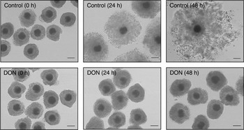
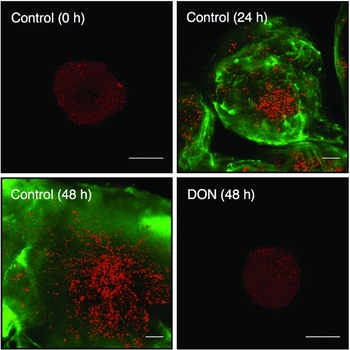
Remarkable cumulus expansion was observed during in vitro maturation without DON. In contrast, complete inhibition of cumulus expansion was observed in the presence of 1.0 mM DON. Even at the end of culturing (48 h), the size of the COCs remained unchanged (Fig. 1). In Fig. 2, considerable accumulation of hyalur onan was observed in the control cultures at 24 h and 48 h. On the other hand, complete inhibition of hyaluronan synthesis was observed when the COCs were exposed to 1 mM DON even after 48 h of culturing.
Microscopic observation of cumulus expansion in porcine COCs during in vitro maturation with and without 6-diazo-5-oxo-1-norleucine (DON). Bars = 100 μm.

Localization of hyaluronan in porcine COCs as detected by immunofluorescence using biotinylated hyaluronan-binding protein. Green = hyaluronan; red = nuclear; bars = 100 μm.

Effects of hyaluronan accumulation on in vitro oocyte maturation
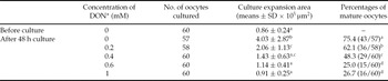
Next, the correlation between the hyaluronan accu mulation within COCs and oocyte maturation was examined (Table 1). The degree of inhibition of cumulus expansion depended on the concentration of DON in the culture medium. The area of the COCs before culturing was 0.86 ± 0.24 × 105 μm2. On the other hand, the areas showing cumulus expansion for the COCs cultured with 0, 0.2, 0.4, 0.6 and 1.0 mM DON were 4.03 ± 2.87, 2.06 ± 1.13, 1.43 ± 0.63, 1.14 ± 0.41 and 0.91 ± 0.25 × 105 μm2, respectively. The rate of oocyte maturation was correspondingly suppressed in a dose-dependent manner by the addition of DON to the culture medium. After culturing the COCs for 48 h in the absence of DON, we observed that 75.4 % of the oocytes had reached metaphase II. The rates of oocyte maturation in the presence of 0.2, 0.4, 0.6 and 1.0 mM DON were 62.1, 48.3, 25.0 and 26.7 %, respectively.
Effects of 6-diazo-5-oxo-1-norleucine on cumulus expansion and oocyte maturation.

a–dValues within the same column with different superscripts are significantly different (P < 0.05).
*6-diazo-5-oxo-1-norleucine.
Discussion
We examined the influence of hyaluronan accumulation within the COCs that expanded during porcine oocyte maturation by using DON, which is an inhibitor of hyaluronan synthesis. DON prevented hyaluronan accumulation within the COCs and completely inhibited cumulus expansion. Furthermore, it suppressed the rate of in vitro oocyte maturation. These results imply that hyaluronan accumulation is not only involved in expanding the volume of the COCs but also actively involved in the induction of oocyte maturation. We propose that the function of hyaluronan during cumulus expansion is an important step in porcine oocyte maturation.
In this study, we confirmed the gradual increase in the volume of COCs during in vitro maturation. It has been reported that the synthesis and accumulation of hyaluronan within the COCs result in cumulus expansion in mice (Chen et al., Reference Chen, Wert, Hendrix, Russell, Cannon and Larsen1990; Salustri et al., Reference Salustri, Yanagishita and Hascall1990; Zhuo et al., Reference Zhuo, Yoneda, Zhao, Yingsung, Yoshida, Kitagawa, Kawamura, Suzuki and Kimata2001). Previous reports on pigs have shown the relationship between cumulus expansion and hyaluronan accumulation (Nagyova et al., Reference Nagyova, Prochazka and Vanderhyden1999; Nemcova et al., Reference Nemcova, Nagyova, Petlach, Tomanek and Prochazka2007). In this experiment, remarkable accumulation of hyaluronan was observed in porcine expanded COCs by the immunostaining method, but hyaluronan accumulation was completely inhibited by treatment with DON. These results showed that cumulus expansion of porcine COCs is dependent on hyaluronan synthesis and accumulation.
We demonstrated that the inhibition of cumulus expansion by DON resulted in a decrease in the rate of oocyte maturation. In the presence of 1 mM DON, cumulus expansion and hyaluronan synthesis were completely inhibited. However, 26.7 % of the oocytes could reach at metaphase II even in the presence of 1 mM DON. This result may suggest that spontaneous oocyte maturation occurs under this culture condition, because the inhibitors of spontaneous maturation (e.g. hypoxanthine) were not used in this study. Therefore, these results suggested that hyaluronan accumulation within the COCs not only contributes to cumulus expansion but also plays an active role in inducing oocyte maturation. In our previous study, we showed that the hyaluronan receptor CD44 is expressed in cumulus cells during oocyte maturation in pigs (Kimura et al., Reference Kimura, Konno, Miyoshi, Matsumoto and Sato2002; Yokoo et al., Reference Yokoo, Miyahayashi, Naganuma, Kimura, Sasada and Sato2002a, Reference Yokoo, Tientha, Kimura, Niwa, Sato and Rodriguez-Martinezb). Additionally, we found that the interaction between hyaluronan and the hyaluronan receptor CD44 during oocyte maturation is important for inducing oocyte maturation in pigs (Yokoo et al., Reference Yokoo, Shimizu, Kimura, Tunjung, Matsumoto, Abe, Sasada, Rodriguez-Martinez and Sato2007). Considering these results, it is reasonable that the inhibition of cumulus expansion by DON would cause insufficient hyaluronan–CD44 interaction during cumulus expansion, resulting in the suppression of the rate of in vitro oocyte maturation. Therefore, we propose that hyaluronan synthesized and accumulated in the COCs has an important function in oocyte maturation.
In previous studies in which the culture conditions prevented cumulus expansion, i.e. under conditions in which, it is possible that the hyaluronan–CD44 interaction could not function sufficiently, meiotic resumption of the oocytes was suppressed (Petr et al., Reference Petr, Zetova, Fulka and Jilek1989; Nagai et al., Reference Nagai, Ding and Moor1993). Moreover, we showed that the inhibition of hyaluronan–CD44 interaction suppressed germinal vesicle breakdown of the oocytes (Yokoo et al., Reference Yokoo, Shimizu, Kimura, Tunjung, Matsumoto, Abe, Sasada, Rodriguez-Martinez and Sato2007). While we have not studied the details of the nuclear phase of the oocytes after culturing in this study, considering these results, there is a strong possibility that during cumulus expansion, hyaluronan is closely involved in meiotic resumption. In contrast to the present study, however, in the study by Gutnisky et al. (Reference Gutnisky, Dalvit, Pintos, Thompson, Beconi and Cetica2007), DON was added to bovine COCs in an in vitro maturation culture and it was observed that the inhibition of cumulus mucification with DON did not alter the oocyte maturation rate. Although this reason for the difference is unclear, the functional role of hyaluronan in oocyte maturation may differ among animal species.
Unlike other glycosaminoglycans, hyaluronan is neither sulphated nor linked to a core protein. Despite its structural simplicity, hyaluronan is known to play a wide range of biological roles; for example, it is a structural component of connective tissue (Lee & Spicer, Reference Lee and Spicer2000) and is involved in cellular movement (Wallach-Dayan et al., Reference Wallach-Dayan, Grabovsky, Moll, Sleeman, Herrlich, Alon and Naor2001) and the activation of intracellular signal transmission (Zhang et al., Reference Zhang, Chang, Zylka, Turley, Harrison and Turley1998). Hence, in general, hyaluronan needs hyaluronan-binding protein(s) to function. It has been reported that some hyaluronan-binding proteins, e.g., TNFIP6 or ITI, may be associated with cumulus expansion. Interestingly, it has been reported that ITI promotes hyaluronan–CD44 interaction in lymphoma tumor cells (Zhuo et al., Reference Zhuo, Kanamori, Kannagi, Itano, Wu, Hamaguchi, Ishiguro and Kimata2006). ITI may also play the role of inducing oocyte maturation by accelerating hyaluronan–CD44 interaction.
In conclusion, our results demonstrated that hyaluronan accumulation within the COCs during in vitro oocyte maturation in pigs contributes to cumulus expansion and oocyte maturation. We propose that the function of hyaluronan during cumulus expansion is a necessary step in the porcine oocyte maturation process. Although details of the mechanism of oocyte maturation in mammals are not yet fully understood, cumulus expansion can be observed during oocyte maturation in nearly all mammals. Therefore, the results of this study include important findings that help elucidate the mechanism of oocyte maturation in mammals.
Acknowledgements
We are grateful to Prof. K. Kimata and Dr L. Zhuo, Aichi Medical University, for their advice on the immunostaining of hyaluronan, T. Nagahara for her precious help, Dr H. Sasada and Dr H. Matsumoto for helpful discussion and the staff of the Meat Inspection Office, Sendai City, for supplying the porcine ovaries. This research was partially supported by Grant-in-Aid for Young Scientists (B), No. 18780209.