Introduction
Our interest in the secondary mineralization in the uranium mines of Red Canyon in White canyon district of south-eastern Utah (USA) began several years ago at the Blue Lizard mine. That mine has now proven to be a prolific source for new mineral species, having already yielded 16, with several others still under study. We have expanded our efforts to several nearby mines including the Green Lizard, Giveaway-Simplot and Markey mines. All share similar geology and mineralogy, and some new species are found at more than one mine. We have provided a brief synopsis of the history of uranium mining in the area in our descriptive paper on shumwayite, a new mineral found at both the Green Lizard and Giveaway-Simplot mines (Kampf et al., Reference Kampf, Plášil, Kasatkin, Marty, Čejka and Lapčák2017). Herein, we describe greenlizardite, the second new mineral from the Green Lizard mine.
Greenlizardite is named for the locality, the Green Lizard mine. The new mineral and its name were approved by the Commission on New Minerals, Nomenclature and Classification of the International Mineralogical Association (IMA2017-001). The description is based on one holotype and one cotype specimen, which are deposited in the collections of the Natural History Museum of Los Angeles County, 900 Exposition Boulevard, Los Angeles, CA 90007, USA, catalogue numbers 66558 and 66559, respectively.
Occurrence
Greenlizardite was discovered on specimens collected underground in the Green Lizard mine (37°34'37.10”N, 110°17'52.80”W) in the White Canyon mining district, San Juan County, Utah, USA. The Green Lizard mine is near the head of Low Canyon on the east side of Red Canyon, 2.1 km north of the Blue Lizard mine. The geology is similar to that of the Blue Lizard mine (Kampf et al., Reference Kampf, Kasatkin, Čejka and Marty2015a; Chenoweth, Reference Chenoweth1993). The Green Lizard mine is also a cotype locality for shumwayite (Kampf et al., Reference Kampf, Plášil, Kasatkin, Marty, Čejka and Lapčák2017).
Mineralized channels are in the Shinarump member of the Chinle Formation. The Shinarump member consists of medium- to coarse-grained sandstone, conglomeratic sandstone beds and thick siltstone lenses. Ore minerals were deposited as replacements of wood and other organic material and as disseminations in the enclosing sandstone. Since the mine closed, oxidation of primary ores in the humid underground environment has produced a variety of secondary minerals, mainly sulfates, as efflorescent crusts on the surfaces of mine walls.
Greenlizardite is a rare mineral, found in association with ammoniozippeite, boussingaultite, dickite and a potentially new Mg uranyl sulfate mineral, currently under study. Other secondary minerals found in the Green Lizard mine assemblage include beshtauite, calcite, gypsum, fermiite, johannite, natrozippeite, oppenheimerite, plášilite, rozenite, shumwayite, sulfur and wetherillite.
Physical and optical properties
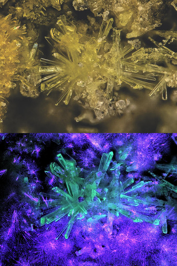
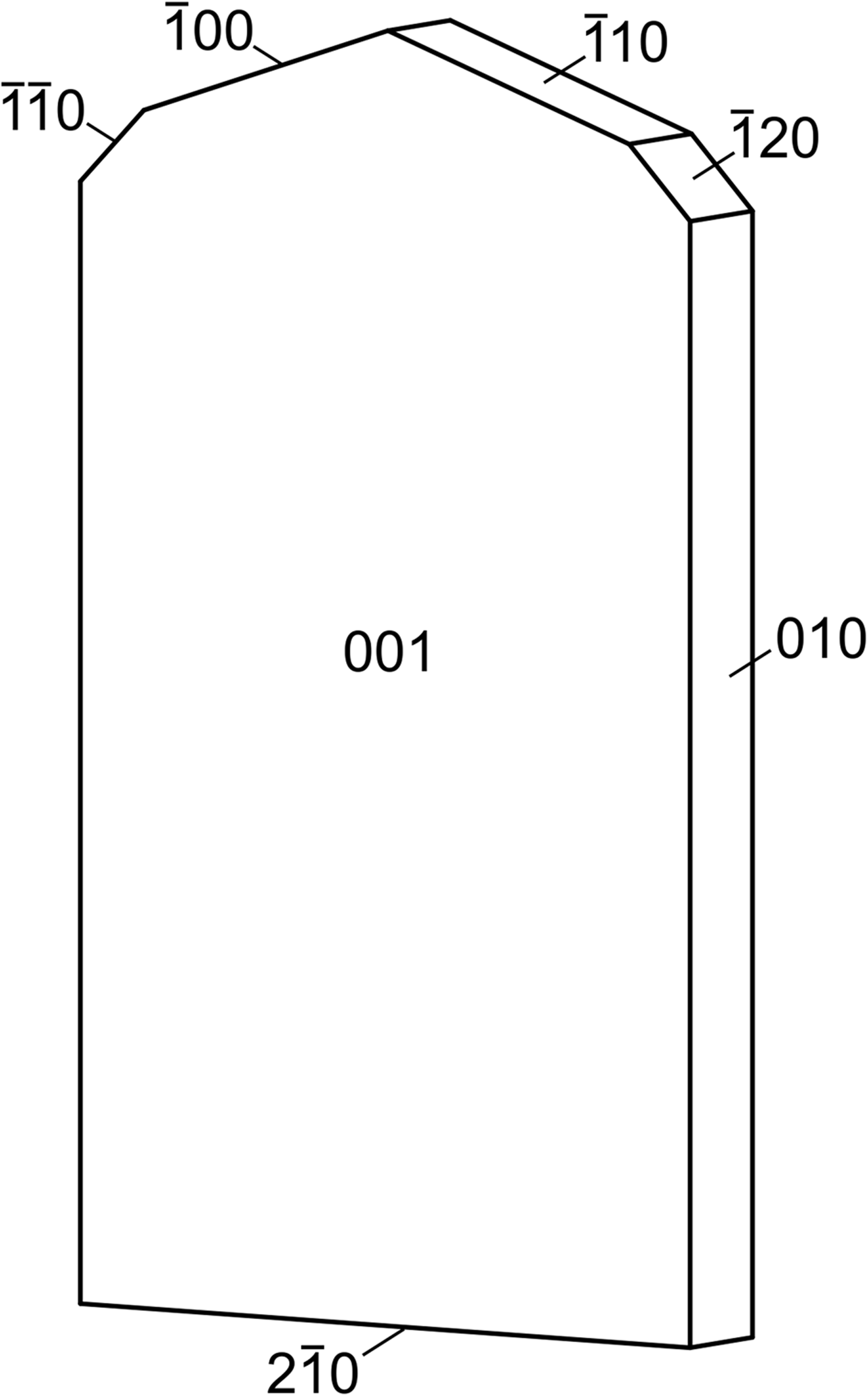
Greenlizardite occurs as light green-yellow blades, up to ~0.3 mm long, forming irregular sprays (Fig. 1). Blades are flattened on {001}, elongated on [100] and exhibit the forms { $\bar 1$00}, {010}, {0
 $\bar 1$0}, {001}, {00
 $\bar 1$}, {
 $\bar 1$10}, {
 $\bar 1\bar 1$0}, {
 $\bar 1$20} and {2
 $\bar 1$0} (Fig. 2). Twinning, probably by reflection on {001}, is observed under crossed polars. The mineral is transparent with a vitreous lustre and has a white streak. It fluoresces greenish-blue when illuminated by a 50 mW 405 nm blue-violet laser (Fig. 1). The Mohs hardness is ~2. Crystals are brittle with irregular fracture and two cleavages: perfect on {001} and good on {2
 $\bar 1$0}. The mineral is easily soluble in room temperature H2O. The density could not be measured because the mineral is soluble in Clerici solution and there is insufficient material for physical measurement. The calculated density is 3.469 g cm–3 for the empirical formula and 3.507 g cm–3 for the ideal formula.
Greenlizardite with ammoniozippeite (left) in incandescent light (top) and 405 nm ultraviolet light (bottom). The field of view is 0.84 mm across.

Crystal drawing of greenlizardite, clinographic projection in nonstandard orientation.

Optically, greenlizardite is biaxial (+), with α = 1.559(1), β = 1.582(1) and γ = 1.608(1), measured in white light. The 2V measured directly on a spindle stage is 88(1)°; the calculated 2V is 87.8°. Dispersion is r < v, moderate. The mineral is weakly pleochroic: X = very pale yellow green, Y = pale yellow green and Z = light yellow green; X < Y < Z. The optical orientation is X ≈ c, Y ≈ a and Z ≈ b*.
Raman spectroscopy
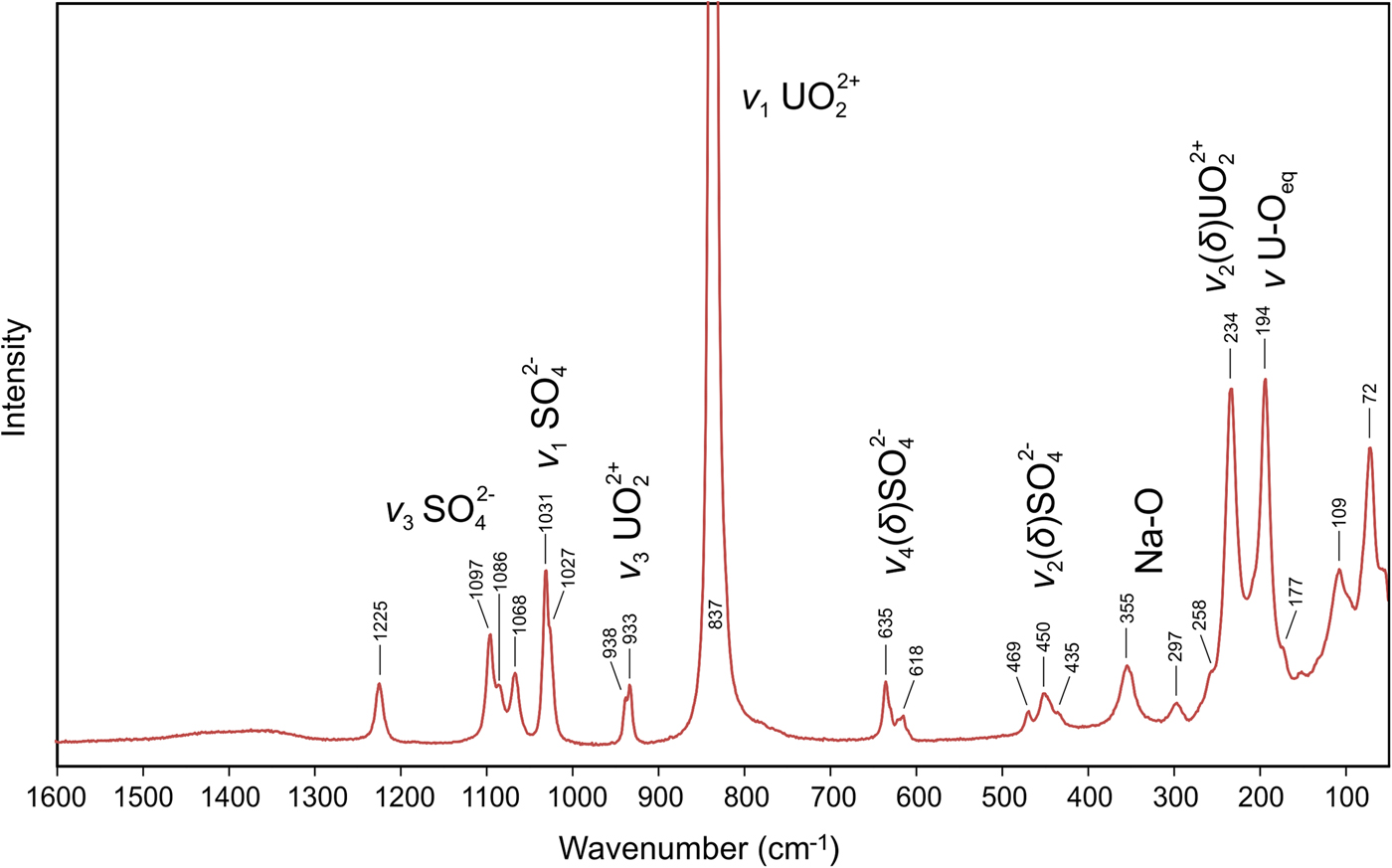
Raman spectroscopy was conducted on a Horiba XploRA PLUS. Pronounced fluorescence was observed using a 532 nm diode laser; consequently, a 785 nm diode laser was utilized. The spectrum was recorded from 3000 to 50 cm–1; however, only the portion from 1600 to 50 cm–1 is shown in Fig. 3 because no features were observed between 3000 and 1600 cm–1.
Raman spectrum of greenlizardite recorded using a 785 nm diode laser.
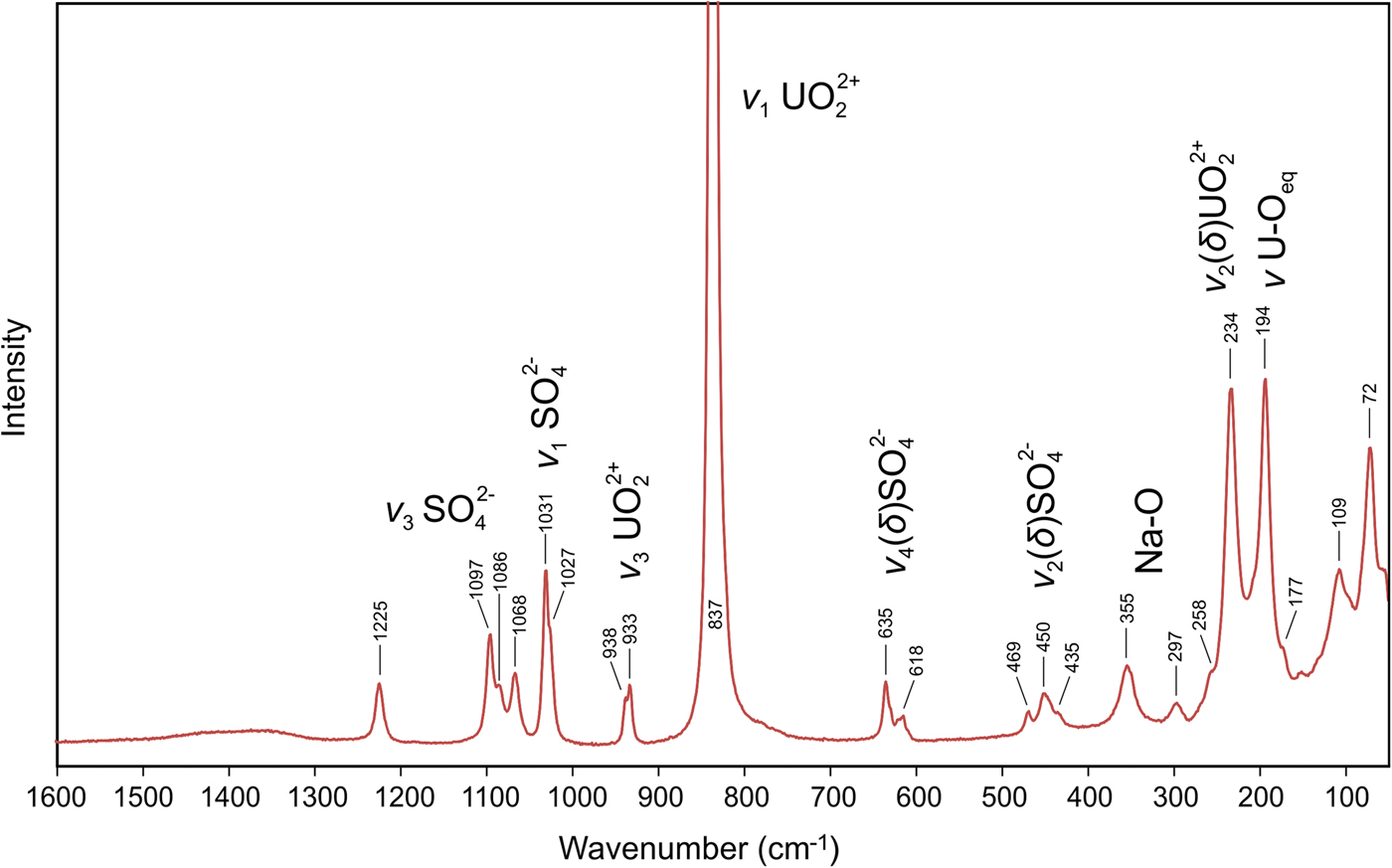
Quite intense Raman bands at 1225 cm–1 and at 1097, 1086 (sh) and 1068 cm–1 are attributed to split triply degenerate ν3 antisymmetric stretching vibrations of the SO4 tetrahedra. The number of split bands is in line with the presence of two symmetrically non-equivalent SO4 tetrahedra in the structure of greenlizardite (cf. bluelizardite or synthetic klaprothite, Plášil et al., Reference Plášil, Meisser and Čejka2016). Bands at 1031 and 1027 (sh) cm–1, having the highest intensity within the region of SO4 stretching modes, are assigned to the ν1 symmetric stretching vibrations of SO4 tetrahedra. A weak, two-component band at 938 and 933 cm–1 is assigned to the ν3 antisymmetric stretching vibration of the uranyl ion, UO2 2+; a very strong band at 837 cm–1 is assigned to the ν1 symmetric U–O stretching vibration of the UO2 2+. The inferred U–O bond lengths (after Bartlett and Cooney, Reference Bartlett and Cooney1989) in the uranyl ion of ~1.77 Å (from ν1) and 1.76 (from ν3) are in line with those derived from the present X-ray study.
Weak Raman bands at 635 and 618 cm–1 are attributed to the ν4 (δ) triply degenerated antisymmetric stretching vibrations of SO4 tetrahedra. Weak Raman bands 469, 450 and 435 (sh) cm–1 are related to the split ν2 (δ) doubly degenerate bending vibrations of the SO4.
The band at 355 cm–1 and possibly also that at 297 cm–1 can be attributed to Na–O stretching vibrations (cf. Kampf et al., Reference Kampf, Plášil, Kasatkin and Marty2015b,Reference Kampf, Plášil, Kasatkin, Marty and Čejkac; Plášil et al., Reference Plášil, Buixaderas, Čejka, Sejkora, Jehlička and Novák2010, Reference Plášil, Meisser and Čejka2016; Volkovich et al., Reference Volkovich, Griffiths, Fray and Fields1998). The two-component strong Raman band at 234 and 194 cm–1, with shoulders at 258 cm–1 and 177 cm–1 is most probably related to the ν2 (δ) doubly degenerate bending vibration of the UO22+ and U–Oeq–ligand stretching modes (cf. Bullock and Parret, Reference Bullock and Parret1970; Ohwada, Reference Ohwada1976; Brittain et al., Reference Brittain, Ansari, Toivonen, Niinisto, Tsao and Perry1985; Plášil et al., Reference Plášil, Buixaderas, Čejka, Sejkora, Jehlička and Novák2010). Weak bands at the lowest energies can be assigned to unclassified lattice modes, most probably skeletal vibrations of the whole sheets of polyhedra.
Composition
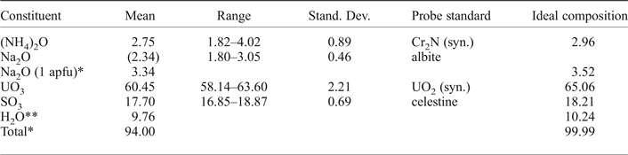
Chemical analyses (6 points on 4 crystals) were performed at the University of Utah on a Cameca SX-50 electron microprobe with four wavelength dispersive spectrometers and using Probe for EPMA software. Analytical conditions were 15 kV accelerating voltage, 10 nA beam current and a beam diameter of 5 µm. Counting times were 30 s on peak and 30 s on background for each element. Raw X-ray intensities were corrected for matrix effects with a PAP algorithm (Pouchou and Pichoir, Reference Pouchou, Pichoir, Heinrich and Newbury1991). A time-dependent intensity correction was applied to Na; the N intensity was time independent. Na and N exhibited significant variability, and a negative relationship was apparent. Greenlizardite was very challenging to analyse. It was not possible to obtain a good polish and some beam damage was noted. The low analysis total and lower than expected Na are attributed to the irregular surfaces of mounted crystals. Preliminary analyses on unpolished crystal faces using an Oxford SEMEDX200EDS on a Hitachi S-3000N SEM employing internal standards, while only semi-quantitative, were consistent with stoichiometric Na atom per formula unit (apfu). Because insufficient material is available for a direct determination of H2O, it has been calculated based upon the structure determination. Analytical data are given in Table 1.
Chemical composition (in wt.%) for greenlizardite.

* The low analysis total and lower than expected Na are attributed to the irregular surfaces of mounted crystals. The Na2O wt.% corresponding to 1 Na apfu is used in the empirical formula.
** Based on the structure.
The empirical formula calculated on the basis of 18 O apfu is (NH4)0.98Na1.00U1.96S2.04O18.00H10.02. The ideal formula is (NH4)Na(UO2)2(SO4)2(OH)2·4H2O. The Gladstone-Dale compatibility index 1 – (KP/KC) is –0.016 based on the empirical formula and –0.008 based on the ideal formula, in both cases in the range of superior compatibility (Mandarino, Reference Mandarino2007).
X-ray crystallography and structure refinement
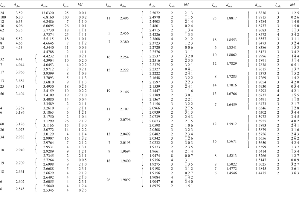
The powder X-ray study was carried out using a Rigaku R-Axis Rapid II curved imaging plate microdiffractometer, with monochromatized MoKα radiation (λ = 0.71075 Å). A Gandolfi-like motion on the φ and ω axes was used to randomize the sample. Observed d values and intensities were derived by profile fitting using JADE 2010 software (Materials Data, Inc.). The powder data presented in Table 2 show good agreement with the pattern calculated from the crystal-structure determination.
Powder X-ray data (d in Å) for greenlizardite. Only calculated lines with I ≥ 2 are listed.

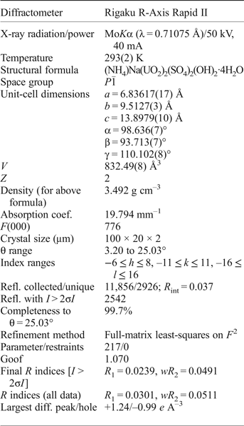
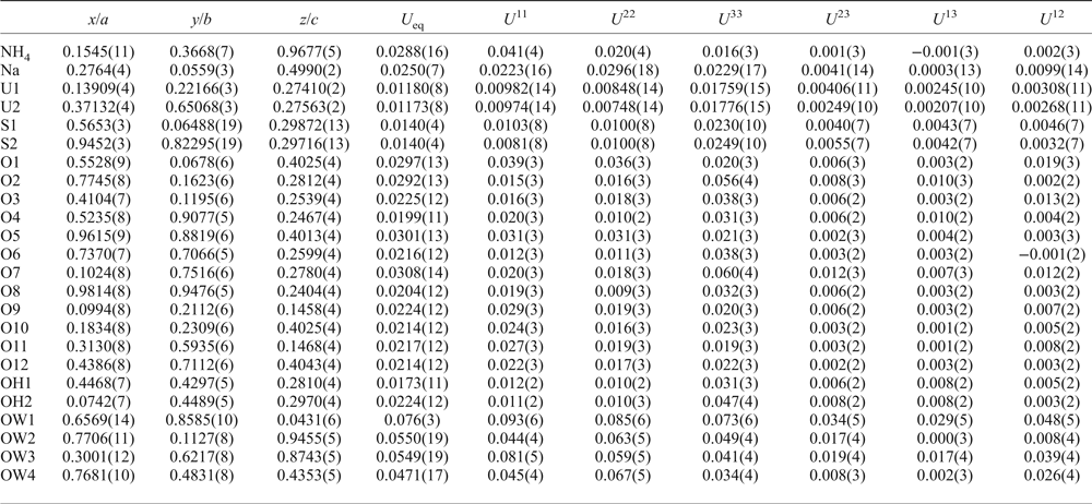
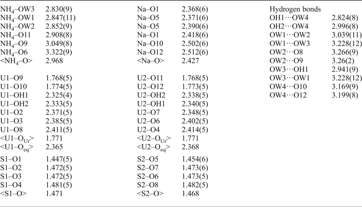
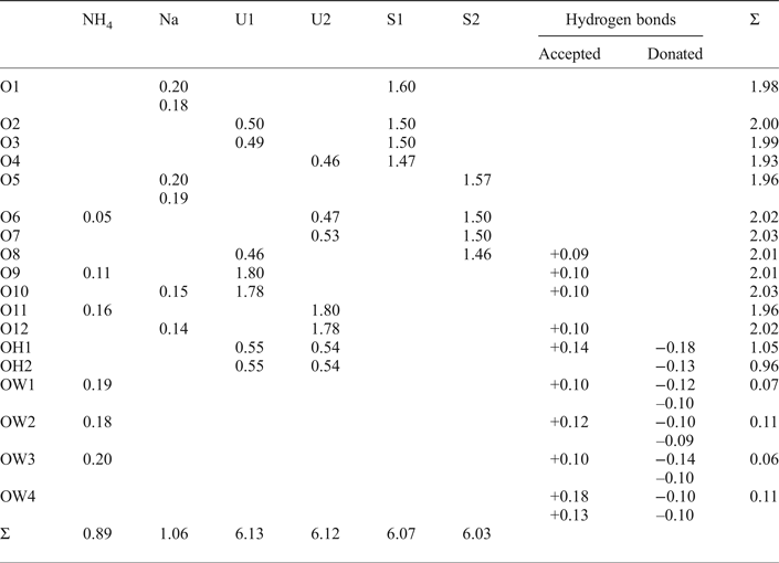
The single-crystal reflection data were collected using the same diffractometer and with the same radiation as noted above. The Rigaku CrystalClear software package was used for processing the data, including the application of both shape-based and empirical absorption corrections, the latter using the multi-scan method with ABSCOR (Higashi, Reference Higashi2001). The structure was solved using SIR2011 (Burla et al, Reference Burla, Caliandro, Camalli, Carrozzini, Cascarano, Giacovazzo, Mallamo, Mazzone, Polidori and Spagna2012). SHELXL-2013 (Sheldrick, Reference Sheldrick2015) was used for the refinement of the structure. Difference-Fourier syntheses suggested possible positions for some of the H atoms; however, their reliability was not judged sufficiently high to report. Consequently, they have not been included in the refined structure. The proposed hydrogen-bonding scheme is based upon bond geometries and bond-valence balance, but must be considered highly conjectural. Data collection and refinement details are given in Table 3, atom coordinates and displacement parameters in Table 4, selected bond distances in Table 5 and a bond-valence analysis in Table 6.
Data collection and structure refinement details for greenlizardite.

R int = Σ|F o2–F o2(mean)|/Σ[F o2]. Goof = S = {Σ[w(F o2–F c2)2]/(n–p)}1/2. R 1 = Σ||F o|–|F c||/Σ|F o|. wR 2 = {Σ[w(F o2–F c2)2]/Σ[w(F o2)2]}1/2; w = 1/[σ2(F o2)+(aP)2+bP] where a is 0.0151, b is 5.461 and P is [2F c2+Max(F o2,0)]/3.
Atom coordinates and displacement parameters (Å2) for greenlizardite.

Selected bond distances (Å) for greenlizardite.

Bond-valence analysis for greenlizardite. Values are expressed in valence units.*

* NH4+–O bond valence parameters from Garcia-Rodriguez et al. (Reference García-Rodríguez, Rute-Pérez, Piñero and González-Silgo2000); all other cation–O bond valence parameters from Gagné and Hawthorne (Reference Gagné and Hawthorne2015); hydrogen-bond strengths based on O–O bond lengths from Ferraris and Ivaldi (Reference Ferraris and Ivaldi1988).
Description of the crystal structure
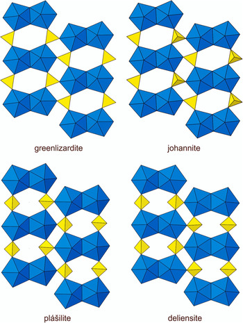
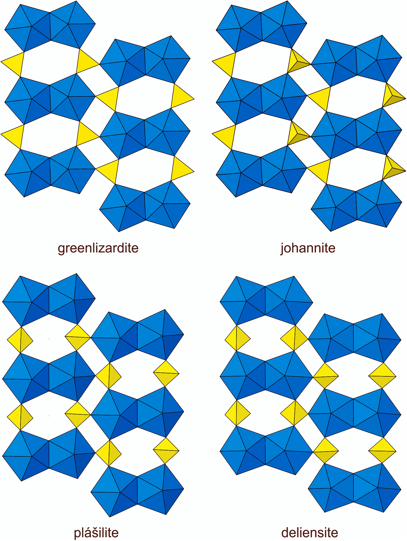
The U sites in the structure of greenlizardite are surrounded by seven O atoms forming a squat UO7 pentagonal bipyramid. This is the most typical coordination for U6+, particularly in uranyl sulfates, where the two short apical bonds of the bipyramid constitute the UO2 uranyl group. In the structure of greenlizardite, pairs of pentagonal bipyramids share a common edge, forming dimers. The dimers are linked by sharing corners with SO4 groups, yielding a [(UO2)2(SO4)2(OH)2]2– sheet parallel to (001) (Fig. 4). This sheet is based on the phosphuranylite anion topology (Burns, Reference Burns2005), with a ring symbol 61524232 (Krivovichev and Burns, Reference Krivovichev, Burns, Krivovichev, Burns and Tananaev2007). The sheets in johannite, Cu[(UO2)2(OH)2(SO4)2](H2O)8, (Mereiter, Reference Mereiter1982), deliensite, Fe(UO2)2(SO4)2(OH)2·7H2O, (Plášil et al., Reference Plášil, Hauser, Petříček, Meisser, Mills, Škoda, Fejfarová, Čejka, Sejkora, Hloušek, Johannet, Machovič and Lapčák2012) and plášilite, Na(UO2)(SO4)(OH)·2H2O (Kampf et al., Reference Kampf, Kasatkin, Čejka and Marty2015a) are topologically identical to that in greenlizardite; however, these sheets are geometrical isomers, differing in the orientation of the SO4 groups (Fig. 4). In greenlizardite, within the same [(UO2)2(SO4)2(OH)2]2– sheet, the corner of each SO4 group that is not shared with UO7 pentagonal bipyramids points in the same direction.
The uranyl sulfate sheets in greenlizardite, johannite, plášilite and deliensite. UO7 bipyramids are blue and SO4 tetrahedra are yellow. Note the differing orientations of SO4 tetrahedra.
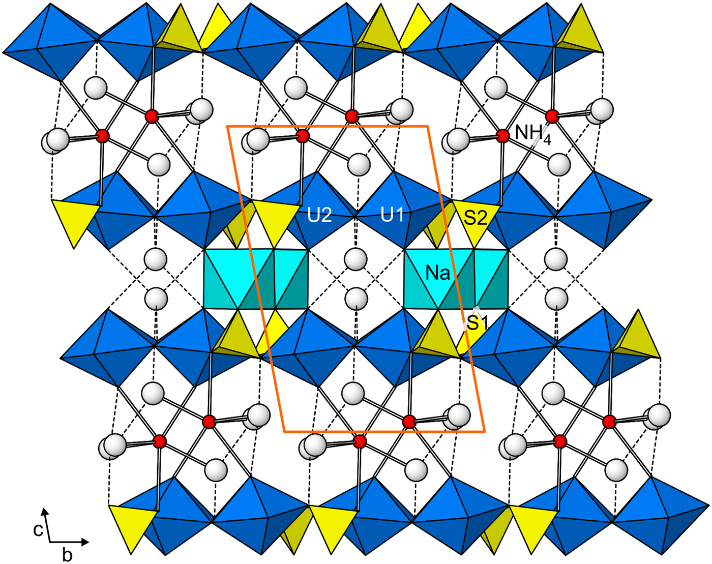
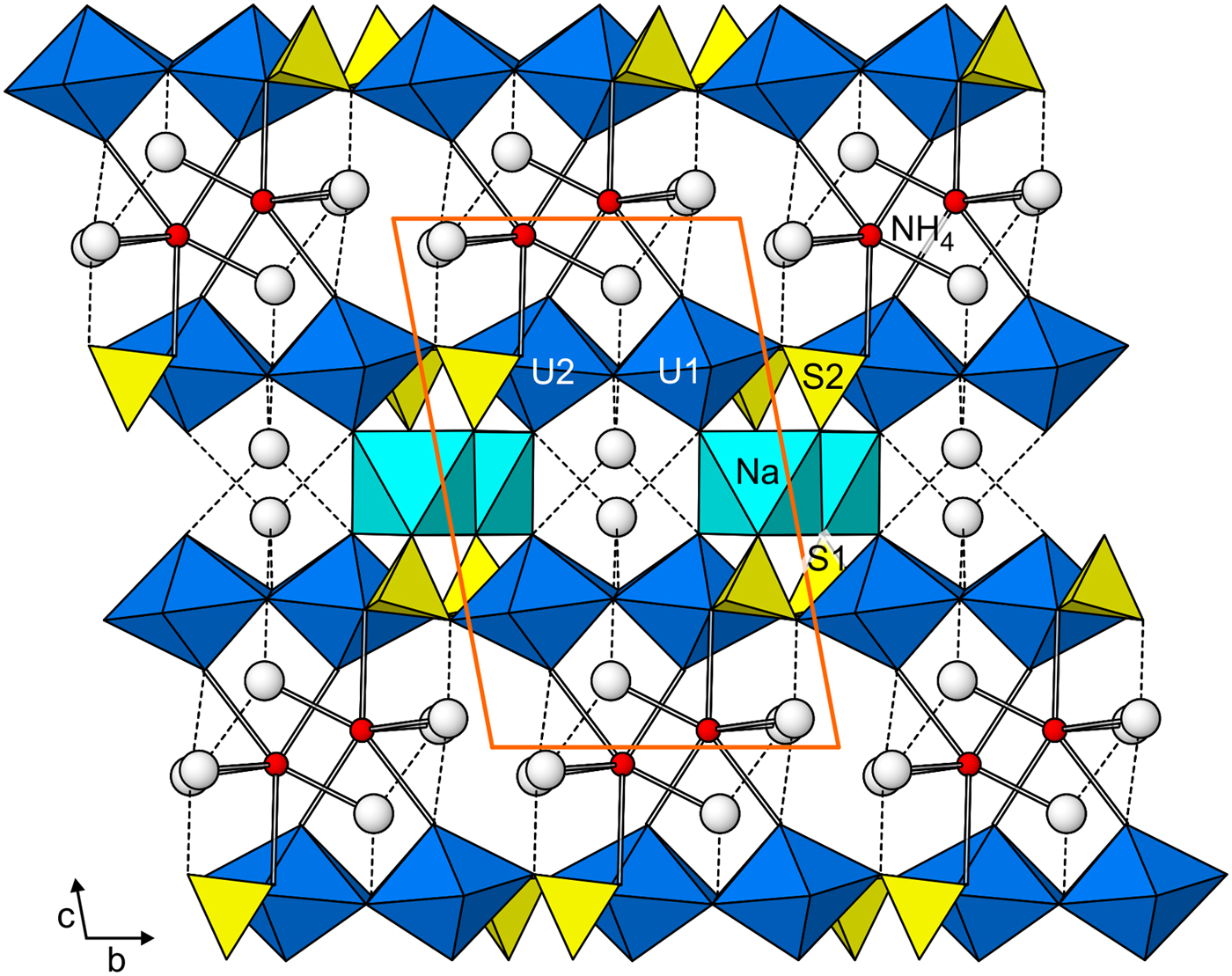
In the structure of greenlizardite (Fig. 5), the Na bonds to two uranyl O atoms (one in each of two adjacent sheets) and four sulfate O atoms (two in each of two adjacent sheets) yielding an approximately octahedral coordination. NaO6 octahedra share edges to form zig-zag chains along [100] and these chains link two adjacent [(UO2)2(SO4)2(OH)2]2– sheets forming a thick slab. This slab has channels along [100] that contain isolated H2O groups (OW4). The NH4 group and three H2O groups (OW1, OW2 and OW3) are located in the region between adjacent slabs. The NH4 bonds to each of the three interlayer H2O groups (OW1, OW2 and OW3) and links adjacent slabs together by bonding to two uranyl O atoms (O9 and O11) in one slab and one of the equatorial O atoms (O6) of a UO7 bipyramid in another slab (which is also part of the S2O4 tetrahedron). The NH4–O6 bond is quite long (3.322 Å), which would be expected to result in very weak linkage between slabs; however, the NH4–O6 linkage is augmented by extensive hydrogen bonding from the interlayer H2O groups (OW1, OW2 and OW3).
The structure of greenlizardite viewed down [100]. O atoms of isolated H2O groups are large white spheres. NH4–O bonds are shown as sticks. Hydrogen bonds are shown as dashed lines. The unit-cell outline is in orange.
The linkages between the sheets in the structure of greenlizardite are quite different than those in the structures of johannite, deliensite and plášilite. Most notably, there are two different intersheet regions in greenlizardite, one containing edge-sharing chains of NaO6 octahedra and the other containing irregular (NH4)O3(H2O)3 polyhedra and they link the sheets together as described above. The structures of johannite, deliensite and plášilite all contain only one type of intersheet region. In deliensite, an individual Fe2+O(H2O)5 octahedron links to one uranyl O atom in one sheet and links to the adjacent sheet only through hydrogen bonding. In johannite, an individual Jahn-Teller distorted CuO2(H2O)4 octahedron shares its apical O atoms with uranyl O atoms in two adjacent sheets. In plášilite, NaO2(H2O)4 octahedra form a cis edge-sharing straight chain and each octahedron shares one O atom with a uranyl group in one sheet and one O atom with a sulfate group in the adjacent sheet.
Acknowledgements
The manuscript benefitted from constructive comments by reviewers Travis Olds and Andrew Locock. This study was funded by the John Jago Trelawney Endowment to the Mineral Sciences Department of the Natural History Museum of Los Angeles County. JP was supported through the project No. 17-09161S of the Grant agency of the Czech Republic.