Introduction
Anomalies of the first branchial cleft commonly present as fistulas and cysts, and frequently occur in the practice of otolaryngology. WorkReference Work1 described a classification system for first branchial cleft anomalies, which separated them according to embryological origin. Type I anomalies are associated with the first cleft and arise from ectoderm; they are usually located medial to the concha and pass parallel to the external auditory canal. Type II anomalies are derived from the first cleft as well as the first and second arches, and are found inferior to the angle of the mandible, with the tract of the lesion extending superiorly over the ramus of the mandible and running either medial or lateral to the trunk of the facial nerve.
Here, we present a case of interesting anomalies of the first branchial cleft, including two different tracts manifesting in the same patient, which occurred together with an unusual otomicroscopic finding: a fibrous band of tissue in the external auditory canal that terminated on the tympanic membrane.
Clinical case
A seven-year-old girl presented to the emergency department after three days of progressive swelling and erythema over the left parotid area, together with decreased oral intake due to restricted jaw movement. C-reactive protein concentration was found to be elevated, at 68.3 mg/l, and her white blood cell count was 103/µl. Computed tomography (CT) scanning of the neck with contrast showed a 3.7 × 2.3 cm, left, level two neck abscess with rim enhancement, posterior and lateral to the angle of the mandible. Intravenous clindamycin was administered and the patient underwent incision and drainage.
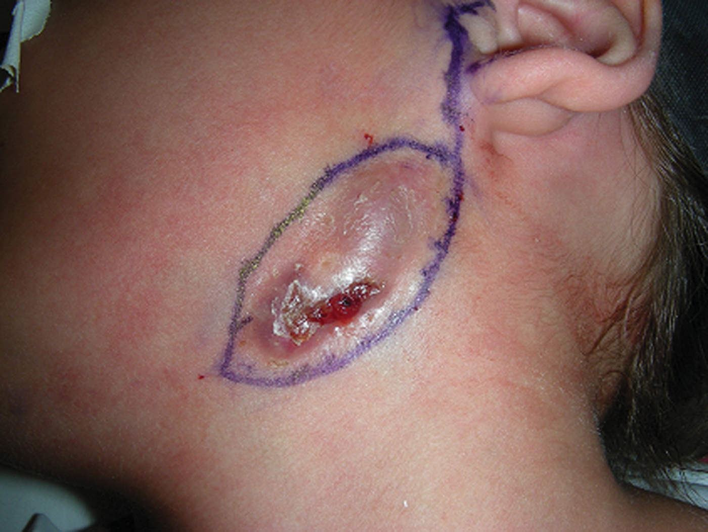
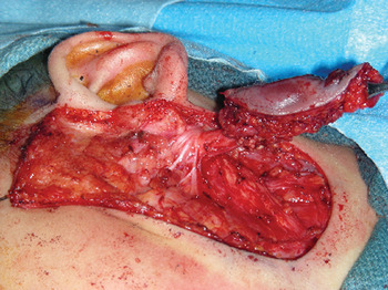
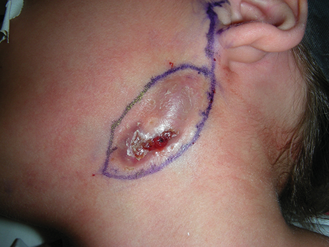
Recurrent episodes of drainage from a fistulous tract prompted a repeated CT scan several weeks later, and a diagnosis of type II first branchial cleft cyst was made. A superficial parotidectomy with excision of the branchial cleft anomaly was performed. At the time of surgery, the fistulous tract was found to be located inferior to the ramus of the mandible (Figure 1) and to travel medial to the pes anserinus of the facial nerve (Figure 2), and to extend towards the ear, communicating with the anterior auricular cartilage. The tract was removed with total preservation of the facial nerve. Histological examination of the sinus tract revealed benign cartilage, glands and squamous epithelium.
Surgical photograph showing the branchial cleft anomaly, inferior to the ramus of the mandible.
Surgical photograph showing the fistulous tract passing medially to the pes anserinus of the facial nerve.

Three months post-operatively, the patient developed an area of fluctuance in the pre-auricular space. She was returned to the operating theatre for excision of a second fistulous tract, which was noted to be travelling medially and inferiorly along the cartilage of the external auditory canal. It was excised to the level of the junction between the cartilaginous and bony canal wall. This finding was consistent with a duplication of the external auditory canal.
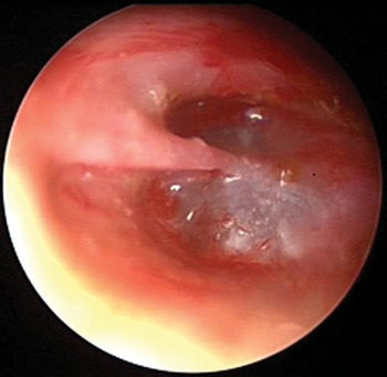
Histological examination showed a sinus tract lined with keratinising squamous epithelium contained within a cartilaginous wall. At the time of the second surgery, the left external auditory canal was examined via otomicroscopy. A fibrous band was noted in the canal, beginning at the junction of the cartilaginous and bony canal and extending medially to attach at the umbo (Figure 3). This band appeared to be a continuation of the duplicated external auditory canal. The fibrous band was excised, and a small tympanic membrane perforation was repaired using a lateral paper patch. The patient had an uneventful post-operative course without recurrence.
Otomicroscopic view showing the membranous band found in the external auditory canal, which extended from the junction of the cartilaginous and bony canal wall to attach to the tympanic membrane at the umbo.

Further review of the patient's medical history showed that this fibrous band had been identified by a primary care provider four years previously. At this time, it had been evaluated via a CT scan of the temporal bone, showing no abnormalities.
Discussion
The first tract resected in our patient appeared medial to the facial nerve and had a histological pattern showing both squamous epithelium and cartilage, consistent with both ectodermal and mesodermal origins. This clearly fits Work'sReference Work1 criteria for a type II anomaly.
The second tract resected does not fit as neatly into Work'sReference Work1 classification system. It had elements of a type I anomaly, in that it ran medial to the concha and parallel to the external auditory canal, but histological examination revealed keratinised epithelium (deriving from ectoderm) and cartilage (deriving from mesoderm). The fibrous band, noted on otomicroscopy to connect with the tympanic membrane, appeared to be related to this duplication anomaly, as the tract ended at the level where the fibrous band arose inside the external auditory canal.
Since the tympanic membrane is derived from all three germ cell layers, it is possible that our patient's fibrous band represented an extension of mesoderm into the sinus tract, explaining the cartilaginous origins in her second branchial cleft tract. Martinez Del Pero et al. Reference Martinez Del Pero, Majumdar, Bateman and Bull2 noted that many specimens classified anatomically as Work type I contained cartilaginous tissue. Their series reported that three of 18 patients with first branchial cleft anomalies were found to have fibrous bands in the external auditory canal that connected to the tympanic membrane. Triglia et al. Reference Triglia, Nicollas, Ducroz, Koltai and Garabedian3 described four of 39 patients with similar findings. Other authors have provided photographic documentation.Reference Martinez Del Pero, Majumdar, Bateman and Bull2–Reference Ramirez, Galofre, Domenech and Martinez6
• Branchial cleft anomalies have traditionally been divided into type I and type II lesions
• It is possible to have more than one lesion present in a single patient, and such tracts may not fit neatly into Work's classification system
• As first branchial cleft lesion diagnosis is often delayed, otomicroscopic findings can be important in early detection
A fibrous band can be an important clinical finding when diagnosing branchial cleft anomalies, especially in an asymptomatic patient. This anomaly was noted in our patient four years prior to her current presentation with an infected branchial cleft cyst. Series documented by Triglia et al. Reference Triglia, Nicollas, Ducroz, Koltai and Garabedian3 and Ford et al. Reference Ford, Balakrishnan, Evans and Bailey7 have shown that these cases often present in a delayed fashion, with the mean delay in diagnosis reported as 3.5 years in Triglia and colleagues' series and four years in Ford and colleagues' series. In our patient, earlier recognition of the fibrous anomaly could have enabled further exploration of the possibility of a branchial cleft cyst upon her initial presentation.
It is generally accepted that total excision of these lesions is the only curative form of treatment. In many of these cases, this will involve identification of the facial nerve and careful dissection and preservation. In their series, Triglia et al. Reference Triglia, Nicollas, Ducroz, Koltai and Garabedian3 noted that 92 per cent of these patients required exposure of the facial nerve. Other authorsReference Martinez Del Pero, Majumdar, Bateman and Bull2 have noted the variable nature of the lesion location in respect to the facial nerve. Our case demonstrates a tract that lay medial to the main trunk of the facial nerve; identification of the nerve facilitated safe removal of the pathology.