Stress researchers require interdisciplinary frameworks to study the effects and consequences of chronic stress. One major obstacle in understanding and defining stress is that it is a multidimensional construct involving interactions among inputs (environmental stressors), processes (subjective psychological distress), and outputs (objective biological stress responses; Levine, Reference Levine2005; Levine & Ursin, Reference Levine, Ursin, Brown, Koobb and Rivier1991) that are often investigated separately from each other. Over the last two decades, the concepts of allostasis (Sterling & Eyer, Reference Sterling, Eyer, Fisher and Reason1988) and of allostatic load (AL; McEwen & Stellar, Reference McEwen and Stellar1993) have served as the impetus for hundreds of publications that go far beyond the perspective that “stress” is an exclusively social, psychological, or biological construct, but rather all constructs studied together in an integrative framework (for an earlier review, see Lupien et al., Reference Lupien, Ouellet-Morin, Hupbach, Walker, Tu, Buss and Cicchetti2006).
Originating as a biopsychosocial model, AL refers to the “wear and tear” the body experiences when organisms are subjugated to chronic stress. Central to this biological damage is altered stress hormone functioning that inexorably strains interconnected biomarkers that eventually collapse like domino pieces trailing toward stress-related endpoints. The traditional operational definition of the AL index is the sum number of dysregulated neuroendocrine, immune, metabolic, and cardiovascular biomarkers (Seeman, Singer, Rowe, Horwitz, & McEwen, Reference Seeman, Singer, Rowe, Horwitz and McEwen1997). This aggregate or AL index has then been used to tease apart the psychosocial determinants of disordered, diseased, and deceased outcomes. An emerging body of literature clearly demonstrates that the AL index is associated with numerous physical outcomes throughout life span development and even psychopathological outcomes (for an earlier review, see Juster, McEwen, & Lupien, Reference Juster, McEwen and Lupien2010).
With the aim of refining the AL model further in application to psychopathological phenomena throughout the life cycle, we offer the following review of the causes and effects of AL. The first part of this article will review literature on powerful antecedents of AL: (a) early adversity that predisposes individuals to lifelong disease susceptibilities; (b) genetic and epigenetic factors; (c) mediation or moderation by environmental toxins (ETs); as well as (d) interactions among biological sex, sociocultural gender, and sex hormones. We follow secondly with an exploration of pathophysiological manifestations that could be used as human models of AL: (e) living with schizophrenia or bipolar disorder; (f) the dementia spectrum; and, finally, (g) the experience of caregiving. Our hope is to transcend the boundaries of existing theory, research, and practice by proposing that the AL model can evolve into a transdisciplinary framework that ushers a holistic approach to understand and help distressed individuals. Sharing key similarities with developmental psychopathological models, a transdisciplinary AL model likewise would seek to address the causes, mechanisms, and heterogeniety manifested among diverse stress-related disease trajectories.
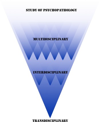
Transdisciplinarity is an approach to scientific inquiry aimed at solving complex research questions by transcending traditional disciplinary boundaries (Kessel & Rosenfield, Reference Kessel and Rosenfield2008; Rosenfield, Reference Rosenfield1992). The complexities inherent among numerous interacting factors that contribute to an individual's experience of health or disease necessarily calls for transdisciplinary models that do more than integrate knowledge (Albrecht, Freeman, & Higginbotham, Reference Albrecht, Freeman and Higginbotham1998; Kessel, Rosenfield, & Anderson, Reference Kessel, Rosenfield and Anderson2008). In transdisciplinary health models, our concepts of health can evolve beyond the absence of disease, and even beyond the sum of biopsychosocial parts toward more holistic ones.
In this view, health is an emergent property of individuals taken as a whole (Picard, Sabiston, & McNamara, Reference Picard, Sabiston and McNamara2011; see Figure 1). All factors discussed throughout this review interact in unique ways to influence one's risk of or protection against the noxious effects of chronic stress. In this spirit, we encourage the reader to contextualize the presented information within their respective area(s) of expertise in partnership with others and especially regarding complementary perspectives espoused in the field of developmental psycholopathology.
The term transdisciplinarity was coined by Jean Piaget in the 1970s to describe integrative, multilevel approaches to scientific inquiry. In more modern perspectives (Kessel & Rosenfield, Reference Kessel and Rosenfield2008; Rosenfield, Reference Rosenfield1992), emergent properties stem from the dynamic interactions among health domains at biological, psychological, social, behavioral, and spiritual levels, that collectively shape global health. A transdisciplinary approach to health research is consistent with the allostatic load model whereby health and disease are believed to emerge as the totality of life domains that are best understood when studied in synergy. Adapted from “The need for a transdisciplinary, global health framework,” by M. Picard, C. M. Sabiston, and J. K. McNamara, 2011, Journal of Alternative and Complementary Medicine, 17, 179–184. Copyright 2011 by Mary Ann Liebert, Inc. Adapted with permission. [A color version of this figure can be viewed online at www.journal.cambridge.org/dpp]

Briefly, the defining features of developmental psychopathology are (a) understanding causal processes, (b) elucidating developmental mechanisms, and (c) emphasis on diverse conceptualizations of continuities and discontinuities between spectrums of normality and abnormality in mental health (Rutter & Sroufe, Reference Rutter and Sroufe2000). Such integrative perspectives focus on how multiple causes and effects contribute to probabilistic rather than deterministic pathways leading to diverse outcomes (Cichetti & Toth, Reference Cicchetti, Rogosch, Gunnar and Toth2009). In particular, “developmental analyses” highlight the dynamic nature of individual differences in vulnerabilities and resiliencies shaped by interacting factors studied from the micro- to macolevel (Cichetti & Toth, Reference Cicchetti, Rogosch, Gunnar and Toth2009). The AL model shares much in this diverse spirit of analysis bridging numerous disciplines together. Yet, empirical substantiations of AL theory remain largely based on population-level analyses.
Specifically, the AL model has been primarily applied to detect physical morbidities and all-cause mortality in epidemiological analyses. Although promising, it has been challenging to propose applications toward more individual-level analyses pertinent to understanding complex psychopathological phenomena. Notable exceptions have emerged; however, there remains some resistance to extensively investigate AL in humans in the neurosciences, psychiatry, and psychology. To address this, our principal objectives in this article are twofold: (a) to review the AL model from a developmental psychopathology perspective in relation to current and future research areas; and (b) to outline transdisciplinary interactions between key AL factors and psychopathological conditions. We begin with a brief introduction of key terms and concepts.
Stress and the Neurobiology of Stress Responses
Stress is defined here as a real or interpreted threat to an individual's physiological and psychological integrity that results in adaptive biological and behavioral responses (McEwen & Seeman, Reference McEwen and Seeman1999). Situations that are novel, unpredictable, threatening to the ego/self, and/or uncontrollable additively contribute to physiological stress responses (Dickerson & Kemeny, Reference Dickerson and Kemeny2004; Lupien et al., Reference Lupien, Ouellet-Morin, Hupbach, Walker, Tu, Buss and Cicchetti2006; Mason, Reference Mason1968). Under these circumstances, stress responses are activated by the autonomic nervous system and neuroendocrine system to mobilize energy necessary to adapt to the demands of the situation (McEwen, Reference McEwen1998b; Sapolsky, Romero, & Munck, Reference Sapolsky, Romero and Munck2000).
At first, perceived and interpreted threats trigger the sympathetic–adrenal–medullary (SAM) axis to release catecholamines (e.g., adrenalin) within seconds from the adrenal medulla surrounding the kidneys. Second, the hypothalamic–pituitary–adrenal (HPA) axis is activated within minutes to produce glucocorticoids. The hypothalamus activates the HPA axis by stimulating corticotrophin-releasing factor. Corticotrophin-releasing factor then travels through a portal system linking the hypothalamus to the pituitary, where it signals the secretion of adrenocorticotrophin hormone (ACTH) from the anterior pituitary rich with blood capillaries. ACTH then travels viscerally to the adrenal cortex surrounding the kidneys where it binds to receptor tissue and precipitates cellular activities in the zona fasticullata region of the adrenal cortex to produce glucocorticoids (GCs) such as cortisol in humans (Sapolsky et al., Reference Sapolsky, Romero and Munck2000). The resulting surges in catecholamines and GCs alter homeostasis by appropriating biological functions that permit fight or flight responses.
The brain's ultimate role during stress responses is to detect threat and adapt. In addition to the pituitary and hypothalamic activities, there are three major brain structures involved in the regulation of stress responses: (a) the hippocampus linked to memory and cognition, in addition to being implicated in negative feedback regulation of the HPA axis; (b) the amygdala responsible for fear conditioning and emotional processing with outputs to autonomic and neuroendocrine regulatory systems; and (c) the prefrontal cortex involved in cognition and coping strategies and exerting top-down control over subcortical structures (Gray & Bingaman, Reference Gray and Bingaman1996; McEwen, Reference McEwen2004; McEwen, Weiss, & Schwartz, Reference McEwen, Weiss and Schwartz1968; Reul & de Kloet, Reference Reul and de Kloet1985; Sanchez, Young, Plotsky, & Insel, Reference Sanchez, Young, Plotsky and Insel2000; Thayer & Lane, Reference Thayer and Lane2009). In concert with the autonomic nervous system, short-term adaptations of organisms during stress responses are dynamic biological processes that ensure survival.
Concept of Allostasis
SAM and HPA axis stress responses are examples of allostasis, defined as adaptive biological processes that preserve “stability through change” (Sterling & Eyer, Reference Sterling, Eyer, Fisher and Reason1988). This concept was coined by an epidemiologist and a neurobiologist to describe how the body maintains homeostasis by allocating metabolic energy to cope with the demands of the environment (Sterling, Reference Sterling and Schulkin2004). In contrast to homeostasis that focuses on static set points (e.g., Ph levels, core body temperature), the concept of allostasis focuses on how continuous reevaluation and readjustments create new set points that maximize the organism's resources (e.g., increased cardiac output when running). Likewise, homeostatic mechanisms cannot adequately account for how the brain appropriates itself beyond rigidly enforced biological mechanisms. For instance, when increased needs create an error signal, negative feedback mechanisms may try to correct the error and cause more error (e.g., rapidly rising causing head rush and dizziness). Allostasis can drive behaviors explaining how anticipation of physiological change can rectify these errors (e.g., getting up slowly to prevent dizziness) and in this way, considers an unusual parameter value not as a failure to defend a set point, but rather as a response to some prediction (Goldstein & Kopin, Reference Goldstein and Kopin2007; Sterling, Reference Sterling and Schulkin2004). This perspective allows for a greater understanding of the relation between physiological needs, behavior, and pathology (Sterling & Eyer, Reference Sterling, Eyer, Fisher and Reason1988).
Homeostatic models generally define health as an internal milieu whereby all physiological parameters have relatively normal values. Disease, on the other hand, occurs when normal ranges fall into abnormal but pharmaceutically correctable ranges. In this view, Sterling and Eyer (Reference Sterling, Eyer, Fisher and Reason1988) argued that the medical model is in danger of iatrogenesis, that is, the deleterious effects that certain treatments can inadvertently induce. Iatrogenic effects can arise when restoring one parameter to a normal range causes other parameters to become “inappropriately” high or low and cause altogether separate health issues (e.g., hypertension medications can negatively alter interconnected biological systems). In contrast, the allostatic model defines health as a state of responsiveness. A parameter with values outside the normal range is not necessarily considered inappropriate as each parameter is controlled by a multitude of mutually reinforcing signals (Sterling, Reference Sterling and Schulkin2004).
In summary, allostasis fundamentally differs from homeostasis because of its emphasis on dynamic rather than static biological set points, considerations of the brain's role in feedback regulation (for a review of allostasis and the brain, see Ganzel, Morris, & Wethington, Reference Ganzel, Morris and Wethington2010), as well as its view of health as a whole-body adaptation to contexts (Schulkin, Reference Schulkin2003b). According to allostatic theory, pathology itself is a form of adaptation where, for instance, an individual develops hypertension in order to conciliate environmental demands, even if these processes are pathophysiological. Health in allostatic theory is therefore defined as optimal predictive fluctuation, whereby shifts in the probability of demand should shift the response, and when the prediction reverses, so should the response (Sterling, Reference Sterling and Schulkin2004). In this view, the allostasis concept is consistent with a transdisciplinary definition of health and disease emphasizing holistic relationships among individuals and environments. It is, however, when these adaptive systems become chronically dysregulated that maladaptive physiological consequences emerge. The concept of allostasis therefore represents a multifaceted mechanism of regulation applicable to psychopathological as well as pathophysiological research by uniting mind and body in explanations of dynamic stability that can go astray.
The AL Model
McEwen and Stellar (Reference McEwen and Stellar1993) expanded upon allostasis by proposing that AL is the price that the body incurs when bombarded by stressful situations and prolonged stress responses. The principal points of the AL model are twofold: (a) AL involves multiple interconnected biomarkers that can falter primarily as a function of stress hormone dysregulations and (b) that the health sciences should not wait idly by for individuals to reach clinical levels on these biomarkers but should instead identify subclinical states before they lead to pathology.
To substantiate these arguments, the AL model synthesizes a rich, interdisciplinary literature on stress and disease by elucidating how numerous factors converge to predispose individuals to stress-related pathologies over the life cycle (McEwen, Reference McEwen2000a). This endeavor necessitates fusion of multiple knowledge bases, as coordination of allostasis depends on the brain's evaluation of threat and execution of physiological responses (Herman, Ostrander, Mueller, & Figueiredo, Reference Herman, Ostrander, Mueller and Figueiredo2005; McEwen, 2007, 2008). This coordination is fundamentally shaped by an individual's constitution (genetics, development), history (trauma/abuse, major life events, early adversity), and behaviors (coping and health habits; McEwen, Reference McEwen1998a). Akin to developmental psychopathological emphasis on how experiences can alter developmental pathways, understanding how these diverse factors are integrated at the individual level to yield altered expressions of normative functioning can provide innovative ways of treating ill individuals and fostering positive health.
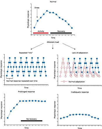
In contrast to healthy allostatic responses whereby individuals react and recover to stressors effectively, allostatic states (Koob & Le Moal, Reference Koob and Le Moal2001) can occur when chronic dysregulation of regulatory systems lead to pathophysiological operating ranges. There are four postulated physiological profiles that represent allostatic states that contribute to AL: (a) repeatedly activated responses, (b) nonhabituating responses, (c) prolonged responses, (d) and inadequate responses (McEwen, Reference McEwen1998a; see Figure 2). Detecting these different allostatic states is possible by investigating biomarker(s) dynamics in transdisciplinary manner regarding health outcomes. Because allostatic states prompt compensatory remediation among numerous biomarkers, each of which is shaped by numerous factors, delineating time courses of dysregulation in AL involves the challenge of determining which players falter first in the cascade (McEwen, Reference McEwen2008).
Four types of allostatic states. The top panel illustrates normal allostatic responses, in which a response is initiated by a stressor, sustained for an appropriate interval, and then turned off. The remaining panels illustrate four conditions that lead to allostatic load, namely, repeated “hits,” lack of adaptation, prolonged response, and inadequate response (McEwen, Reference McEwen2006a) . [A color version of this figure can be viewed online at www.journal.cambridge.org/dpp]

Multiple biomarkers are hypothesized to function as part of a nonlinear network that contributes to a tripartite sequence leading to AL (McEwen, Reference McEwen1998a). At first, overactivation of primary mediators such as stress hormones and pro- and anti-inflammatory cytokines exact primary effects on cellular functions such as enzyme, receptor, ion channel, and genomic activities. Synergistic effects of several primary mediators then leads to tissue and organ-specific changes in several systems. Subsidiary systems, in turn, recalibrate their own activities to compensate for the over- and/or underproduction of primary mediators and effects.
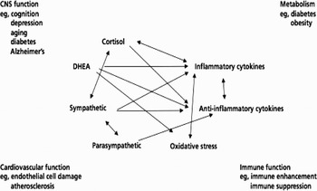
This leads to secondary outcomes, whereby metabolic, cardiovascular, immune, and reproductive biomarkers become imbalanced. Multiple pathways interact in a nonlinear fashion whereby oscillations in biomarker levels drive compensatory recalibrations of other intertwined biomarkers (see Figure 3). For example, inflammatory cytokine production is negatively regulated via anti-inflammatory cytokines as well as via parasympathetic and GC pathways, whereas sympathetic activity increases inflammatory cytokine production and parasympathetic activity restrains sympathetic activity (McEwen, Reference McEwen2006a). The synergistic effects of multiple primary mediators and primary effects exerting themselves with secondary outcomes progressively strains the organism until a breaking point. AL becomes overloading when tertiary outcomes emerge with the manifestation of clinical end points such as cardiovascular disease and mortality.
Nonlinear network of mediators of allostasis and allostatic load. Arrows indicate that each system regulates the others in a reciprocal manner, creating a nonlinear network (McEwen, Reference McEwen2006a).

In an evolutionary biological perspective, allostatic overload (McEwen & Wingfield, Reference McEwen and Wingfield2003) occurs in two variants. First, Type 1 allostatic overload occurs when energy demands exceed energy income, representing a negative energy balance. For example, lack of food because of habitat changes results in emergency life-sustaining responses that pit the organism into a survival mode whereby energy stores are uncompromisingly mobilized from stores as GCs take charge (Goymann & Wingfield, Reference Goymann and Wingfield2004; Korte, Koolhaas, Wingfield, & McEwen, Reference Korte, Koolhaas, Wingfield and McEwen2005). Second, Type 2 allostatic overload occurs if energy demands are not exceeded and the organism continues to take in or store as much or even more energy than it needs, representing a positive energy balance. For example, today's industrialized agricultural products allow for massive food supplies that contribute to troubling obesity and metabolic problems (McEwen & Wingfield, Reference McEwen and Wingfield2003). Transcending theory, AL, and allostatic overload have been the subject of a growing empirical literature that holds as central tenants the advantages of assessing multiple biomarkers, particularly primary mediators (McEwen, Reference McEwen2003b), at subclinical levels as a function of different contexts (e.g., socioeconomic status [SES], stressful life events, toxic workplaces).
Measuring AL
Work by Teressa Seeman and colleauges has been instrumental in developing an AL index that encompasses primary mediators and secondary outcomes to predict tertiary outcomes. The traditional count-based AL index was based on analyses from the MacArthur Studies of Successful Aging, a cohort of high-functioning American older adults ages 70 to 79 followed longitudinally. The original AL index included 10 biomarkers from neuroendocrine (cortisol, adrenalin, noradrenalin, and dehydroepiandrosterone-sulfate [DHEA-S]), metabolic (total cholesterol to high density lipoprotein [HDL] cholesterol, HDL cholesterol, glycosylated hemoglobin [HbA1c], and waist to hip ratio), and cardiovascular (systolic and diastolic blood pressure) systems (Seeman, Singer, et al., 1997). Once all biomarker values were obtained from the sample, participants' values fall within the highest risk with respect to the sample's biomarker distributions. This has traditionally been the 75th percentile for biomarkers whereby high levels denote disease vulnerabilities (e.g., glucose, low density lipoprotein [LDL] cholesterol), whereas the 25th percentile is for biomarkers whereby low levels are unhealthy (e.g., DHEA, HDL cholesterol). The AL index is then calculated by adding together the number of biomarkers that reach these cut offs. Using this multisystemic index that has subsequently included diverse biomarkers (see Table 1) and alternative calculations, numerous studies have assessed the relationship between antecedents of AL and the functional significance of high AL.
By applying this AL index in analyses of the MacArthur cohort, it was shown that increased AL levels are related to cognitive and physical impairments at baseline and associated declines over 3 years, as well as an increased risk of having cardiovascular disease (CVD; Seeman, Singer, et al., 1997). At 7-year follow-up, those with the highest AL levels were at greatest risk of cognitive and physical decline as well as risk of CVD and even all-cause mortality. Note that although differences were found when using neuroendocrine (stress hormones and DHEA-S) versus metabolic syndrome biomarkers (glycolipids, blood pressure, adiposity), the multisystemic AL index was better than using only single biomarkers or the aforementioned clusters to predict outcomes (Seeman, McEwen, Rowe, & Singer, Reference Seeman, McEwen, Rowe and Singer2001). In a similar cross-sectional analysis, it was shown that primary mediators contribute independently of cardiovascular and metabolic risk factors in predicting cognitive and physical functioning declines (Karlamangla, Singer, McEwen, Rowe, & Seeman, Reference Karlamangla, Singer, McEwen, Rowe and Seeman2002). Fortunately, even in old age and regardless of sex, decrements in AL over 2 and 4 years diminishes one's risk of dying (Karlamangla, Singer, & Seeman, Reference Karlamangla, Singer and Seeman2006).
Individual biomarkers commonly incorporated into the allostatic load index

Note: Adapted from Juster et al. (Reference Juster, McEwen and Lupien2010a). HPA, hypothalamic–pituitary–adrenal.
These results were cross-culturally complemented using the Social Environment and Biomarkers Study of Aging with Taiwanese aged 54 to 71. Consistent with the MacArthur cohort, increased AL was associated with increased 3-year mortality risk (Goldman, Turra, Glei, Seplaki, et al., Reference Seplaki, Goldman, Weinstein and Lin2006). Seplaki and collaborators have contributed several important methodological studies using the Taiwanese cohort, showing that increased AL is related to poorer health status (self-rated health, activities of daily living, mobility, temporal orientation), depressive symptoms, and cognitive impairments (Seplaki, Goldman, Glei, & Weinstein, Reference Seplaki, Goldman, Glei and Weinstein2005; Seplaki, Goldman, Weinstein, & Lin, Reference Seplaki, Goldman, Weinstein and Lin2004, 2006). Furthermore, 3-year mortality risk, mobility limitations, cognitive decline, and depressive symptomatology were predicted by higher AL, but driven differentially by traditional clinical risk factors versus neuroendoimmunological mediators (Goldman, Turra, Glei, Lin, & Weinstein, Reference Goldman, Turra, Glei, Lin and Weinstein2006).
Seplaki and Goldman's analyses are the first to link geriatric depression to AL. About 8% to 20% of community-dwelling older adults will experience depression (Brundtland, Reference Brundtland2001), which is currently the second leading cause of invalidity in the Western world after cardiovascular disease (WHO, 2001) and will further effect an estimated 1.2 to 2 billion people over the ages of 60 from 2025 to 2050 (WHO, 2002a). Adverse psychosocial factors throughout life lead to higher AL levels, and by implication, to greater risk of psychopathologies as a whole. Individuals with increased perceived stress are more likely to have increased AL levels (Glei, Goldman, Chuang, & Weinstein, Reference Glei, Goldman, Chuang and Weinstein2007; Goldman, Glei, Seplaki, Lui, & Weinstein, Reference Goldman, Glei, Seplaki, Lui and Weinstein2005) and risk of developing physical and psychiatric symptoms. It is important to note that such disease manifestations are rooted in past stressful experiences such as during working years.
As one of the most noxious stressors adults can experience before retirement and transitions into older age, job insecurities and adverse working conditions powerfully effect disease trajectories (Taylor, Repetti, & Seeman, Reference Taylor, Repetti and Seeman1997) and have been the subject of numerous empirical AL studies (Bellingrath, Weigl, & Kudielka, Reference Bellingrath, Weigl and Kudielka2009; Fischer et al., Reference Fischer, Kudielka, von Kanel, Siegrist, Thayer and Fischer2009; Johansson, Huang, & Lindfors, Reference Johansson, Huang and Lindfors2007; Juster et al., in press; Kinnunen, Kaprio, & Pulkkinen, Reference Kinnunen, Kaprio and Pulkkinen2005; Langelaan, Bakker, Schaufeli, van Rhenen, & van Doornen, Reference Langelaan, Bakker, Schaufeli, van Rhenen and van Doornen2007; Li et al., Reference Li, Zhang, Sun, Ke, Dong and Wang2007; Schnorpfeil et al., Reference Schnorpfeil, Noll, Schulze, Ehlert, Frey and Fischer2003; Sun, Wang, Zhang, & Li, Reference Sun, Wang, Zhang and Li2007; von Thiele, Lindfors, & Lundberg, Reference von Thiele, Lindfors and Lundberg2006). Taken together, work-related factors (e.g., job demands, limited decision making, effort–reward imbalance, burnout symptoms) exert an adverse effect on AL levels. These workplace studies have turned the focus onto the psychosocial antecedents of AL at younger ages; however, due to their cross-sectional design, can give only limited prospective indications of how the AL index might be associated with disease outcomes in later life.
Decomposing the biopsychosocial factors that contribute to specific disease trajectories is the next step in substantiating the prospective utility of the AL model in detecting problems of the mind and body alike. Moreover, AL indices in conjunction to triangulation of methods may represent an increasingly accurate means of evaluating an individual's global health (Picard et al., Reference Picard, Sabiston and McNamara2011). This represents a holistic state emerging from the integration of multiple life domains that ultimately determine one's resilience, longevity, and life satisfaction. One must bear in mind that AL levels steadily increase with age (Juster et al., Reference Juster, McEwen and Lupien2010) and because of this, it is increasingly important to assess AL at earlier ages, particularly during major developmental stages that shape our neurocognitive systems and patterns of responding to life's stressors (Cicchetti, Reference Cicchetti2002). Of particular interest are the “biological signatures” (Gruenewald, Seeman, Ryff, Karlamangla, & Singer, Reference Gruenewald, Seeman, Ryff, Karlamangla and Singer2006) that might characterize unique psychopathological manifestations. Such biological signatures might give indications of an individual's probability of developing particular ailments in the future. In this vein, we turn now to literature on the development of AL and psychopathology as they begin in early life.
Biological Embedding in Early Life
Early life adversities can unfavorably alter allostatic mechanisms (McEwen, Reference McEwen2003a). Hertzman introduced the term biological embedding to characterize the processes whereby cumulative disadvantages and stressful and/or traumatic experiences during youth lead to calibrations of the individual's lifelong response patterns to stressors that predispose them to morbidities and premature mortality (Hertzman, Reference Hertzman1999; Hertzman & Wiens, Reference Hertzman and Wiens1996). Based on groundbreaking studies of maternal handling in rodents (Meaney, Aitken, van Berkel, Bhatnagar, & Sapolsky, Reference Meaney, Aitken, van Berkel, Bhatnagar and Sapolsky1988), exemplary parenting in rhesus monkeys (Suomi, Delizio, & Harlow, Reference Suomi, Delizio and Harlow1976), and social dominance hierarchies in free-ranging baboons (Sapolsky, Reference Sapolsky2005), Hertzman further argues that biological embedding occurring across the animal kingdom parallels socioeconomic gradients in humans.
Early adversity and low SES
Before birth and thereafter, a child's development is heavily influenced by factors related to SES and linked to detectable AL levels in preadolescence. Gary Evan's pioneering work has demonstrated that as early as age 9, increased AL indices are associated with greater frequency of factors more prevalent in low SES environments (e.g., crowding, noise, violence, single parenthood; Evans, Reference Evans2003). By age 13, early adversities in combination with low maternal responsiveness exacerbate AL levels further (Evans, Kim, Ting, Tesher, & Shannis, Reference Evans, Kim, Ting, Tesher and Shannis2007). Ultimately, exposure to chronic stress during childhood exacts lifelong health consequences and propels youths to view the world as more threatening and challenging (McEwen, Reference McEwen2003a).
At the core of adverse biological embedding in disadvantaged youths is cortisol's ability to modulate cognitive processes that increase vigilance (Buchanan & Lovallo, Reference Buchanan and Lovallo2001; Lupien et al., Reference Lupien, Ouellet-Morin, Hupbach, Walker, Tu, Buss and Cicchetti2006). Consistently, we reported that low SES school-aged children have increased cortisol levels in the absence of attention and memory impairments (Lupien, King, Meaney, & McEwen, Reference Lupien, King, Meaney and McEwen2001). Lower SES children also report feeling that things in life are more impossible in an emotional plausibility task used in a separate study (Lupien et al., Reference Lupien, Fiocco, Wan, Maheu, Lord and Schramek2005), perhaps explaining how individuals born into low SES often remain less educated or wealthy. This intriguing result does not underlie a cognitive deficit per se, but nevertheless represents a cognitive divergence that might impact how low SES children approach and/or avoid certain life situations and opportunities. By age 17, adolescents that have lived in chronic poverty not only have increased AL levels but also working memory impairments (Evans & Schamberg, Reference Evans and Schamberg2009). Another study found that 9th- to 12th-grade adolescents coming from families with lower education (Goodman, McEwen, Huang, Dolan, & Adler, Reference Goodman, McEwen, Huang, Dolan and Adler2005) also had higher AL indices, which suggests that SES-related vulnerabilities are partly intergenerationally passed on.
Conversely, childhood SES does not determine health by itself, as life trajectories in social mobility, social relationships, and parental bonding interactively predict AL levels in adulthood (Singer, Friedman, Seeman, Fava, & Ryff, Reference Singer, Friedman, Seeman, Fava and Ryff2005; Singer & Ryff, Reference Singer and Ryff1999). Alhough resiliency factors like good parenting behavior can help buffer the effects of economic hardships (Houston & Vavak, Reference Houston and Vavak1991), the absence of facilitation when facing early stressful situations only contributes further to earlier signs of AL (Hamil-Luker & O'Rand, Reference Hamil-Luker and O'Rand2007; Repetti, Taylor, & Seeman, Reference Repetti, Taylor and Seeman2002). Low SES, in addition to factors such as dysfunctional familial dynamics and abuses, will shape a child's ability to discriminate threatening from nonthreatening information that consequently distinguishes whether cognitive processes will lead to hardiness against or heightened sensitivity to stressors (Lupien et al., Reference Lupien, Ouellet-Morin, Hupbach, Walker, Tu, Buss and Cicchetti2006).
The “risky” family, interparental conflict, child abuse, and cognitive adaptations
The risky family model describes family systems marked by chronic interpersonal dysfunction (Repetti et al., Reference Repetti, Taylor and Seeman2002). Children growing up in risky families with high conflict and aggression as well as cold, unsupportive, and/or neglectful parenting are more likely to experience marked impairments in AL biomarker functioning, social competencies, impaired growth, and sexual development that can precipitate disease outcomes in adulthood (Repetti et al., Reference Repetti, Taylor and Seeman2002; Taylor et al., Reference Taylor, Repetti and Seeman1997). Even when family dysfunction leads to dissolution, symptoms of high chronic stress exposure can be pervasive for offspring. For example, parental divorce can represent a major stressful life event for some youths that can lead to geographic relocations (Braver, Ellman, & Fabricius, Reference Braver, Ellman and Fabricius2003), altered circadian HPA axis functioning (Kelly, Young, Sweeting, Fischer, & West, Reference Kelly, Young, Sweeting, Fischer and West2008), increased AL levels (Evans, Reference Evans2003; Evans et al., Reference Evans, Kim, Ting, Tesher and Shannis2007), and increased risk of depressive symptoms (Chase-Lansdale, Cherlin, & Kiernan, Reference Chase-Lansdale, Cherlin and Kiernan1995; Gore, Aseltine, & Colton, Reference Gore, Aseltine and Colton1992; Huurre, Junkkari, & Aro, Reference Huurre, Junkkari and Aro2006). Essentially, youths that must attend to constant barrages of stressful circumstances in the absence of coping resources are more vulnerable to numerous stress-related diseases throughout their lifetimes.
Unfortunately in some cases, risky family climates sometimes transform into direct verbal, emotional, physical, and sexual abuse, and/or neglect of youths that can further contribute to deregulations in HPA axis function and increased AL (Cicchetti & Rogosch, Reference Cicchetti and Rogosch2001; McEwen, Reference McEwen2000b). Furthermore, children with blunted morning cortisol levels who have been emotionally maltreated and/or neglected, but not physically and/or sexually abused make more false recognition memory errors (Cicchetti, Rogosch, Howe, & Toth, Reference Cicchetti, Rogosch, Howe and Toth2010), as do adults who were similarly maltreated when young (for a review, see Howe, Toth, & Cicchetti, Reference Howe, Toth, Cicchetti, Cicchetti and Cohen2006). That specific forms of early life traumas have differential impacts on HPA axis functioning represents an important development toward the identification of neurobiological signatures linked to psychopathological vulnerabilities (Cicchetti, Rogosch, Gunnar, & Toth, Reference Cicchetti, Rogosch, Gunnar and Toth2010; Heim, Newport, Mletzko, Miller, & Nemeroff, Reference Heim, Newport, Mletzko, Miller and Nemeroff2008; Heim, Plotsky, & Nemeroff, Reference Heim, Plotsky and Nemeroff2004). A review by Bauer and Boyce (Reference Bauer and Boyce2004) suggests that child abuse and intrafamilial dysfunctions are also associated with numerous tertiary outcomes of AL. This literature collectively suggests that early adversities and familial dysfunctions contribute to biological embedding that alters how youths perceive and cognitively interpret past, present, and future stressors.
It is important to highlight how physiological dysregulations are conceptually linked to eventual cognitive deficits, but developing cognitive capacities in early life can also function as perpetuators of vicious circles that amplify the effects of chronic stress in promotion of impending psychopathology. Such cognitive processes have been touched upon by the concept of preservative cognitive appraisals (Brosschot, Gerin, & Thayer, Reference Brosschot, Gerin and Thayer2006; Brosschot, Pieper, & Thayer, Reference Brosschot, Pieper and Thayer2005; Brosschot & Thayer, Reference Brosschot and Thayer2003) defined as overactivated cognitive representations of stressors (e.g., excessive worrying, rumination, and anticipatory stress) that prolong stress responses and promote allostatic states and AL. Just as individual differences in exposure to early adversities can modulate cognitive schemas that impel preservative cognitions, so too can the reversal of these propensities by psychotherapeutic means and broader social policies.
As prevention is not always possible and children must often endure familial challenges until the end of adolescence and thereafter, the reversal of AL and psychopathological sequelae might be ideally implemented through early interventions (Shonkoff, Boyce, & McEwen, Reference Shonkoff, Boyce and McEwen2009). Community outreach programs and psychotherapies that aim to provide family education, moral, and social support to both parents and youths can lessen the impact of early childhood adversity (Bauer & Boyce, Reference Bauer and Boyce2004) and effectively reduce, for instance, delinquency rates (Gomby, Larner, Stevenson, Lewit, & Behrman, Reference Gomby, Larner, Stevenson, Lewit and Behrman1995). Notwithstanding, public education is crucial for garnering the support and the resources necessary to sustain and refine these programs (Bauer & Boyce, Reference Bauer and Boyce2004). Recognizing that the problem is transdisciplinary in nature is essential and has the potential to contribute to the success of these programs (Kessel et al., Reference Kessel, Rosenfield and Anderson2008). It is during these formative years that positive factors can counteract the biological embedding impelled by negative circumstances. As the following will illustrate, buffering the effects of AL during critical periods can facilitate resilience.
Life cycle model of stress
The timing and duration at which major stressors and/or traumas are experienced profoundly effects the neurological development and the vulnerabilities or resistance individuals have toward experiencing specific psychopathologies (Lupien, McEwen, Gunnar, & Heim, Reference Lupien, McEwen, Gunnar and Heim2009). With regard to brain regions central to HPA axis regulation in humans, the hippocampus attains complete maturation first at age 2, the prefrontal cortex (PFC) next between ages 8 and 14, and finally the amygdala continues to grow slowly until the late 20s (Giedd et al., Reference Giedd, Snell, Lange, Rajapakse, Casey and Kozuch1996; Yakovlev, Reference Yakovlev1967). The life cycle model of stress proposed by Lupien et al. (Reference Lupien, McEwen, Gunnar and Heim2009) states that exposure to adversity and chronic stress during key critical periods can impair the development of these brain regions, particularly those that have not fully matured. In this manner, smaller regional volumes in conjunction to biological signatures (e.g., hypercortisolism vs. hypocortisolism) can be used to predict differential risk profiles for specific psychopathologies (e.g., depression vs. posttraumatic stress disorder) in adulthood as well as inform when certain traumas might have occurred (Lupien et al., Reference Lupien, McEwen, Gunnar and Heim2009).
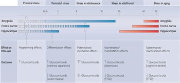
The growth and decline of these brain regions depends on the developmental phases at which the stressors are experienced, which leads to differential effects on HPA axis functioning: (a) prenatal stress leads to programming effects, (b) postnatal stress leads to differentiation effects, (c) stress in adolescence leads to potentiation/incubation effects, and (d) stress in adulthood and aging culminate with maintenance/manifestation effects (Figure 4). For example, if an individual experiences major stressors in early adulthood, there is less chance that this will structurally affect their already fully matured hippocampus or frontal lobes, but could instead lead to hypertrophy of the amygdala as this region is still maturing. This may then lead to maintenance/manifestation effects and psychopathologies later on in adulthood.
The life cycle model of stress (Lupien et al., Reference Lupien, McEwen, Gunnar and Heim2009). How the effects of chronic or repeated exposure to stress (or a single exposure to severe stress) at different stages in life depend on the brain areas that are developing or declining. Stress in the prenatal period affects the development of many of the brain regions that are involved in regulating the hypothalamus–pituitary–adrenal (HPA) axis such as the hippocampus, the frontal cortex, and the amygdala (programming effects). Postnatal stress has varying effects: exposure to maternal separation during childhood leads to increased secretion of glucocorticoids, whereas exposure to severe abuse is associated with decreased levels of glucocorticoids. Thus, glucocorticoid production during childhood differentiates as a function of the environment (differentiation effects). From the prenatal period onward, all developing brain areas are sensitive to the effects of stress hormones (broken blue bars); however, some areas undergo rapid growth during a particular period (solid blue bars). From birth to 2 years of age the hippocampus is developing; it might therefore be the brain area that is most vulnerable to the effects of stress at this time. By contrast, exposure to stress from birth to late childhood might lead to changes in amygdala volume, as this brain region continues to develop until the late 20s. During adolescence the hippocampus is fully organized, the amygdala is still developing, and there is an important increase in frontal volume. Consequently, stress exposure during this period should have major effects on the frontal cortex. Studies show that adolescents are highly vulnerable to stress, possibly because of a protracted glucocorticoid response to stress that persists into adulthood (potentiation/incubation effects). In adulthood and during aging the brain regions that undergo the most rapid decline as a result of aging (red bars) are highly vulnerable to the effects of stress hormones. Stress during these periods can lead to the manifestation of incubated effects of early adversity on the brain (manifestation effects) or to maintenance of chronic effects of stress (maintenance effects). For complete information on the life cycle model of stress, see Lupien et al. (Reference Lupien, McEwen, Gunnar and Heim2009). [A color version of this figure can be viewed online at www.journal.cambridge.org/dpp]

This model brings together the hithero opposing neurotoxicity hypothesis (Sapolsky, Krey, & McEwen, Reference Sapolsky, Krey and McEwen1986) and vulnerability hypothesis (Gilbertson et al., Reference Gilbertson, Shenton, Ciszewski, Kasai, Lasko and Orr2002): the former argues that prolonged exposure to GCs causes neurological damage, whereas the latter argues that neurological impairments are the cause of abnormal GCs dysregulations, and as such, represent a risk factor for the development of psychopathologies rooted in genetics and early life events (Lupien et al., Reference Lupien, McEwen, Gunnar and Heim2009).
In this vein, early adversities are associated with neurological impairments at the level of the hippocampus, amygdala, and PFC that render individuals more vulnerable to developing numerous psychopathologies (McEwen & Gianaros, Reference McEwen and Gianaros2010; Teicher et al., Reference Teicher, Andersen, Polcari, Anderson, Navalta and Kim2003). Animal studies reveal that both prenatal and postnatal exposures biologically embed the HPA axis by shaping cerebral regions like the hippocampus (Champagne & Meaney, Reference Champagne and Meaney2001; McEwen, Reference McEwen2003a) that leads to programming and differentiation effects. In humans, children who have experienced severe abuse show decreased overall brain volumes and are more susceptible to earlier onsets of posttraumatic stress disorder (PTSD) with longer disease durations (De Bellis et al., Reference De Bellis, Keshavan, Clark, Casey, Giedd and Boring1999).
In contrast to adult studies that consistently report smaller hippocampal volume (HV) in PTSD patients (Bremner et al., Reference Bremner, Randall, Vermetten, Staib, Bronen and Mazure1997; Karl et al., Reference Karl, Schaefer, Malta, Dorfel, Rohleder and Werner2006), most child PTSD studies find no evidence for structural hippocampal aberrations (Carrion et al., Reference Carrion, Weems, Eliez, Patwardhan, Brown and Ray2001; De Bellis, Hall, Boring, Frustaci, & Moritz, Reference De Bellis, Hall, Boring, Frustaci and Moritz2001; De Bellis et al., Reference De Bellis, Keshavan, Clark, Casey, Giedd and Boring1999). A recent meta-analysis confirmed that children with PTSD do not necessarily experience smaller HV, although adults with histories of childhood maltreatment often do (Woon & Hedges, Reference Woon and Hedges2008). For instance, individuals with a history of childhood sexual abuse have decreased HVs (Vythilingam et al., Reference Vythilingam, Heim, Newport, Miller, Anderson and Bronen2002) as well as neuropsychological deficits (Bremner, Vermetten, Afzal, & Vythilingam, Reference Bremner, Vermetten, Afzal and Vythilingam2004; Navalta, Polcari, Webster, Boghossian, & Teicher, Reference Navalta, Polcari, Webster, Boghossian and Teicher2006). A recent study uncovered a sex disparity in relation to these early exposures, whereby low birth weight predicted smaller HV in adulthood only in women reporting low parental care but not men (Buss et al., Reference Buss, Lord, Wadiwalla, Hellhammer, Lupien and Meaney2007). These findings suggest that the effects of the prenatal environment combined with childhood maltreatment may not appear until adulthood or late adolescence; however, it is possible that these children had smaller HV to begin with due to prenatal factors.
One's risk of developing major depression is also modulated by early adversity. Individuals with major depression often have heightened basal cortisol levels (Burke, Davis, Otte, & Mohr, Reference Burke, Davis, Otte and Mohr2005; Porter & Gallagher, Reference Porter and Gallagher2006), hippocampal-dependent declarative memory impairments (Burt, Zembar, & Niederehe, Reference Burt, Zembar and Niederehe1995; Fossati et al., Reference Fossati, Harvey, Le Bastard, Ergis, Jouvent and Allilaire2004) and encoding deficits (Bremner, Vythilingam, Vermetten, Vaccarino, & Charney, Reference Bremner, Vythilingam, Vermetten, Vaccarino and Charney2004). In addition, they have decreased HV (Campbell, Marriott, Nahmias, & MacQueen, Reference Campbell, Marriott, Nahmias and MacQueen2004; Videbech & Ravnkilde, Reference Videbech and Ravnkilde2004) that actually predicts the frequency and duration of subsequent major depressive episodes (Campbell et al., Reference Campbell, Marriott, Nahmias and MacQueen2004; MacQueen et al., Reference MacQueen, Campbell, McEwen, MacDonald, Amano and Joffe2003; Sheline, Sanghavi, Mintun, & Gado, Reference Sheline, Sanghavi, Mintun and Gado1999; Videbech & Ravnkilde, Reference Videbech and Ravnkilde2004). When investigating the association between major depression and reduced HV, it is important to take into account whether participants have a history of abuse or maltreatment during childhood (Heim & Nemeroff, Reference Heim and Nemeroff2001). For example, women with a history of childhood sexual or physical abuse are more likely to have hyperactive HPA axis functioning (Heim et al., Reference Heim, Newport, Heit, Graham, Wilcox and Bonsall2000) as well as smaller HV, whereas women without a history of abuse often show similar HV to healthy controls (Vythilingam et al., Reference Vythilingam, Heim, Newport, Miller, Anderson and Bronen2002). That differences in history of abuse lead to variant neuroendocrine functioning suggest that the nature and especially the timing of stressors and/or traumas lead to alternative forms of biological embedding with predispositions to specific diseases (Cicchetti, Rogosch, Gunnar, et al., 2010).
Consistent with this idea, patients with PTSD often show decreased levels of basal cortisol (Yehuda, Golier, & Kaufman, Reference Yehuda, Golier and Kaufman2005) instead of the hypercortisolemic profile often found in depression. These alterations in HPA axis functioning may commence in childhood following abuse or neglect, subsequently increasing the risk for PTSD in adulthood. Combat veterans with PTSD are more likely to have histories of childhood abuse in comparison to veterans who do not develop PTSD (Bremner, Southwick, Johnson, Yehuda, & Charney, Reference Bremner, Southwick, Johnson, Yehuda and Charney1993). Although adult PTSD patients have decreased HV (Karl et al., Reference Karl, Schaefer, Malta, Dorfel, Rohleder and Werner2006; Smith, Reference Smith2005), hippocampal atrophy may be either a consequence of PTSD, or instead a form of vulnerability toward developing PTSD (Gilbertson et al., Reference Gilbertson, Shenton, Ciszewski, Kasai, Lasko and Orr2002; Lupien et al., Reference Lupien, Evans, Lord, Miles, Pruessner and Pike2007). Supporting this is a classic monozygotic twin study, whereby combat veterans who went to war and developed PTSD as well as their twins who did not go to combat both had smaller HV in comparison to twin pairs that did not develop PTSD (Gilbertson et al., Reference Gilbertson, Shenton, Ciszewski, Kasai, Lasko and Orr2002). By contrast, when HV was measured soon after exposure to trauma and before the development of PTSD symptoms, no differences were found between those that later developed PTSD and those that did not (Bonne et al., Reference Bonne, Brandes, Gilboa, Gomori, Shenton and Pitman2001). This chicken–egg phenomenon will only be better understood with prospective life-spanning studies.
Dysregulations of HPA axis activity in psychiatric conditions such as depression and PTSD may alter other physiological biomarkers and consequently increase AL (McEwen & Gianaros, Reference McEwen and Gianaros2010). In the only study of its kind in the AL literature, it was found that mothers of childhood cancer survivors who experienced increased distress and PTSD symptoms also had increased AL levels driven in part by lower cortisol levels (Glover, Stuber, & Poland, Reference Glover, Stuber and Poland2006) in addition to smaller right HVs (Glover, Garcia-Arcena, & Mohlman, Reference Glover, Garcia-Arcena and Mohlman2008) when compared to control mothers. These analyses utilized both high and low ends of cortisol variation since hypocortisolism characterizes certain diseases (Fries, Hellhammer, & Hellhammer, Reference Fries, Hellhammer and Hellhammer2005). Hypocortisolism has been empirically substantiated regarding AL and symptoms of chronic fatigue syndrome (Goertzel et al., Reference Goertzel, Pennachin, de Souza Coelho, Maloney, Jones and Gurbaxani2006; Hellhammer, Schlotz, Stone, Pirke, & Hellhammer, Reference Hellhammer, Schlotz, Stone, Pirke and Hellhammer2004; Maloney, Boneva, Nater, & Reeves, Reference Maloney, Boneva, Nater and Reeves2009; Maloney et al., Reference Maloney, Gurbaxani, Jones, de Souza Coelho, Pennachin and Goertzel2006; Smith, Maloney, Falkenberg, Dimulescu, & Rajeevan, Reference Smith, Maloney, Falkenberg, Dimulescu and Rajeevan2009), PTSD (Glover, Reference Glover2006; Glover et al., Reference Glover, Stuber and Poland2006, Reference Glover, Garcia-Arcena and Mohlman2008), and burnout (Juster et al., Reference Juster, McEwen and Lupienin press). More research is needed to disentangle specific pathways emergent from earlier life experiences that shape the neurobiological substrates associated with these differential biological signatures (e.g., hypo- vs. hypercortisolism). In addition to furthering our understanding of causal mechanisms, such knowledge would guide diagnostic approaches and improve the efficacy of early interventions.
Conclusion and future directions
Early adversities such as low SES (for a review, see Dowd, Simanek, & Aiello, Reference Dowd, Simanek and Aiello2009), living in risky family environments, suffering neglect and/or abuses, can all lead to biological embedding that increases AL and lifelong psychopathological susceptibilities. We believe that these deleterious effects on the hippocampus, amygdala, PFC, and overall brain functioning depend on the developmental timing and duration of elevated GC exposure (Lupien et al., Reference Lupien, McEwen, Gunnar and Heim2009). The balance between adaptive neuroplasticity and neurodegeneration suggests an important dynamic, whereby marked brain changes occur even after maturation and persist throughout the life cycle. Therefore, dynamic parameters of brain changes in childhood and adolescent neurodevelopment, as well as later life alterations should be taken into account when analyzing processes mediating the consequences of stress exposure on the brain. An important additional element of this puzzle that has received limited substantiation in the AL literature are the genetic vulnerabilities we inherit. Genetic and epigenetic interactions can function as strings of fate that can protract biological embedding effects throughout a lifetime. Yet, genetic vulnerabilities and early adversity can also be reversed in certain enriching contexts, underlining a probabilistic rather than deterministic perspective.
For decades, diathesis–stress models have dominated theoretical discourse on how genetic predispositions to diseases can be amplified when stressors reach an intolerable threshold. This concept has many merits; however, it limits science's ability to go beyond hypotheses that fixate on vulnerability and which often cannot account for how diatheses sometimes reverse into a resilient trait. In this spirit, the Swedes have an expression called maskrosbarn (dandelion child) and a neoglism called orkidebarn (orchid child) that offers an empowering, alternative perspective to append into diathesis–stress models (Boyce & Ellis, Reference Boyce and Ellis2005). Quoting David Dobbs, “most of us have genes that make us as hardy as dandelions: able to take root and survive almost anywhere. A few of us, however, are more like the orchid: fragile and fickle, but capable of blooming spectacularly if given greenhouse care” (Dobbs, Reference Dobbs2009, p. 60).
This perspective states that inherited genotypes with potentially negative consequences can be modulated to generate a multitude of potentially positive phenotypes. In terms of evolution, natural selection has permitted certain “bad” genes to remain in the gene pool if, under certain circumstances, they could enhance fitness and survival (Dobbs, Reference Dobbs2009). With this in mind, it is important to recognize that diverse interactions among nature (genes) and nurture (environment) are unremittingly codependent.
Human Genetics: A Common Missing Piece of the Model
A comprehensive review on the antecedents of AL must incorporate genetic underpinnings involved in diathesis–stress models. Genetics provide insights into the interaction between stress exposure and the regulation of internal biological processes across an individual's life span and across generations. A rich literature on the genetics involved in stress-related diseases is now reviewed to encourage future investigations.
Twin studies
Twin studies have demonstrated significant genetic contribution to basal cortisol secretion (Linkowski et al., Reference Linkowski, Van Onderbergen, Kerkhofs, Bosson, Mendlewicz and Van Cauter1993; Steptoe, van Jaarsveld, Semmler, Plomin, & Wardle, Reference Steptoe, van Jaarsveld, Semmler, Plomin and Wardle2009), stress reactivity (Steptoe et al., Reference Steptoe, van Jaarsveld, Semmler, Plomin and Wardle2009), and other biomarkers commonly employed in the AL model (De Geus, Kupper, Boomsma, & Snieder, Reference De Geus, Kupper, Boomsma and Snieder2007; Williams, Blanche, & Krauss, Reference Williams, Blanche and Krauss2005). In a study assessing salivary cortisol in unaffected twins discordant for affective disorders (Vinberg, Bennike, Kyvik, Andersen, & Kessing, Reference Vinberg, Bennike, Kyvik, Andersen and Kessing2008), it was found that high-risk twins displayed elevated evening cortisol levels compared to low-risk twins. Notably, the high-risk twins in this study previously reported signs of discrete cognitive dysfunction (Christensen, Kyvik, & Kessing, Reference Christensen, Kyvik and Kessing2006) and a tendency for higher levels of neuroticism (Vinberg, Mortensen, Kyvik, & Kessing, Reference Vinberg, Mortensen, Kyvik and Kessing2007). Overall, twin studies suggest that genetic factors control the expression of HPA axis activity.
Although findings are mixed (Kirschbaum, Wust, Faig, & Hellhammer, Reference Kirschbaum, Wust, Faig and Hellhammer1992), quantitative genetic modeling has shown significant heritability for cortisol reactivity to a stressor (Steptoe et al., Reference Steptoe, van Jaarsveld, Semmler, Plomin and Wardle2009). In a recent meta-analysis that evaluated all published twin studies that assessed heart rate or blood pressure reactivity to stress exposure, the pooled heritability estimates ranged from 26% to 43% for reactivity to psychosocial stress (i.e., mental stressor) and 21% to 55% for reactivity to physiological stress (i.e., cold pressor test; Wu, Snieder, & de Geus, Reference Wu, Snieder and de Geus2010). These studies highlight the importance of genetics as an underlying mechanism in individual differences in stress reactivity.
In uncovering the molecular pathway between genetic variability and AL, twin studies have examined the genetic underpinnings of additional biological variables found within the AL model. In a study of Swedish elderly twins, a dominant genetic component was reported for LDL, glucose, HbA1C, and C-reactive protein (CRP; Rahman et al., Reference Rahman, Bennet, Pedersen, de Faire, Svensson and Magnusson2009). In a study assessing genetic and environmental contributions to cardiovascular disease and metabolic syndrome (Elder et al., Reference Elder, Lichtenstein, Pittas, Roberts, Fuss and Greenberg2009), it was reported that cholesterol and triglycerides were highly heritable (56% to 77%), and waist circumference, glucose, insulin, insulin resistance, and blood pressure were moderately heritable (43% to 57%). Common environmental factors contributed to 23% to 40% of the variables of waist circumference, systolic blood pressure, and glucose, suggesting a dominant genetic influence on total variance of factors associated with disease. Atlhough twin studies provide important information, the magnitude of heritability reveals little about the genetic structure of particular endophenotypes and plausible number of genes involved.
Genetic association studies in humans
Association studies have provided essential information on how specific genes contribute to the regulation of key biological processes involved in how sensitive individuals are likely to respond to stressors. Reports of gene–environment interactions demonstrate just how variation in genetic profiles interact with environmental stimuli, resulting in differential health outcomes or multifinality (Cichetti & Toth, Reference Cicchetti, Rogosch, Gunnar and Toth2009). In accordance with the stress–diathesis model, individual differences at the genetic level may alter primary effects and secondary outcomes, resulting in varied susceptibilities to tertiary outcomes. Over the years, a number of genetic polymorphisms have been assessed in association with stress and stress-related disease in humans, including the serotonin transporter linked polymorphism (5-HTTLPR, short/long; Caspi et al., Reference Caspi, Sugden, Moffitt, Taylor, Craig and Harrington2003), brain-derived neurotrophic factor (BDNF, Met66Val; Vinberg et al., Reference Vinberg, Trajkovska, Bennike, Knorr, Knudsen and Kessing2009), apolipoprotein E (APOE, epsilon e4; Fiocco, Poirier, Joober, Nair, & Lupien, Reference Fiocco, Poirier, Joober, Nair and Lupien2008), and the catechol-O-methyltransferase gene (COMT, Met158Val; Yavich, Forsberg, Karayiorgou, Gogos, & Mannisto, Reference Yavich, Forsberg, Karayiorgou, Gogos and Mannisto2007).
The 5-HTTLPR gene has been widely investigated for its role in affective disorders. Studies show that individuals with the short allele display greater stress reactivity to psychosocial stress, with the greatest response in those homozygotic for the short allele (Gotlib, Joormann, Minor, & Hallmayer, Reference Gotlib, Joormann, Minor and Hallmayer2008; Way & Taylor, Reference Way and Taylor2010). Further, it is reported that the short allele interacts with stressful life events to increase risk of depression (Caspi et al., Reference Caspi, Sugden, Moffitt, Taylor, Craig and Harrington2003; Kendler, Kuhn, Vittum, Prescott, & Riley, Reference Kendler, Kuhn, Vittum, Prescott and Riley2005) and neuronal activation of brain regions involved in depression (Canli et al., Reference Canli, Qiu, Omura, Congdon, Haas and Amin2006). 5-HTTLPR has also been shown to moderate the association between lipid profiles and depression prevalence in older adults, with the association apparent only in homozygotic carriers of the short allele (Kim et al., Reference Kim, Stewart, Kim, Shin, Yang and Yoon2009). On the other hand, a recent meta-analysis failed to conclude that the 5-HTTLPR genotype alone or in interaction with stressful life events is associated with an elevated risk of depression regardless of sex (Risch et al., Reference Risch, Herrell, Lehner, Liang, Eaves and Hoh2009). Genetic polymorphisms may thus affect the magnitude of AL acquired over time as an individual faces and responds to environmental challenges, which then determines risk of adverse psychological and health sequelae associated with biological concomitants of the AL model.
COMT catalyzes the degradation of catecholamines like dopamine in the brain (Yavich et al., Reference Yavich, Forsberg, Karayiorgou, Gogos and Mannisto2007) and is, as will be elaborated in a separate section, linked to psychotic disorders (Craddock, Owen, & O'Donovan, Reference Craddock, Owen and O'Donovan2006). The gene that codes for COMT contains a functional common Val158Met polymorphism, with the Val allele exhibiting a three- to fourfold increase in enzyme activity compared to the Met allele (Chen et al., Reference Chen, Lipska, Halim, Ma, Matsumoto and Melhem2004). Recent studies show that, compared to Val/Val carriers, healthy adults with the Met/Met genotype display a diminished opiod system response and greater sensory and affective ratings of pain following a sustained pain challenge (Zubieta et al., Reference Zubieta, Heitzeg, Smith, Bueller, Xu and Xu2003) and increased limbic and prefrontal activation following the presentation of unpleasant stimuli (Smolka et al., Reference Smolka, Schumann, Wrase, Grusser, Flor and Mann2005). These studies suggest that the Met allele lowers physiological and psychological resilience against negative environmental challenges, which may increase the risk of stress-induced disease.
In a study that assessed reactivity to a psychosocial stressor in healthy controls in risk probands and major depressive disorder patients, an association between COMT genotype and stress reactivity was reported (Jabbi et al., Reference Jabbi, Kema, van der Pompe, te Meerman, Ormel and den Boer2007). This association differed depending on group status: Met/Met carriers displayed greater cortisol and adrenalin reactivity following the stressor. Yet, in at-risk probands, the Val/Val genotype group displayed greater reactivity. No association was found in the patient group; however, this was attributed to current medication use. This study presents the possibility that epigenetic mechanisms are at play: risk exposure alters the function of the gene, which then biases an individual's response to subsequent stressors.
Epigenetics
Epigenetics examines changes in DNA methylation and histone modifications produced by perturbations in the environment (e.g., early adversity),which in turn alter the function, but not the structure, of a gene. Research shows that changes incurred through epigenetics are stable and can even be transmitted intergenerationally (Meaney & Szyf, Reference Meaney and Szyf2005). Although a majority of epigenetic findings stem from animal research (Francis, Diorio, Liu, & Meaney, Reference Francis, Diorio, Liu and Meaney1999; Szyf, Weaver, Champagne, Diorio, & Meaney, Reference Szyf, Weaver, Champagne, Diorio and Meaney2005; Weaver et al., Reference Weaver, Cervoni, Champagne, D'Alessio, Sharma and Seckl2004), two studies have thus far evaluated the epigenetic model in humans.
A twin study that examined the genetic and environmental contributions to cortisol reactivity in a cohort of 19-month-old twins from high and low family adversity found that high family adversity has a differentiation developmental effect on cortisol reactivity (Ouellet-Morin et al., Reference Ouellet-Morin, Boivin, Dionne, Lupien, Arseneault and Barr2008). Specifically, both genetic and environmental factors explained the variance in cortisol reactivity to a mild stressor (i.e., unfamiliar situation) for twins from low family adversity. By contrast, variance in cortisol reactivity was only explained by shared and unique environmental factors in twins who had experienced high family adversity (characterized as having four of the following: maternal smoking during pregnancy, low birth weight, low family income, low maternal education level, single parenthood, young motherhood, and maternal hostile or reactive behaviors). As discussed earlier, infants exposed to adverse environments likely undergo alterations in their biological embedding over and above any genetic contributions, and yet the pathways driving stress responsivity can differ markedly from one family context to the other. Until more research is conducted, the precise manner whereby such mechanisms promote resilience or vulnerability is speculative, but might involve differential neurodevelopmental processes.
Epigenetic differentiation precipitated by early adversity might effect GC gene promotor expression in the hippocampus via DNA methylation. A recent postmortem study (McGowan et al., Reference McGowan, Sasaki, D'Alessio, Dymov, Labonte and Szyf2009) assessing the epigenetic regulation of the GC receptor in suicide victims with or without a history of childhood abuse reported differential neuron-specific GC receptor promotor in postmortem hippocampi. Specifically, decreased levels of GC receptor mRNA and increased cytosine methylation of the GC receptor promotor was found in abused suicide victims. Early adversity can also induce modifications in the methylation status of the BDNF gene, which can be transmitted from one generation to the next (Roth, Lubin, Sodhi, & Kleinman, Reference Roth, Lubin, Sodhi and Kleinman2009). Although these studies are tips of the iceberg, they nonetheless suggest that genetic effects and epigenetic interactions might influence AL susceptibilities via physiological recalibrations of the HPA axis and regulatory neural substrates.
Genetic study of AL
To date, only one study has directly assessed the association between genetic variance and AL (Smith et al., Reference Smith, Maloney, Falkenberg, Dimulescu and Rajeevan2009). In a population-based study of chronic fatigue syndrome, 182 Caucasian participants were genotyped and an AL index was calculated, based on measures representing metabolic, cardiovascular, inflammatory, HPA axis, and autonomic activities. Polymorphisms in 32 candidate genes related to HPA axis activity, neurotransmitter systems, inflammation, cardiovascular, and metabolic functions were analyzed. After correcting for covariates and multiple comparisons, genetic polymorphism of the gene that codes for angiotensin-I converting enzyme (ACE C/T: rs4968591) remained significantly associated with AL status. Specifically, participants with the testosterone allele of the ACE gene were more likely to display a higher AL index than participants with the C allele.
ACE catalyses the conversion of angiotensin-I (Ang-I) to Ang-II, which is both pro-inflammatory and pro-oxidant and thus plays an important role in cardiovascular, endocrine, and immune functions (Garg & Yusuf, Reference Garg and Yusuf1995; Malik et al., Reference Malik, Williamson, Abbott, Carrington, Iqbal and Schady1998; Maschio et al., Reference Maschio, Alberti, Janin, Locatelli, Mann and Motolese1996). The ACE enzyme further converts Aβ42 to Aß40 in the brain (Zou et al., Reference Zou, Maeda, Watanabe, Liu, Liu and Oba2009), which is implicated in the development of neuronal plaques of particular concern in older life dementias. Polymorphism of the ACE gene has been associated with Type II diabetes mellitus (Stephens et al., Reference Stephens, Dhamrait, Cooper, Acharya, Miller and Hurel2005), obstructive pulmonary disease (Tkacova et al., Reference Tkacova, Joppa, Stancak, Salagovic, Misikova and Kalina2005), and Alzheimer's disease (AD; Kolsch et al., Reference Kolsch, Jessen, Freymann, Kreis, Hentschel and Maier2005). As the common ACE polymorphism in these studies (ACE I/D) is not in linkage disequilibrium with the ACE T/C gene, additional prospective research is needed to confirm the association between ACE and AL and any functional, long-term consequences.
Conclusion and future directions
Future studies should go beyond the basic stress–diathesis structure and consider genetic and epigenetic influences on biological systems that are integral to the AL model. Future investigations of genetic mechanisms contributing to AL such as telomere length as tertiary outcomes would be immensely informative (Epel, Reference Epel2009; Epel et al., Reference Epel, Blackburn, Lin, Dhabhar, Adler and Morrow2004; Sapolsky, Reference Sapolsky2004). In addition, studies incorporating multiple dimensions of outcomes (biological, psychological, social, behavioral, spiritual) and global self-perceptions of health (Jylha, Reference Jylha2009) would help us determine how genetic and epigenetic factors interact with other systems and with individual's health in general to promote vulnerability or resilience. A more comprehensive understanding of the (epi)genetic regulatory mechanisms and their relationship with chronic stress should help inform individualized prevention strategies for reducing or minimizing the underpinnings of AL.
Like the case of the orchid child discussed previously, it is imperative to understand how protective factors can be harnessed to dampen the effects of risk factors like harmful genetic makeups and/or early adversities. For instance, an exemplary review of gene–stress–behavior interactions in adolescent alcohol use outlines how to intervene early to minimize the likelihood that inherited vulnerabilities toward alcoholism and associated psychopathologies before morbidities manifest themselves (Zimmermann, Blomeyer, Laucht, & Mann, Reference Zimmermann, Blomeyer, Laucht and Mann2007). The interface between individuals and foreign substances that endanger health (e.g., cigarettes) is an important avenue to pursue with respect to stress research, and yet, these have had little place in the AL literature other than as behavioral covariates in statistical equations. As numerous noxious factors knowingly and unknowingly enter our bodies to exert deleterious effects, we would do well to determine the precise mechanisms of their actions on the brain and body. To address this lacuna, we now turn to how ETs can directly or indirectly affect AL throughout life.
ETs: Mediators or Moderators of AL?
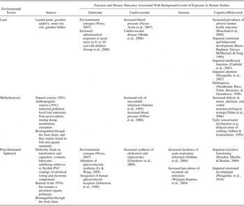
Genetic variance exists in an individual's ability to absorb, distribute, metabolize, and excrete ETs. For example, genotypes associated with heavy metal absorption and toxicity include hemochromatosis (Wright et al., Reference Wright, Silverman, Schwartz, Tsaih, Senter and Sparrow2004), vitamin D receptor (Chuang et al., Reference Chuang, Yu, Ho, Wu, Lin and Wu2004), and apolipoprotein-E (Godfrey, Wojcik, & Krone, Reference Godfrey, Wojcik and Krone2003). ETs are principally industrial chemicals like lead (Pb), methylmercury (MeHg), and polychlorinated biphenyls (PCB) that can contribute to functional disruptions, neurodevelopmental delays, behavioral, as well as cognitive impairments throughout the life span (Bradley & Corwyn, Reference Bradley and Corwyn2002; Furumoto-Dawson, Gehlert, Sohmer, Olopade, & Sacks, Reference Furumoto-Dawson, Gehlert, Sohmer, Olopade and Sacks2007). Unfortunately, ETs have received little attention in the AL model, and yet they have the propensity to potently effect AL biomarkers by means of mediation or moderation effects at differential periods throughout life span development (see Table 2).
Identified environmental toxins frequently studied

Note: Lead has been recognized as a poison for millennia and has been the focus of public health regulation in much of the developed world for the better part of the past century. Most clinicians and researchers consider 10–15 µg/dl the blood level at which lead is neurotoxic for children. Even so, the prevalence of blood lead levels >10 µg/dL among US children 1–5 years old was 2.2% in 2000, representing over 310,000 children with high exposure (Gilbert & Weiss, Reference Gilbert and Weiss2006). At an ecological level, inorganic mercury is converted into methylmercury (MeHg) by microbial action, primarily in sediments of fresh and ocean waters. MeHg readily enters the aquatic food chain and is biomagnified as it accumulates in predatory fish such as swordfish, pike, and ocean tuna. Larger and longer living fish therefore contain more accumulated MeHg levels. Fish consumption, while not the only source of MeHg exposure, is thus a significant vector for human exposure, particularly for populations dependent on fish as a primary food source. Polychlorinated biphenyls are another family of ubiquitous environmental toxins that were once used as dielectric fluids in transformers and capacitors, coolants, lubricants, stabilizing additives in flexible plastic coatings of electrical wiring, and electronic components.
Vulnerable populations to ETs exposure
Although everyone is vulnerable to ETs, children and older adults are particularly sensitive to exposure. Extensive studies of Pb, MeHg, and PCBs reveal multiple mechanisms by which these compounds disrupt normal brain development (Landrigan, Reference Landrigan1999; Stein, Schettler, Wallinga, & Valenti, Reference Stein, Schettler, Wallinga and Valenti2002). Children also have greater exposure to ETs than adults in terms proportioned body weight by way of water consumption, food, and air, as well as through increased hand to mouth and playground behaviors.
Aging individuals are also highly vulnerable to a lifetime's worth of accumulated ETs that can exacerbate senescence. For instance, toxicokinetic studies have demonstrated that 95% of Pb is stored in the skeleton only to then be released into circulation during age-related bone demineralization (Rabinowitz, Reference Rabinowitz1991). Data from the National Health and Nutrition Examination Survey (NHANES) confirm that adults over the age of 50 have the highest blood Pb levels (Pirkle et al., Reference Pirkle, Kaufmann, Brody, Hickman, Gunter and Paschal1998). Moreover, our elders have much higher exposure to ETs than younger generations benefitting from recent bans and/or restrictions. In sum, older adults and youths are extremely vulnerable to ETs that can, like chronic stress, increase sensitivities toward disrupted development and accelerated brain declines (Lupien et al., Reference Lupien, McEwen, Gunnar and Heim2009).
ETs as endocrine disruptors
One plausible pathway whereby ETs impact the body and the brain is through interactions with the endocrine system. Early observations of reproductive organ malformations and aberrant sexual behaviors in wildlife first unveiled the ability of ETs to interfere with steroid hormones. This led to their infamous classification as endocrine disrupting chemicals (Hotchkiss et al., Reference Hotchkiss, Rider, Blystone, Wilson, Hartig and Ankley2008; Zala & Penn, Reference Zala and Penn2004), which the World Health Organization defines as “an exogenous substance or mixture that alters function(s) of the endocrine system and consequently causes adverse health effects in an intact organism, or its progeny, or (sub)populations” (WHO, 2002b). Identified environmental estrogens and antiestrogens include certain heavy metals (Pb, cadmium, MeHg), industrial chemicals (PCBs, dioxins, and furans), pesticides (DDT, organophosphates), herbicides (Atrazine), and plasticizers (bisphenol A, surfactants; Darbre, Reference Darbre2006; Weiss, Reference Weiss2007) that are dangerous even at very low doses (Denham et al., Reference Denham, Schell, Deane, Gallo, Ravenscroft and DeCaprio2005; Jacobson & Jacobson, Reference Jacobson and Jacobson1996).
As ETs are ubiquitous, affect particularly children and elders, and interact with endocrine functioning, they should be addressed by the AL model. In the following paragraph, we will examine whether ETs might (a) directly impact the HPA axis, (b) potentiate other chronic stressors, and/or (c) effect individual AL biomarkers.
ETs and AL
Although scarcely studied in humans, ETs are believed to function as GC receptor antagonists (Johansson, Nilsson, & Lund, Reference Johansson, Nilsson and Lund1998) and can alter GC biosynthesis in adrenocortical cells (Li & Wang, Reference Li and Wang2005). Animal studies reveal that dysregulated HPA axis responses to acute stressors are related to heavy metal exposure (cadmium, lead, mercury) and organic environmental contaminants (PCBs, solvents, pesticides) in fish (Brodeur, Sherwood, Rasmussen, & Hontela, Reference Brodeur, Sherwood, Rasmussen and Hontela1997; Hontela, Dumont, Duclos, & Fortin, Reference Hontela, Dumont, Duclos and Fortin1995), amphibians (Gendron, Bishop, Fortin, & Hontela, Reference Gendron, Bishop, Fortin and Hontela1997), birds (Love et al., Reference Love, Shutt, Silfies, Bortolotti, Smits and Bird2003), rodents (Meserve, Murray, & Landis, Reference Meserve, Murray and Landis1992; Vyskocil, Fiala, Ettlerova, & Tenjnorova, Reference Vyskocil, Fiala, Ettlerova and Tenjnorova1990), and large mammals (Oskam et al., Reference Oskam, Ropstad, Lie, Derocher, Wiig and Dahl2004; Zimmer et al., Reference Zimmer, Gutleb, Lyche, Dahl, Oskam and Krogenaes2009). In humans, even low levels of Pb exposure correspond to increased cardiovascular (Gump et al., Reference Gump, Stewart, Reihman, Lonky, Darvill and Matthews2005) and cortisol reactivity to the cold pressor task in 9- to 10-year-old children (Gump et al., Reference Gump, Stewart, Reihman, Lonky, Darvill and Parsons2008). As the cold pressor task is primarily a physical stressor, it will be important to replicate these findings using psychosocial stressors that elicit social–evaluative threats known to induce greater HPA axis activations than other laboratory tasks (Dickerson & Kemeny, Reference Dickerson and Kemeny2004).
ETs may directly impair the HPA axis or alternatively act as mediators of chronic stressors like poverty. ET exposures are demographically constrained, occurring most frequently in socioeconomically disadvantaged environments (Sexton, Reference Sexton1997). For instance, children living in older houses containing Pb paint (Pirkle et al., Reference Pirkle, Kaufmann, Brody, Hickman, Gunter and Paschal1998) and/or living in the center of large cities are exposed to greater Pb levels than those living more peripherally (Brody et al., Reference Brody, Pirkle, Kramer, Flegal, Matte and Gunter1994; Pirkle et al., Reference Pirkle, Brody, Gunter, Kramer, Paschal and Flegal1994; Sexton et al., Reference Sexton, Adgate, Fredrickson, Ryan, Needham and Ashley2006). Impoverished individuals are also more likely than affluent ones to live near environmental hazards like industrial plants, reside in urban areas where ambient levels of many air pollutants tend to be higher, eat significantly greater amounts of contaminated fish, and be employed in potentially dangerous occupations (Sexton et al., Reference Sexton, Greaves, Church, Adgate, Ramachandran and Tweedie2000).
In addition, high Pb levels could be a mechanism whereby family income effects HPA axis functioning in children (Gump et al., Reference Gump, Reihman, Stewart, Lonky, Granger and Matthews2009). Using data from the Oswego study, increased early childhood blood Pb levels significantly mediated the association between decreased family income and augmented salivary cortisol levels following acute stress. Although this might denote a meditational effect, the direction of the association is indiscernible, as reverse causality remains a possibility: adrenocortical reactivity may have effects on the toxicokinetics of blood to bone transfers of Pb (Mushak, Reference Mushak2003).
ETs might also indirectly synergize HPA axis dysregulations and increase vulnerabilities toward intergenerational transmission of psychopathologies. A notable American population study recently found that background levels of Pb were associated with increased odds of developing major depression in 20- to 40-year-old adults (Bouchard et al., Reference Bouchard, Bellinger, Weuve, Matthews-Bellinger, Gilman and Wright2009). Of interest, this age range corresponds to the average reproductive years of humans. Our group has previously shown that children with increased cortisol levels are more likely to have depressed mothers and come from disadvantaged neighborhoods (Lupien et al., Reference Lupien, King, Meaney and McEwen2000). It follows that living in lower socioeconomic strata with increased Pb exposure could directly impact children's HPA functioning or indirectly via the mother's mental health, or perhaps a mix of both. At a transdisciplinary level, such complex interactions among environmental, biological, and psychosocial factors in developing children could result in an altered state of global health that could lead to psychopathologies and concomitant comorbidities. The interactions of ETs with other types of factors (e.g., early adversity, genetic endowment) may thus lead to complex dysregulations at multiple levels that consequently render the individual more susceptible to the effects of chronic stress by enhancing the toll of AL.
In mechanistically considering ETs in interaction with AL, only one group have systematically assessed the combined effects of ETs and stressors using the rodent model (Cory-Slechta et al., Reference Cory-Slechta, Virgolini, Rossi-George, Thiruchelvam, Lisek and Weston2008). Based on the premise that Pb exposure acts on the mesocorticolimbic dopamine pathway (Zuch, O'Mara, & Cory-Slechta, Reference Zuch, O'Mara and Cory-Slechta1998) in semblance to products of the HPA axis, Cory-Schleta and colleagues (2008) confirmed that Pb and GCs interact in this tract. By assigning dams to four experimental groups as a function of Pb exposure (waterborne 2 months before breeding and into lactation vs. not) and maternal stress (restraint at gestational day 16 and 17 vs. not), they observed behaviors dependent on this pathway (e.g., locomotion, impulsivity) in 60-day-old offspring. Results showed that the dams exposed to combined Pb and restraint had female offspring with higher corticosterone levels than contrast groups (Cory-Slechta, Virgolini, Thiruchelvam, Weston, & Bauter, Reference Cory-Slechta, Virgolini, Thiruchelvam, Weston and Bauter2004). In a subsequent study that limited the timing of Pb exposure to postweaning periods, it was shown that interactions among Pb and stressors are not restricted to maternally mediated exposure (Virgolini, Chen, Weston, Bauter, & Cory-Slechta, Reference Virgolini, Chen, Weston, Bauter and Cory-Slechta2005). Even at low levels (10 µg/dl), Pb exposure interacts with prenatal stress to further modify both behavioral and corticosterone responses to stressors (Virgolini, Rossi-George, Weston, & Cory-Slechta, Reference Virgolini, Rossi-George, Weston and Cory-Slechta2008). These findings translate into a major danger for population health. In the United States, the acceptable childhood blood Pb threshold is currently 10 µg/dl. Yet amidst public health efforts to restrict exposure, 1.6% of American children ages 1 to 5 have levels greater than or equal to 10 µg/dL (Centers for Disease & Prevention, 2005; Jones et al., Reference Jones, Homa, Meyer, Brody, Caldwell and Pirkle2009).
As far as we know, only one epidemiological study has reported interactive effects of stress and Pb in the general population (Peters et al., Reference Peters, Kubzansky, McNeely, Schwartz, Spiro, Sparrow and Wright2007). In the Normative Aging Study, individuals with increased psychological stress, in addition to cumulative Pb exposure, had an increased risk of developing hypertension independent of standard covariates. Specifically, those reporting high stress levels had a 2.65 times greater chance of developing hypertension per standard deviation increase in Pb. These results strongly suggest that the effect of Pb on hypertension interacts with chronic stress levels independent of demographic and behavioral risk factors. Apart from Pb, ETs may exert their influence on the HPA axis through various pathways: (a) directly by affecting the HPA axis, (b) indirectly by mediating the effects of environmental factors on the HPA axis functioning, and (c) indirectly by potentiating the effects of maternal stress on offspring HPA axis functioning and behavioral outcomes. These pathways may represent an additional form of AL that can increase the probability of developing physical and mental disease. Although ETs have been proposed as mediators or moderators of AL (Loucks, Juster, & Pruessner, Reference Loucks, Juster and Pruessner2008), no corresponding studies exist and so we must therefore turn to studies of individual AL biomarkers.
ETs as disrupters of individual AL biomarkers
In addition to neuroendocrine effects, ETs can impair cardiovascular, immune, and metabolic functions. A positive correlation between Pb exposure and blood pressure has been identified in numerous studies (Fewtrell, Pruss-Ustun, Landrigan, & yuso-Mateos, Reference Fewtrell, Pruss-Ustun, Landrigan and yuso-Mateos2004; Gump et al., Reference Gump, Stewart, Reihman, Lonky, Darvill and Matthews2005; Nash et al., Reference Nash, Magder, Lustberg, Sherwin, Rubin and Kaufmann2003; Navas-Acien, Guallar, Silbergeld, & Rothenberg, Reference Navas-Acien, Guallar, Silbergeld and Rothenberg2007). Increased vulnerabilities for cardiovascular morbidities and mortality have even been shown at lead levels <5 µg/dl in large epidemiological studies (Menke, Muntner, Batuman, Silbergeld, & Guallar, Reference Menke, Muntner, Batuman, Silbergeld and Guallar2006; Muntner, Menke, DeSalvo, Rabito, & Batuman, Reference Muntner, Menke, DeSalvo, Rabito and Batuman2005). Concerning MeHg toxicity, most studies have been conducted in populations dependant on fish as a primary food source, such as in the Seychelles study (Davidson et al., Reference Davidson, Myers, Cox, Axtell, Shamlaye and Sloane-Reeves1998; Myers et al., Reference Myers, Davidson, Shamlaye, Axtell, Cernichiari and Choisy1997) or the Faroe Island study (Debes, Budtz-Jorgensen, Weihe, White, & Grandjean, Reference Debes, Budtz-Jorgensen, Weihe, White and Grandjean2006; Grandjean et al., Reference Grandjean, Weihe, Jorgensen, Clarkson, Cernichiari and Videro1992).
Although controversial (Chan & Egeland, Reference Chan and Egeland2004; Stern, Reference Stern2005), several reports substantiate an adverse association between MeHg exposure and relative risk of myocardial infarction (Salonen et al., Reference Salonen, Seppanen, Nyyssonen, Korpela, Kauhanen and Kantola1995) and hypertension (Fillion et al., Reference Fillion, Mergler, Sousa Passos, Larribe, Lemire and Guimaraes2006). Likewise, PCB 126 (a dioxinlike compound) exposure in female rats increases cardiovascular risk factors like hypercholesterolemia and hypertension (Lind, Orberg, Edlund, Sjoblom, & Lind, Reference Lind, Orberg, Edlund, Sjoblom and Lind2004). One human study even showed that Native Americans with increased PCB levels had an increased risk of developing cardiovascular disease through increased synthesis of cholesterol and triglycerides (Goncharov et al., Reference Goncharov, Haase, Santiago-Rivera, Morse, McCaffrey and Rej2008).
Although the immunotoxic mechanism of ETs is not well understood, deleterious effects have been observed in animals (Esser & Welzel, Reference Esser and Welzel1993) and humans (Baccarelli et al., Reference Baccarelli, Mocarelli, Patterson, Bonzini, Pesatori and Caporaso2002; Heilmann, Grandjean, Weihe, Nielsen, & Budtz-Jorgensen, Reference Heilmann, Grandjean, Weihe, Nielsen and Budtz-Jorgensen2006). An accumulating body of evidence suggests that in utero ET exposure might increase susceptibility to infections in young children (Dallaire et al., Reference Dallaire, Dewailly, Vezina, Muckle, Weber and Bruneau2006; Weisglas-Kuperus, Vreugdenhil, & Mulder, Reference Weisglas-Kuperus, Vreugdenhil and Mulder2004). In the NHANES, exposure to monohydroxy polycyclic aromatic hydrocarbons (derived from incomplete combustion of organic matter and fossil fuels including diesel engines, domestic heating, and pyrolysis of coal or tobacco smoke) was associated with increased CRP (Everett et al., Reference Everett, King, Player, Matheson, Post and Mainous2010). In the previous phase of the NHANES study, exposure to cadmium, which is used as a pigment and plastic stabilizer, was also positively associated with increased plasma CRP and fibrinogen levels (Lin, Rathod, Ho, & Caffrey, Reference Lin, Rathod, Ho and Caffrey2009), whereas Pb exposure was associated to inflammatory biomarkers (Songdej, Winters, McCabe, & van Wijngaarden, Reference Songdej, Winters, McCabe and van Wijngaarden2010).
As mentioned before, ETs also increase lipid profiles (Goncharov et al., Reference Goncharov, Haase, Santiago-Rivera, Morse, McCaffrey and Rej2008). Specifically, PCBs and other dioxinlike components are associated with increased risk factors representative of metabolic syndrome in Americans (Lee, Lee, Porta, Steffes, & Jacobs, Reference Lee, Lee, Porta, Steffes and Jacobs2007) and Japanese (Uemura et al., Reference Uemura, Arisawa, Hiyoshi, Kitayama, Takami and Sawachika2009) alike. Notwithstanding, differential ET exposures are based largely on socioeconomic as well as cultural differences both within and between populations that should be considered by future investigations.
Conclusion and future directions
Because several AL biomarkers are effected by or interact with ETs, we believe that they should be explored as mediators and/or moderators in the AL model. In particular, elucidating how ETs effect developmental trajectories toward pathological outcomes from childhood until old age will be important to consider. We have limited this review to the best characterized ETs (Pb, MeHg, and PCBs), but many other chemicals currently used in numerous commercial goods are identified or suspected endocrine disrupters with neurotoxic effects. Furthermore, the majority of ETs reviewed are endocrine disrupting chemicals that alter HPA axis and sex hormone functions in humans (Agusa et al., Reference Agusa, Kunito, Iwata, Monirith, Chamnan and Tana2007).
ETs are likely also effected by numerous other factors (psychosocial, biological, behavioral). Studying the impact of ETs on psychopathology could be improved by adopting a transdisciplinary approach that focuses on the interaction of ETs with multiple levels of outcomes existing in parallel. For instance, are the sexes exposed differently to ETs based on gender-typed work environments (e.g., factories, primary industries), and how can ETs interact with sex hormones to disrupt physiology? We are far from being able to accurately respond to such questions. Just in terms of sex differences in ET exposures, genetic interactions, and early adversities, much remains unknown, as the concept of “sex” is complex but powerful in determining patterns of health and disease. In order to facilitate answers for future inquiries in ETs research and all the aforementioned litarature, we next explore nuances among one's biological sex, one's socioculturally generated gender identity and behaviors, and the modulating effects of sex hormones on AL levels.
Sex, Gender, and Sex Hormones
One's sex confers a unique set of vulnerabilities to specific disease pathways. Women are more prone to affective and autoimmune disorders, whereas men are more at risk of early mortality, substance abuse, conduct/antisocial disorder, and infectious diseases. Although men are more stress responsive to acute stressors (Kajantie & Phillips, Reference Kajantie and Phillips2006; Kirschbaum, Wust, Hellhammer, 1992; Kudielka & Kirschbaum, Reference Kudielka and Kirschbaum2005), women experience greater subjective distress, perhaps due to sociocultural factors. As this section will elucidate, biological sex, gender roles, and sex hormones are elusively linked to AL levels.
The term sex refers to the biological differences between men and women such as genetic sex (female XX and male XY chromosomes) and hormonal milieu. For instance, men have higher testosterone levels and women have higher estrogen and progesterone levels. These sex steroids have both organizational and activational effects that shape sex differences ranging from morphology to behavior. Organizational effects refer to permanent actions during a critical early developmental period that modulate brain responses to certain stimuli later in life. In contrast, activational effects refer to transient physiological and/or behavioural responses triggered by the activation of the matured gonadal axis occuring at any age thereafter.
Lifelong dynamics and exposure to sex steroids differ greatly between sexes: men experience an age-related linear decline in testosterone levels, whereas women's lives are characterized by constant periodic fluctuations including menarche, the menstrual cycle, pregnancies, and menopause. Consequently, when it comes to AL, the biological sex differences observed might depend on sex steroids (i.e., testosterone vs. estrogens), levels of sex steroids (i.e., phase of the menstrual cycle, pregnancy, menopause), and previous or lifelong exposures (i.e., age at menarche/puberty, number of pregnancies, age at menopause, exogenous administration such as hormonal contraceptive and replacement therapy). As differences in the hormonal milieu vary greatly between individuals throughout a lifetime, a simple dichotomized perspective of sex differences might be oversimplified. Incorporating validated measures of the most important dynamic variables not homogenously distributed across sexes could refine the current dichotomous classification of sex.
Men and women also differ in terms of their socialization, identity, and subjective roles that are expected of them within given sociocultural periods (Lips, Reference Lips2008). Gender or sex roles refer to the implicit and explicit dissimilarities in an array of socioculturally constructed roles and relationships as well as sex-typical personality factors (e.g., assertiveness). To date, little is known of the impact of gender on stress, although various gender-specific and health-related behaviors confer a differential vulnerability or resilience to various diseases within and between societies (Courtenay, Reference Courtenay2000). In terms of stress responsivity, a study found that men tend to benefit from their partner's social support by secreting decreased cortisol levels when facing an acute stressor whereas women had an increased cortisol response in the presence of their partner but not in the presence of a stranger (Kirschbaum, Klauer, Filipp, & Hellhammer, Reference Kirschbaum, Klauer, Filipp and Hellhammer1995).
Gender might also be linked to diverse patterns of threat processing and coping mechanisms. Women demonstrate increased cortisol levels when facing social rejection challenges, whereas men tend to be more reactive when confronted with achievement-based stressors (Stroud, Salovey, & Epel, Reference Stroud, Salovey and Epel2002). As tasks that elicit social–evaluative threat induce HPA axis activations (Dickerson & Kemeny, Reference Dickerson and Kemeny2004), differences in stress perception might be driven by social constructs and gender-based attributions. Substantiating this is a fascinating evolutionary argument hypothesizing that men and women cope differently to stress. Accordingly, the primary stress response pattern for men is the “fight or flight” response, whereas women might be more prone to engage “tend and befriend” mechanisms, such as nurturing and socializing behaviors that may protect against the demands of pregnancy, nursing, and child care (Taylor et al., Reference Taylor, Klein, Lewis, Gruenewald, Gurung and Updegraff2000). This idea might shed some light on sex differences found in the AL literature that are moderated by social and relational factors. Taken from a developmental psychopathology perspective, interactions among sex and gender force us to reconsider how dichotomous and dimensional variables coexist to affect a wide spectrum of behaviors, cognitions, and lifelong propensities toward certain diseases.
AL sex differences
The MacArthur studies provide evidence for age-specific sex differences in AL (Juster et al., Reference Juster, McEwen and Lupien2010). High cortisol levels are associated with memory impairment in women (Seeman, Singer, et al., 1997), whereas elevated noradrenalin levels relate to cognitive decline in men (Karlamangla, Singer, Chodosh, McEwen, & Seeman, Reference Karlamangla, Singer, Chodosh, McEwen and Seeman2005). Using advanced statistical techniques to tease apart biomarker combinations demarcating 12-year mortality risk between the sexes, Gruenewald and colleagues (2006) found that high risk pathways of biomarker clustering for men included adrenalin, noradrenalin, interleukin-6, C-reaction protein, and fibrinogen, whereas for women included interleukin-6, CRP, HbA1c, and systolic blood presure (Gruenewald et al., Reference Gruenewald, Seeman, Ryff, Karlamangla and Singer2006). Of interest, elevated systolic blood pressure occurred in 100% of female high-risk pathways but only 17% for male high-risk pathways principally dominated by elevated fibrinogen, noradrenline, and adrenalin levels otherwise completely absent for females.
Such “biological signatures” demonstrate that there are multiple sex specific routes whereby exacerbated biomarkers lead to AL and mortality risk. In an earlier analysis, however, cardiovascular biomarkers were more often dysregulated for males, whereas neuroendocrine biomarkers were more often dysregulated for females (Seeman, Singer, Ryff, Dienberg Love, & Levy-Storms, Reference Seeman, Singer, Ryff, Dienberg Love and Levy-Storms2002). These findings from the same cohort measured at different time points reveal that AL pathways did differ between the sexes, although sex-specific biomarker clusters depend on age and perhaps sex hormones levels. Note that AL sex differences may not be as easily detected in younger cohorts. For instance, a recent 15-year follow-up study using the Coronary Artery Risk Development in Young Adults study did not find evidence for sex nor ethnicity differences in forty year old participants on average (Seeman et al., Reference Seeman, Gruenewald, Karlamangla, Sidney, Liu and McEwen2009). Noteworthy is that the authors' application of structural equal modeling of AL biomarkers accounted for as much 84% of explained variance among heart rate variability, blood pressure, inflammatory, metabolic, catecholaminergic, and GC meta-factors. It would be interesting to see how clusters of AL biomarkers differ between the sexes in follow-ups of other cohort studies, and to what extent the social and cultural environment moderates potential effects throughout the life cycle.
Early life experiences, SES, and ethnicity further moderate AL among the sexes. Retrospective reports of positive parental bonding, harmonious later life relationships, and the presence of a spouse are associated with lower AL in elderly men who benefit most from emotional support (Seeman et al., Reference Seeman, Singer, Ryff, Dienberg Love and Levy-Storms2002, Reference Seeman, Glei, Goldman, Weinstein, Singer and Lin2004). On the other hand, low SES results in elevated levels of AL in African American women (Geronimus, Hicken, Keene, & Bound, Reference Geronimus, Hicken, Keene and Bound2006) and female sex leads to higher AL with higher type II diabetes risk in American Samoan women (Crews, Reference Crews2007). Social ties with close friends is protective in both elderly women and men (Seeman et al., Reference Seeman, Glei, Goldman, Weinstein, Singer and Lin2004), whereas concurrent and cumulative perceived stress increases AL in both sexes, but it is most strongly expressed in women (Goldman et al., Reference Goldman, Glei, Seplaki, Lui and Weinstein2005). Of interest, decreased AL levels have been reported in women but not men who regularly attend religious services (Maselko, Kubzansky, Kawachi, Seeman, & Berkman, Reference Maselko, Kubzansky, Kawachi, Seeman and Berkman2007) and who have a greater sense of coherence and meaningfulness in life (Lindfors, Lundberg, & Lundberg, Reference Lindfors, Lundberg and Lundberg2006). These studies highlight the importance of sex in the assessment of AL biomarkers and perhaps as well to the existential and spiritual benefits conferred on particular gender-typed behaviors.
Taken together, such findings corroborate important interactions known to exist between spiritual factors such as sense of purpose, meaningfulness, connectedness, and other health domains (Picard et al., Reference Picard, Sabiston and McNamara2011) within the context of social and cultural elements. Although these findings corrorabrate existing literature (Seeman, Dubin, & Seeman, Reference Seeman, Dubin and Seeman2003), much concerning such multifaceted interactions in relation to psychopathology remain to be elucidated. Most important, we must highlight that no AL studies have measured sex hormones, although their potential implications in various AL stages and disease trajectories is critical, as are their role in these sex differences.
Male sex hormones
Starting at the third to fourth decade of life, testosterone levels in men generally decrease at a proportion of 1% to 2% per year (Feldman et al., Reference Feldman, Longcope, Derby, Johannes, Araujo and Coviello2002). It is now well known that androgen levels have a series of impacts on different biological systems. Beyond reproductive functions, male sex hormones interact and even regulate some of the AL primary mediators and secondary outcomes that could potentially lead to some serious issues, especially in hypogonadal men.
The HPA axis has an inhibitory effect on the hypothalamic–pituitary–gonadal (HPG) axis at the detriment of reproductive behavior and other HPG tract functions (Viau, Reference Viau2002). In humans, increased GCs levels impair the expression of the luteinizing hormone-releasing hormone, the secretion of luteinizing hormones, and the synthesis of sex steroids like testosterone (Rivier & Rivest, Reference Rivier and Rivest1991; Tilbrook, Turner, & Clarke, Reference Tilbrook, Turner and Clarke2000). However, the relationship between these two endocrine systems is not unidirectional. Although cortisol has inhibitory effects at all three levels of the HPG axis, testosterone itself inhibits HPA axis activity at the hypothalamic level (Terburg, Morgan, & van Honk, Reference Terburg, Morgan and van Honk2009), exerting a strong influence on GC regulation that may modulate sex differences in stress-related HPA activity (Kerr & Kerr, Reference Kerr and Kerr2001; Kornstein et al., Reference Kornstein, Schatzberg, Thase, Yonkers, McCullough and Keitner2000). In a variety of species, it is well known that males show lower ACTH and GC levels in both basal and stress-induced HPA axis activity due, in part, to the inhibitory effects of testosterone (Handa, Burgess, Kerr, & O'Keefe, Reference Handa, Burgess, Kerr and O'Keefe1994; Young, Reference Young1998). Moreover, in a male rat model of stress, gonadectomy increases ACTH and corticosterone responses to acute stress while 5-alpha-dihydrotestosterone replacement reverses this effect; this modulatory effect has not, however, been addressed under conditions of chronic stress (Handa, Nunley, et al., 1994).
There is also a strong relation between testosterone and cytokine production. Normal aging is associated with increasing levels of cytokines including interleukin-6 (IL-6) and tumor necrosis factor-alpha (TNF-α) during the same time period as age-related testosterone decreases. There is evidence that both IL-6 and TNF-α have an influence on the three levels of the HPG axis, resulting in impaired synthesis and release of testosterone (Maggio et al., Reference Maggio, Basaria, Ceda, Ble, Ling and Bandinelli2005; Mealy, Robinson, Millette, Majzoub, & Wilmore, Reference Mealy, Robinson, Millette, Majzoub and Wilmore1990). Higher plasma cytokine levels have been observed in men with lower androgen levels (Pugh, English, Jones, & Channer, Reference Pugh, English, Jones and Channer2000). In contrast, testosterone treatment has been reported to reduce the quantity of circulating pro-inflammatory cytokines. In an in vitro and in vivo experimental study, Pugh and coworkers (2000) found that androgens suppress macrophage production of cytokines and that gonadotropin therapy reduces the high levels observed in hypogonadal men. These findings suggest that exogenous testosterone may be used as a potential anti-inflammatory, although further research must be done to confirm the causality between inflammatory markers and testosterone in aging men.
In terms of secondary outcomes, testosterone deficiency has been shown to alter vascular smooth muscle reactivity, which results in vasoconstriction (Traish, Guay, Feeley, & Saad, Reference Traish, Guay, Feeley and Saad2009) and is further shown to result in significant reductions in blood pressure in 66 hypogonadal men with intramuscular testosterone therapy (Zitzmann & Nieschlag, Reference Zitzmann and Nieschlag2007). Several studies have also reported that low testosterone levels are associated with increased total cholesterol and LDL cholesterol, whereas androgen treatment results in a favorable lipid profile. Testosterone in addition has a positive association with HDL cholesterol levels, although there are some discrepancies when it comes to remediation by means of testosterone treatment (Traish, Saad, & Guay, Reference Traish, Saad and Guay2009). Low testosterone levels also correspond to elevated insulin, glucose, and HbA1c levels, which are reversed by androgen therapy (Traish, Saad, Feeley, & Guay, Reference Traish, Saad, Feeley and Guay2009). Several studies demonstrate that testosterone supplementation in hypogonadal middle-aged men leads to a significant reduction of visceral fat, that acts like an endocrine organ promoting cytokine production, and a decrease of plasma insulin levels and increased glucose concentrations (Marin, Holmang, et al., 1992; Marin, Krotkiewski, & Bjorntorp, Reference Marin, Krotkiewski and Bjorntorp1992; Rebuffe-Scrive, Marin, & Bjorntorp, Reference Rebuffe-Scrive, Marin and Bjorntorp1991). A host of studies have come to conclude that clinically reduced testosterone plasma levels lead to a higher risk of developing Type II diabetes, insulin resistance, obesity, atherosclerosis, metabolic syndrome, and cardiovascular disease (Traish, Saad, Freeley, et al., 2009). In sum, androgen deficiency is associated with many pathophysiological states that modulate increased AL.
At a neurological level, testosterone also acts as an endogenous neuroprotective factor in men independently from estrogen. Androgens promote neuron viability during neural development. In adult brains, testosterone treatment enhances nerve regeneration and attenuates neuron loss following injury or disease-related toxicity, especially in motor neurons (Jones, Pugh, Hall, Channer, & Jones, Reference Jones, Pugh, Hall, Channer and Jones2003). In addition, androgens play a protective role in neuron survival in brain areas including the hippocampus and cortical regions particularly vulnerable to neurodegenerative diseases such as AD (Simerly, Chang, Muramatsu, & Swanson, Reference Simerly, Chang, Muramatsu and Swanson1990), functioning as an antiaging agent with beneficial regulation of pathogenic β-amyloid accumulations.
Although there are still some inconsistencies in the literature, endogenous testosterone levels effect cognition with reports of impaired memory and visuospatial functions in middle-aged and elderly men with lower testosterone levels and conversely enhanced attention, executive function, processing speed, and global cognition in individuals with higher testosterone levels (Warren, Serby, & Roane, Reference Warren, Serby and Roane2008). In a cross-sectional study with 400 men between the ages of 40 and 80, a curvilinear association between testosterone and memory performance and processing capacity was observed in the oldest group pointing to an optimal sex hormone level for cognitive functioning, although no explanatory mechanism has been proposed (Muller, Aleman, Grobbee, de Haan, & van der Schouw, Reference Muller, Aleman, Grobbee, de Haan and van der Schouw2005).
A relationship has also been observed between androgen deficiency and symptoms of major depression such as weakness, fatigue, insomnia, irritability, lack of concentration, and memory impairment. Although it is not clear that androgens have a direct effect on the development or maintenance of major depressive disorder, prevalence rates are higher in hypogonadal men (Amore, Reference Amore2005). Furthermore, low testosterone values are associated with male depressive illness, whereas androgen therapy seems to have antidepressant proprieties. It has been proposed that the heterogeneity in testosterone acts on the HPA axis and may explain the variability in individual resistance to antidepressant treatment (Holsboer & Barden, Reference Holsboer and Barden1996). These results identify a potential mechanism for the variability in psychopharmacological responsivity that should be explored further.
Testosterone levels may also play a crucial role in other psychiatric conditions, such as antisocial personality disorder (APD) and psychopathy. A large body of evidence has established the testosterone–cortisol ratio as a potential marker for aggressive tendencies. High testosterone levels combined with low cortisol values lead to more attention toward anger and confrontation, less inhibition of aggressive behavior, and less experienced fear (Terburg et al., Reference Terburg, Morgan and van Honk2009). These factors overlap with APD and psychopathy symptomatology, and the ratio imbalance may be one of the mechanisms explaining social aggression in this personality disorder. Pharmaceutically readjusting and maintaining balanced testosterone and cortisol levels might reduce antisocial and aggressive behaviors.
In summary, there is considerable evidence that testosterone can be beneficial, especially in middle-aged and elderly men as well as individuals suffering from hypogonadism. Testosterone replacement therapy may offer hope in improving numerous clinical conditions, as testosterone influences AL at every level of dysregulation starting with primary mediators like GCs and cytokines, right through to tertiary outcomes including, among others, cardiovascular disease, type-II diabetes, atherosclerosis, and AD. By contrast, we must be very cautious in arriving at any hasty conclusions before further research establishes the specific role androgens exert on many interconnected biomarkers, modulators, and outcomes assessed using the AL model. At a transdisciplinary level, documenting time course patterns of interactions between testosterone with respect to other life domains may yield important insights into the global health implications of abnormal testosterone levels.
Female sex hormones
Like testosterone, estrogens have essential biological effects on many physiological processes. Although estrogens are traditionally connected with female reproduction, the importance of these hormones in nonreproductive processes such as cardiovascular health, immune functioning, bone synthesis, and brain morphology has also been implicated with a plethora of mechanisms toward disease pathways.
There are several interactions between estrogens and stress hormones that potentiate neuroprotection from cumulative stress perception and consequent HPA axis dysregulation. The mechanisms underlying the cross-talk between the gonadal and adrenal axis remain elusive, but evidence suggests that estrogens might counteract the potentially damaging actions exerted on neural integrity and functioning (McEwen, Reference McEwen2002a).
Acute stimulation of the HPA axis by either a physical or psychosocial stressors suggests that estrogen regulation may help explain the differential reactivity often observed between men and women. Men tend to show a higher cortisol increase than women when performing a psychosocial stress task (Kajantie & Phillips, Reference Kajantie and Phillips2006); however, results are mixed when physical stressors and pharmacological challenges are employed (Kirschbaum et al., Reference Kirschbaum, Wust, Faig and Hellhammer1992; Roca et al., Reference Roca, Schmidt, Deuster, Danaceau, Altemus and Putnam2005; Uhart, Chong, Oswald, Lin, & Wand, Reference Uhart, Chong, Oswald, Lin and Wand2006). Another consideration is that HPA axis activities generally increase with aging, sometimes so much so that this effect can be three times stronger in postmenopausal women compared to older men (Otte et al., Reference Otte, Hart, Neylan, Marmar, Yaffe and Mohr2005). Furthermore, during the menstrual cycle, estrogen and progesterone vary greatly and the use of a hormone-based contraceptive pill or menopause-related hormone therapy can impact stress reactivity (Bouma, Riese, Ormel, Verhulst, & Oldehinkel, Reference Bouma, Riese, Ormel, Verhulst and Oldehinkel2009; Kajantie & Phillips, Reference Kajantie and Phillips2006; Kirschbaum, Kudielka, Gaab, Schommer, & Hellhammer, Reference Kirschbaum, Kudielka, Gaab, Schommer and Hellhammer1999; Kirschbaum, Pirke, & Hellhammer, Reference Kirschbaum, Pirke and Hellhammer1995; Komesaroff, Esler, & Sudhir, Reference Komesaroff, Esler and Sudhir1999; Marinari, Leshner, & Doyle, Reference Marinari, Leshner and Doyle1976; Patacchioli et al., Reference Patacchioli, Simeoni, Monnazzi, Pace, Capri and Perrone2006; Prinz, Bailey, Moe, Wilkinson, & Scanlan, Reference Prinz, Bailey, Moe, Wilkinson and Scanlan2001). Although evidence is scarce, particularly concerning progesterone, it does seem that estrogen has beneficial effects on dampening the HPA axis, although much more research is needed before drawing any conclusions (Kajantie & Phillips, Reference Kajantie and Phillips2006).
Differential stress responses observed between sexes and at different hormonal states might depend on the nature of the stressor. One explanation is that each stressor has a different route that triggers the HPA axis. Psychosocial stressors involve the limbic system because it is based on the conscious interpretation of the situation, whereas physical stressors trigger the hypothalamus, although pharmacological challenges can impact all three levels of the HPA axis depending on the agent used (Dedovic, Duchesne, Andrews, Engert, & Pruessner, Reference Dedovic, Duchesne, Andrews, Engert and Pruessner2009).
Of interest, brain structures involved in the interpretation of a stressor and regulation of the HPA axis are also rich in estrogen receptors that are responsive to different estrogen levels and are sex dimorphic in terms of developmental programming. For example, the hippocampus is involved in the appraisal and contextualization of stress, is essential when sorting memories under stress, and is structurally sensitive to the neurotoxic effects of cortisol. As estrogens have neuroprotective effects, hormone therapy (HT) following menopause can protect the integrity of the hippocampus in a duration- and potentially time-dependent manner.
It has been observed that women using HT have larger HVs than nonusers and men (Eberling et al., Reference Eberling, Wu, Haan, Mungas, Buonocore and Jagust2003; Lord, Buss, Lupien, & Pruessner, Reference Lord, Buss, Lupien and Pruessner2008). This sexual dimorphism suggests that sex and/or gender and sex steroids may have a significant impact on HPA axis regulation and AL levels as one ages (for a review, see Wolf & Kudielka, Reference Wolf and Kudielka2008). In order to resolve this issue and hopefully develop individualized approaches to health prevention and care, studies investigating multiple levels of outcomes that interact with the aforementioned variables are needed.
Like testosteron, the primary immunological mediators of AL are particularity sensitive to estrogen regulation. Estrogens play a role in the sex difference characterizing many conditions with inflammatory components including stroke, bone metabolism, Alzheimer disease, Parkinson disease, and even chronic autoimmune diseases such as multiple sclerosis and rheumatoid arthritis. Whether estrogens have protective anti-inflammatory effects or pro-inflammatory properties is uncertain. Translating animal and in vitro results on modulatory effects of estrogens on cytokines to the human condition is particularly difficult and complex because the site of action (cells, organs), estrogen dosage administered, compound studied (17β-estradiol vs. conjugated estrogens), sex, menstrual cycle, pregnancy, menopausal status, exogenous administration (i.e., contraceptive pill, HT), and age all have significant impacts on the effects and interactions observed (Cutolo, Capellino, & Straub, Reference Cutolo, Capellino and Straub2008; Cutolo & Straub, Reference Cutolo and Straub2009; Cutolo et al., Reference Cutolo, Sulli, Capellino, Villaggio, Montagna and Seriolo2004; Hughes & Clark, Reference Hughes and Clark2007; Pfeilschifter, Koditz, Pfohl, & Schatz, Reference Pfeilschifter, Koditz, Pfohl and Schatz2002; Rossouw et al., Reference Rossouw, Cushman, Greenland, Lloyd-Jones, Bray and Kooperberg2008). Of interest, evidence suggests that a time-dependent modulatory effect of estrogens on cytokines occurs with increased risk of CVD around menopause and hot flashes and accelerated phase of bone loss being limited to the first 5–10 years following menopause (Kronenberg, Reference Kronenberg1990; Maxwell, Reference Maxwell1998; Pfeilschifter et al., Reference Pfeilschifter, Koditz, Pfohl and Schatz2002). Other pathways accounting for sex differences in several AL-related conditions involve differential lipidic and glycemic functions, suggesting that estrogens exert a lifetime modulatory role on related pathologies (e.g., diabetes, obesity).
Estrogen levels are notoriously linked to cardiovascular risk at early as well as old age. Sex hormones appear to modulate lipid levels, especially HDL cholesterol and insulin resistance in prepubescent children and adolescents (Agirbasli et al., Reference Agirbasli, Agaoglu, Orak, Caglioz, Ocek and Karabag2010). Moreover, estrogens have beneficial effects on lipid metabolism by decreasing LDL cholesterol and increasing HDL cholesterol. HT studies that examine postmenopausal women converges toward a protective role of estrogens against CVD (O'Brien & Nguyen, Reference O'Brien and Nguyen1997). However, randomized controlled trials such as the Heart and Estrogen/Progestin Replacement Study and the Women's Health Initiative (WHI) study led to unexpected and puzzling results: women with coronary heart disease (CHD) in the hormone group had a higher risk of CHD events during the first year followed by a decrease during year 3 to 5 and no effect after nearly 7 years (Grady et al., Reference Grady, Herrington, Bittner, Blumenthal, Davidson and Hlatky2002).
The WHI results have been widely interpreted as demonstrating that combined HT has no beneficial effect and may even increase CVD. These controversial results were in disagreement with results from large epidemiological studies that showed protection by HT. This led researchers to believe that women in the observational studies must have initiated HT at or near the menopausal transition. Treatment in the WHI studies started 12 years after menopause, such that beneficial effects of estrogens could only be observed if treatment was initiated during the menopausal transition before damages linked to estrogen deficiency were present and potentially irreversible, therefore rendering HT more harmful than beneficial in later life (Harman, Brinton, et al., 2005; Harman, Naftolin, Brinton, & Judelson, Reference Harman, Naftolin, Brinton and Judelson2005; Low et al., Reference Low, Russell, Holman, Shepherd, Hicks and Brown2002; Miller et al., Reference Miller, Black, Brinton, Budoff, Cedars and Hodis2009; Rossouw et al., Reference Rossouw, Prentice, Manson, Wu, Barad and Barnabei2007). It is important that future research address this controversy.
Estrogens are also involved in insulin regulation and glucose metabolism, although mixed results prevent any conclusions. Both endogenous and exogenous estrogens are known to influence insulin sensitivity across the menstrual cycle, during pregnancy, in the menopausal transition, and in relation to hormonal contraception and HT (Bruns & Kemnitz, Reference Bruns and Kemnitz2004). Menopause, in particular, effects body composition in some women, which in turn is associated with impairments in glucose metabolism and insulin sensitivity. Whether estrogen effects on glycemic regulation depend on body composition changes and/or age is unknown (Bruns & Kemnitz, Reference Bruns and Kemnitz2004; Szmuilowicz, Stuenkel, & Seely, Reference Szmuilowicz, Stuenkel and Seely2009).
Several studies have documented estrogen effects on cognitive functions (e.g., learning and memory), on mood states, as well as on neurodevelopmental and neurodegenerative conditions. Estrogens have neuroprotective properties ranging from decreased neuronal loss, neurogenesis, neurotransmission, synaptic plasticity, and dendritic spine density and connectivity (Behl, Reference Behl2002; McEwen, Reference McEwen2002a). Moreover, estrogens may impact brain metabolic states by enhancing glucose transport and cerebral blood flow, which are markedly altered in AD (Eberling, Reed, Coleman, & Jagust, Reference Eberling, Reed, Coleman and Jagust2000; Greene, Reference Greene2000; Maki & Resnick, Reference Maki and Resnick2000). Cognitive functions are sensitive to the effects of estrogen, as differences are found during the menstrual cycle, pregnancy, menopausal transition, and upon use of HT later in life (Ancelin & Ritchie, Reference Ancelin and Ritchie2005; Sherwin, Reference Sherwin2007). Timing of HT initiation following menopause and duration of treatment may also be important factors to consider when delineating effects of estrogens on cognitive functions (MacLennan et al., Reference MacLennan, Henderson, Paine, Mathias, Ramsay and Ryan2006).
Increased endogenous estrogen exposure appear to decrease the risk of AD, with later age at menarche increasing the risk of developing AD later in life (Paganini-Hill & Henderson, Reference Paganini-Hill and Henderson1994). Of interest, females carrying the E4 allele of the ApoE gene with longer reproductive periods are at even greater risk of dementia (Geerlings et al., Reference Geerlings, Ruitenberg, Witteman, van Swieten, Hofman and van Duijn2001). Furthermore, women who undergo menopause surgically are at increased risk of cognitive impairment or dementia, the risk of which increases the earlier the surgery was done (Rocca et al., Reference Rocca, Bower, Maraganore, Ahlskog, Grossardt and de Andrade2007). Several studies have even found that reproductive events in a woman's life, such as age of menarche, the postpartum period, and menopause can increase the risk of developing psychiatric conditions (Bennett, Einarson, Taddio, Koren, & Einarson, Reference Bennett, Einarson, Taddio, Koren and Einarson2004; Brandes, Soares, & Cohen, Reference Brandes, Soares and Cohen2004; Pigott, Reference Pigott2003; Ross & McLean, Reference Ross and McLean2006; Steiner, Dunn, & Born, Reference Steiner, Dunn and Born2003). For instance, an analyses of the NHANES cohort found that earlier age of menarche was associated with increased AL levels (Allsworth, Weitzen, & Boardman, Reference Allsworth, Weitzen and Boardman2005). As women have heightened vulnerability for developing specific mood disorders like depression, it will be important that future AL studies take female sex hormones into consideration. This will help reveal the degree to which female sex hormones interact with other processes to influence key biological and psychological outcomes.
Conclusion and future directions
These results collectively suggest that AL trajectories might be effected by differences stemming from sex, gender, and lifelong interactions to sex steroid exposures. It is hypothesized that if gender inequalities could be decreased or even eliminated, sex differences would no longer exist in these conditions (Lips, Reference Lips2008). To better understand sex differences in AL, special attention must be directed toward the differential sociocultural and biological contexts of individuals. An important challenge will also be to understand the dynamic interactions among sex and gender differences in psychopathologies throughout life span development that change throughout history. For instance, cardiovascular disease, once a predominantly male pathology, now claims at least 41% of all deaths of Canadian women in comparison to 37% for men (Plotnikoff, Reference Plotnikoff1997). This could be partially because women have increased their participation in the workforce by 126% over the last 30 years (World Bank, 2001) and now represent 42% of the entire global labor force (ILO, 2000; WHO, 1999). Perhaps this equitable progress has had a physiological price. Higher levels of stress for working women who must balance multiple work-family roles and responsibilities can further strain their health and well-being (Lagerlof, Reference Lagerlof2005). By applying a transdisciplinary approach that elucidates how sex, gender, and sex hormones interact within the context of society as a whole, we will be adding an important piece to the puzzle addressing AL sex differences.
In the first part of this review article, we have explored how early life adversities, genetics, ETs, and sex differences can influence AL levels throughout life span development. Now in the second part, models of AL will be discussed for three psychological circumstances, namely, severe mental illnesses like schizophrenia and bipolar disorder, the dementia spectrum, and caregiving. These three experiences represent unique “experiments of nature” whereby AL manifests itself respectively by means of pharmacological remediation, pathological aging, or by virtue of helping others at personal detriment. Beginning with severe mental illness, we shall elucidate potential mechanisms whereby healing the distressed mind can be hurtful for the body.
Schizophrenia and Bipolar Disorder
The AL index could be particularly powerful in identifying schizophrenic or bipolar patients at high risk of developing serious comorbidities due in part to pharmacology and behavioral compensation. As this section will elaborate, we believe that the AL index could be a useful monitoring tool to facilitate and implement preventive care in these severe mental illnesses (SMI). The AL index could in addition be used to assess long-term side effects of pharmacological treatment and complex intervention programs, as no tool to achieve this goal is currently available. We first review physiological correlates of schizophrenia and bipolar disorder within an AL framework, followed by pharmacological reversals of these dysregulations, and then conclude with how pharmacology can provoke comorbidities.
Physiological dysregulations in severe mental illness
Individuals with SMI are at increased risk of physiological dysreguations. No studies have assessed AL indices regarding schizophrenic and bipolar patients (for an excellent theoretical review, see Kapczinski et al., Reference Kapczinski, Vieta, Andreazza, Frey, Gomes and Tramontina2008); however, a review of individual biomarkers suggests that it could be a useful and easily constructed measure of multisystemic functioning throughout the disease courses. In considering primary mediators of AL, increased basal HPA axis activity can be found across different pathological stages in schizophrenia and bipolar disorder (Gallagher, Watson, Smith, Young, & Ferrier, Reference Gallagher, Watson, Smith, Young and Ferrier2007; Linkowski et al., Reference Linkowski, Kerkhofs, Van Onderbergen, Hubain, Copinschi and L'Hermite-Baleriaux1994). Across the board, higher baseline cortisol levels have been reported in drug-naive first-episode patients with schizophrenia (Muck-Seler et al., Reference Muck-Seler, Pivac, Mustapic, Crncevic, Jakovljevic and Sagud2004; Walsh, Spelman, Sharifi, & Thakore, Reference Walsh, Spelman, Sharifi and Thakore2005) as well as in medicated (Gallagher et al., Reference Gallagher, Watson, Smith, Young and Ferrier2007; Ritsner et al., Reference Ritsner, Gibel, Maayan, Ratner, Ram and Modai2007) and in chronic schizophrenic patients (Yilmaz et al., Reference Yilmaz, Herken, Cicek, Celik, Yurekli and Akyol2007).
In contrast to basal activity, findings focused on HPA reactivity assessed by the dexamethsone suppression test in schizophrenic patients are less consistent: both increased (Muck-Seler, Pivac, Jakovljevic, & Brzovic, Reference Muck-Seler, Pivac, Jakovljevic and Brzovic1999) and decreased reactivity (Ismail, Murray, Wheeler, & O'Keane, Reference Ismail, Murray, Wheeler and O'Keane1998) are reported. Conversely, in bipolar patients, elevated reactivity has been found (Sher, Reference Sher2006) regardless of the phase of the illness (Watson, Gallagher, Ritchie, Ferrier, & Young, Reference Watson, Gallagher, Ritchie, Ferrier and Young2004). Of interest, the persistence of abnormal HPA axis reactivity has also been observed in a portion of patients in remission who are at higher risk of depressive relapse (Vieta et al., Reference Vieta, Gasto, Martinez de Osaba, Nieto, Canto and Otero1997).
One potential pathophysiological pathway linking cortisol release to schizophrenic symptoms is via its interference with dopamine functioning prominently implicated in psychotic symptoms. Cortisol secretion augments dopamine neurotransmission (Czyrak, Mackowiak, Chocyk, Fijal, & Wedzony, Reference Czyrak, Mackowiak, Chocyk, Fijal and Wedzony2003; Dallman et al., Reference Dallman, Akana, Strack, Scribner, Pecoraro and La Fleur2004; Marinelli, Rudick, Hu, & White, Reference Marinelli, Rudick, Hu and White2006; Moghaddam, Reference Moghaddam2002; Pruessner, Champagne, Meaney, & Dagher, Reference Pruessner, Champagne, Meaney and Dagher2004) and the HPA axis and dopaminergic neurocircuitry appear to reciprocally activate each other (Walker, Mittal, & Tessner, Reference Walker, Mittal and Tessner2008). Consistently, higher cortisol responses to amphetamines are related to more positive subjective drug effects, higher ratings of positive drug effects, and greater dopamine release in normal individuals (Oswald et al., Reference Oswald, Wong, McCaul, Zhou, Kuwabara and Choi2005).
DHEA represents another primary mediator of AL that acts as a functional HPA axis antagonist (Maninger, Wolkowitz, Reus, Epel, & Mellon, Reference Maninger, Wolkowitz, Reus, Epel and Mellon2009). The DHEA:cortisol ratio in chronic schizophrenia patients is negatively correlated with the duration of illness (Maninger et al., Reference Maninger, Wolkowitz, Reus, Epel and Mellon2009). Moreover, decreased DHEA levels are associated with the severity of disease and cognitive impairment, whereas medication might normalize levels. In the bipolar population, research is scarce, with only one report of normal DHEA levels (Gallagher et al., Reference Gallagher, Watson, Smith, Young and Ferrier2007). In sum, abnormalities in HPA axis functioning in SMI patients seem to be characterized by increased basal cortisol levels, abnormal diurnal patterns of secretion, and dampened GC receptor signaling.
Different cytokines also function as complex primary mediators that contribute to AL (Kapczinski et al., Reference Kapczinski, Vieta, Andreazza, Frey, Gomes and Tramontina2008). This is due in part to their interactions with the neuroendocrine system and involvement in mood regulation. For example, IL-2 can elicit positive and negative symptoms of schizophrenia in normal individuals, including hallucinations, distortions, impaired cognition, and fatigue (Denicoff et al., Reference Denicoff, Rubinow, Papa, Simpson, Seipp and Lotze1987).
Individuals with schizophrenia have increased blood levels of IL-6, interleukin 1 receptor agonist (IL-1RA), soluble interleukin 2 receptor (sIL-2R) and decreased secretion of IL-2 by peripheral blood leukocytes (Potvin et al., Reference Potvin, Stip, Sepehry, Gendron, Bah and Kouassi2008). In bipolar patients, elevations in IL1 receptor (IL1-R), IL-2R, IL-4, IL-6, IL-8, and a decrease in IL-2 have been identified (Breunis et al., Reference Breunis, Kupka, Nolen, Suppes, Denicoff and Leverich2003; Kim, Jung, Myint, Kim, & Park, Reference Kim, Jung, Myint, Kim and Park2007; Liu et al., Reference Liu, Yang, Chou, Chen, Shen and Leu2004; O'Brien, Scully, Scott, & Dinan, Reference O'Brien, Scully, Scott and Dinan2006; Ortiz-Dominguez et al., Reference Ortiz-Dominguez, Hernandez, Berlanga, Gutierrez-Mora, Moreno and Heinze2007; Tsai et al., Reference Tsai, Chen, Yang, Chen, Lee and Singh1999). Based on these results, an ongoing “pro-inflammatory syndrome” in both schizophrenic and bipolar patients may manifest itself. However, it is difficult to distinguish between the relative influence of pathophysiological mechanisms inherent to SMI and other factors that influence cytokines levels, such as adiposity and, as will be discussed shortly, interactions with medications and associated biomarkers (Bob et al., Reference Bob, Raboch, Maes, Susta, Pavlat and Jasova2010; Trayhurn & Wood, Reference Trayhurn and Wood2004).
Pharmacotherapeutic factors in severe mental illness
In the vast majority of cases, the treatment of schizophrenia and bipolar disorder relies on acute pharmacological interventions aimed at overt psychotic/affective symptoms. These drugs have considerable potential to modulate ongoing allostatic processes (e.g., HPA axis reactivity, immune regulation, neuroplasticity, cognitive functions, metabolic regulations) by antagonizing some while synergizing others.
The following will discuss commonly established pharmacological classes of drugs, namely, first-generation antipsychotics (FGA or typical), second-generation antipsychotics (SGA or atypical), mood stabilizing agents, and different classes of antidepressants used in the treatment of SMI. SGAs dampen overactivation of the stress axis by reducing ACTH and cortisol levels in schizophrenic patients (Mann et al., Reference Mann, Rossbach, Muller, Muller-Siecheneder, Pott and Linde2006; Markianos, Hatzimanolis, & Lykouras, Reference Markianos, Hatzimanolis and Lykouras1999; Ryan, Sharifi, Condren, & Thakore, Reference Ryan, Sharifi, Condren and Thakore2004; Zhang, Zhou, Cao, Wu, & Shen, Reference Zhang, Zhou, Cao, Wu and Shen2005) as well as in healthy individuals (Cohrs et al., Reference Cohrs, Roher, Jordan, Meier, Huether and Wuttke2006). Higher pretreatment cortisol levels have been related to higher antipsychotic responsiveness (Ritsner et al., Reference Ritsner, Gibel, Maayan, Ratner, Ram and Biadsy2005) and cortisol levels correlate with negative symptoms following withdrawal from SGAs (Zhang et al., Reference Zhang, Zhou, Cao, Wu and Shen2005).
On the other hand, conflicting results exist regarding the effect of mood stabilizing agents such as lithium and carbamazepine on HPA axis responsivity (Bschor et al., Reference Bschor, Adli, Baethge, Eichmann, Ising and Uhr2002; Watson et al., Reference Watson, Gallagher, Ritchie, Ferrier and Young2004). Antidepressant drugs impact HPA axis activation via diverse mechanisms of action (Horstmann et al., Reference Horstmann, Dose, Lucae, Kloiber, Menke, Hennings and Spieler2009; Schule, Reference Schule2007). Of interest, the potential of selective serotonin reuptake inhibitors to restore disturbed HPA axis feedback control could be beneficial not only for patients with affective disorders (Walker et al., Reference Walker, Mittal and Tessner2008) but also adolescents at clinical risk for psychosis. Some selective serotonin reuptake inhibitors appear to attenuate prodromal psychotic symptoms and reduce the risk of converting to Axis I psychosis (Cornblatt et al., Reference Cornblatt, Lencz, Smith, Olsen, Auther and Nakayama2007).
In addition, psychotropic drugs have important immunomodulatory effects. FGAs like haloperidol, chlorpromazine, and flupentixol inhibit certain aspects of lymphocyte proliferation in vitro (Baker, Santalo, & Blumenstein, Reference Baker, Santalo and Blumenstein1977; Schleuning, Duggan, & Reem, Reference Schleuning, Duggan and Reem1989). In vivo data also indicate that antipsychotic medication may be responsible for the increases in IL-2R and decreases of IL-2 (Potvin et al., Reference Potvin, Stip, Sepehry, Gendron, Bah and Kouassi2008; Zhang et al., Reference Zhang, Zhou, Qi, Chen, Cao and Chen da2009). These findings suggest that antipsychotic administration may have immunosuppressive effects (Maes, Meltzer, & Bosmans, Reference Maes, Meltzer and Bosmans1994).
The potential to modulate neuroendocrine and immune regulation illustrates that the effects of psychiatric medications currently in vogue extend far beyond tuning doses in an isolated manner, one neurochemical system at a time. Rather, psychotropic agents can promote neuroprotective and neurotrophic processes involving multiple signaling pathways (Lieberman et al., Reference Lieberman, Bymaster, Meltzer, Deutch, Duncan and Marx2008). For instance, patients treated with antipychotics have reduced oxidative stress (Dakhale, Khanzode, Saoji, Khobragade, & Turankar, Reference Dakhale, Khanzode, Saoji, Khobragade and Turankar2004), lower levels of lipid peroxidation (Kropp et al., Reference Kropp, Kern, Lange, Degner, Hajak and Kornhuber2005), and specific SGAs might even be able to ameliorate gray matter loses in patients in the early stages (Lieberman et al., Reference Lieberman, Tollefson, Charles, Zipursky, Sharma and Kahn2005; van Haren et al., Reference van Haren, Hulshoff Pol, Schnack, Cahn, Mandl and Collins2007).
Likewise, mood stabilizing and antidepressant treatments have also been consistently reported to influence neuroplastic processes (Hunsberger, Austin, Henter, & Chen, Reference Hunsberger, Austin, Henter and Chen2009). Neuroimaging studies in clinical populations reveal that lithium treatment is associated with an increase of gray matter in different limbic regions such as the anterior cingulate (Bearden et al., Reference Bearden, Thompson, Dalwani, Hayashi, Lee and Nicoletti2007), hippocampus (Yucel et al., Reference Yucel, McKinnon, Taylor, Macdonald, Alda and Young2007), amygdala (Foland et al., Reference Foland, Altshuler, Sugar, Lee, Leow and Townsend2008), as well as with regional increases in N-acetyl aspartate, an index of neuronal viability (Moore, Bebchuk, Wilds, Chen, & Manji, Reference Moore, Bebchuk, Wilds, Chen and Manji2000). Chronic administration of antidepressant agents have also been shown to increase hippocampal neurogenesis in animal models and prevent adverse effects of chronic stress exposure on the brain in the animal model (Hunsberger et al., Reference Hunsberger, Austin, Henter and Chen2009). In humans, antidepressant treatment may also increase BDNF levels in depressed patients (Shimizu et al., Reference Shimizu, Hashimoto, Okamura, Koike, Komatsu and Kumakiri2003).
Consistently, the neuroplastic potential of psychiatric medications appears to be paralleled by clinically relevant cognitive improvements. A number of clinical and multicenter trials have reported that SGAs improve various cognitive symptoms in schizophrenic patients (Cornblatt et al., Reference Cornblatt, Carson, Ali, Kern, Luo and Green2002; Harvey et al., Reference Harvey, Meltzer, Simpson, Potkin, Loebel and Siu2004; Potkin, Fleming, Jin, & Gulasekaram, Reference Potkin, Fleming, Jin and Gulasekaram2001; Schuepbach, Keshavan, Kmiec, & Sweeney, Reference Schuepbach, Keshavan, Kmiec and Sweeney2002; Stip et al., Reference Stip, Remington, Dursun, Reiss, Rotstein and MacEwan2003), but effects are less pronounced using FGAs (Bilder et al., Reference Bilder, Goldman, Volavka, Czobor, Hoptman and Sheitman2002; Green et al., Reference Green, Marder, Glynn, McGurk, Wirshing and Wirshing2002). These findings indicate that pharmacological interventions may decelerate, halt, and even reverse the detrimental cognitive processes found in SMI. Unfortunately, where psychiatry can positively take away, it can also negatively give in the form of diverse side effects that can manifest themselves as secondary and tertiary outcomes. Such pharmaceutically induced conditions contribute to distressing rates of comorbidities that exacerbate AL levels further in SMI patients.
Antipsychotics are associated with an increased risk of developing the metabolic syndrome via weight gain, abnormalities in lipids, and glucose metabolism. Antipsychotic agents differ significantly in their potential to cause clinically relevant weight changes (>7% increase; Haddad & Sharma, Reference Haddad and Sharma2007). There are several mechanisms proposed to explain the weight gain related to the antipsychotic treatment. First, a common side effect of antipsychotics is increased appetite (Kroeze et al., Reference Kroeze, Hufeisen, Popadak, Renock, Steinberg and Ernsberger2003; Muller, Muglia, Fortune, & Kennedy, Reference Muller, Muglia, Fortune and Kennedy2004) due to genetic mechanisms (Ellingrod et al., Reference Ellingrod, Perry, Ringold, Lund, Bever-Stille and Fleming2005) and impaired molecular actions (Allison et al., Reference Allison, Mentore, Heo, Chandler, Cappelleri and Infante1999; Elman, Borsook, & Lukas, Reference Elman, Borsook and Lukas2006). Second, due to the their sedative effects, antipsychotics contribute to decreased physical activity and energy expenditure (Lublin, Eberhard, & Levander, Reference Lublin, Eberhard and Levander2005). Third, SGAs can further deteriorate preexisting hedonic interferences with the opioidergic system in schizophrenic patients (Elman et al., Reference Elman, Borsook and Lukas2006).
In contrast, no weight gain is observed in about 25% of cases of metabolic syndrome under antipsychotic treatment, suggesting that other antipsychotic-induced metabolic disturbances might be at play (Newcomer, Reference Newcomer2005). In a Finnish cohort study (Saari et al., Reference Saari, Koponen, Laitinen, Jokelainen, Lauren and Isohanni2004), it was concluded that patients on antipsychotic medications were three times more likely to have elevated cholesterol and triglycerides profiles than nonmedicated control subjects. In addition to lipid profiles, antipsychotics may directly affect pancreatic functions and induce glucose abnormalities (Haddad & Sharma, Reference Haddad and Sharma2007). Literature regarding adverse metabolic effects of mood stabilizers and antidepressants is currently less comprehensive. Nonetheless, treatment with major mood stabilizers like lithium (Vendsborg, Bech, & Rafaelsen, Reference Vendsborg, Bech and Rafaelsen1976), valproate, and carbamazepine (Swann, Reference Swann2001) are associated with weight gain. That pharmacotherapy can act as both antidote and venom represents the difficulties of altering the biological functioning of selected systems without contaminating others. The reader will remember that these effects are a central criticism addressed by the allostasis concept that cautions against the dangers of iatrogenically inducing illness in patients.
Iatrogenic effects and pharmaceutical AL
In addition to negative physiological effects, several psychotropic agents seem to interact with illness-related predispositions to synergistically worsen some unhealthy lifestyle behaviors and ultimately induce a “pharmacological” AL. We define pharmaceutical AL (PAL) as the adverse iatrogenic (“brought on by the healer”) effects medications can exert that then inadvertently prompt individuals to remediate the biological system(s) effected by unhealthy means (e.g., smoking, see Figure 5, substance abuse). Current evidence points to three critical neurochemical systems that influence PAL: the (a) acetylcholinergic, (b) dopaminergic, and (c) opioidergic systems.
Smoking as an example of pharmaceutical allostatic load. In this illustration, smoking is used to automedicate against symptoms iatrogenically brought forth by prescribed antipsychotic medications in interaction with constitutional factors. [A color version of this figure can be viewed online at www.journal.cambridge.org/dpp]

First, activity of cholinergic circuitry (e.g., nicotinic and muscarinic receptors) is related to memory and attention capabilities. Many psychotropic agents are known to antagonize muscarinic receptors (Minzenberg, Poole, Benton, & Vinogradov, Reference Minzenberg, Poole, Benton and Vinogradov2004) that can inadvertently induce cognitive perturbations for countless schizophrenic patients (Minzenberg et al., Reference Minzenberg, Poole, Benton and Vinogradov2004). Patients are therefore very likely to engage in behaviors that temporarily reregulate these impairing symptoms. Such iatrogenic cognitive impairments only add to the already preexisting perturbations of the cholinergic system found in schizophrenic patients. Of importance and unfortunately, nicotine can counteract these PAL induced cognitive dysfunctions (Levin, Wilson, Rose, & McEvoy, Reference Levin, Wilson, Rose and McEvoy1996), which might explain why psychiatric patients extensively self-medicate when treated with anticholinergics.
Second, dopamine D2 receptor antagonists central to antipsychotic dampening of positive symptoms (i.e., delusions, hallucinations) due to excessive dopamine levels are also linked to well-known extrapyramidal (i.e., akathisia, dyskinesia, dystonia), endocrine (i.e., hyperprolactinemia), and a host of other side effects. As evidenced by pharmacological studies in a healthy sample, haloperidol (an FGA and potent D2 receptor antagonist) administration causes psychomotor slowing and impaired executive function (Peretti et al., Reference Peretti, Danion, Kauffmann-Muller, Grange, Patat and Rosenzweig1997) as well as sustained attention deficits and decreased self-reported quality of life (Saeedi, Remington, & Christensen, Reference Saeedi, Remington and Christensen2006). This latter negative impact on affective regulation is consistent with reports of dysphoric responses occurring during antipsychotic treatment in psychiatric populations (Voruganti & Awad, Reference Voruganti and Awad2004).
Differential potency of FGA and SGA to induce dopamine deficits seems to be clinically important, as illustrated by lower rates of substance abuse (Noordsy & Green, Reference Noordsy and Green2003) in patients treated by clozapine (an SGA) compared to FGAs. Furthermore, D2 receptor blocking in the striatum might increase risk of addictive behaviors (Krystal et al., Reference Krystal, D'Souza, Gallinat, Driesen, Abi-Dargham and Petrakis2006). Pharmacological D2-receptor modulation worsens preexisting vulnerabilities toward increased tobacco smoking in psychiatric patients. In a prospective study, the potent D2-receptor blocking FGAs were associated with an increase in nicotine dependence, but not in SGAs (Kim, Han, Joo, & Min, Reference Kim, Han, Joo and Min2010). Of interest, treatment with aripiprazol, a partial agonist of the D2 receptor, reduces both nicotine dependence and cigarette cravings (Kim et al., Reference Kim, Han, Joo and Min2010). Similarly, patients treated with clozapine display lower rates of smoking and higher rates of smoking cessation compared to those treated with FGA (McEvoy et al., Reference McEvoy, Freudenreich, McGee, VanderZwaag, Levin and Rose1995). It is noteworthy that nicotine has the potential to alleviate several physical side effects related to D2 receptor blocking, such as akathisia (Yang, Nelson, Kamaraju, Wilson, & McEvoy, Reference Yang, Nelson, Kamaraju, Wilson and McEvoy2002), a syndrome of unpleasant restlessness. Schizophrenic patients consistently self-report higher desires for “sensorimotor manipulation,” “need for stimulation,” and score lower on “sociability” than normals (Barr, Procyshyn, Hui, Johnson, & Honer, Reference Barr, Procyshyn, Hui, Johnson and Honer2008), suggesting that their motivations toward maladaptive behaviors represents PAL related to antipsychotic medications.
Third, SGAs may increase opioidergic neurotransmission and subsequently alter the “liking” processes of reward systems, leading to hedonic preference for sweet and fatty foods (Elman et al., Reference Elman, Borsook and Lukas2006). Once again, these inclinations toward diets that counteract the effects of medications contribute to increased rates of obesity and metabolic problems. Although we must be cautious in deriving any conclusions given differences among specific agents, dosage regiments, and an overall paucity of evidence, it can nevertheless be postulated that interactions between psychotropic drugs and several important neurobiological mechanisms might drive PAL effects. At the heart of these three neurochemical systems involved in PAL are interactions to regulate HPA axis activity by dampening cortisol dynamics associated with SMI and medications.
In sum, pharmacological agents have neuroprotective and neurotrophic effects, but they can also aggravate preexisting susceptibilities by PAL, which in turn contribute further to dysregulation of primary mediators, secondary outcomes, and comorbid tertiary AL outcomes (e.g., substance abuse, cardiovascular disease, somatic complaints; see Figure 6).
Antipsychotic effects on allostatic load levels. Pharmacological allostatic load prompts schizophrenic and bipolar patients to remediate themselves against the side effects brought forth by antipsychotics with maladaptive behaviors. This leads to increased or decreased levels for biomarkers representing the primary mediators and secondary outcomes, as well as increased or decreased risk of developing comorbid tertiary outcomes. Conversely, neuroprotective and neurotrophic effects of antipsychotics can concordantly ameliorate these parameters of allostatic load. Arrows correspond to increases or decreases as a function of antipsychotics, while equal signs represent no change. [A color version of this figure can be viewed online at www.journal.cambridge.org/dpp]

Conclusion and future directions
The concept of PAL is formulated here in the hopes of contributing greater understanding to the complexities of pharmacological remediation. That pharmacotherapy can act as a form of AL in interaction with behavioral compensation is an idea that could be explored further in line with current thinking coming from research on addiction (e.g., Koob, Reference Koob2008, Reference Koob2009; Koob & Le Moal, Reference Koob and Le Moal2008a, 2008b). In so doing, we do not wish to minimize the important achievements of psychiatry and pharmacotherapy in general. On the contrary, the aims of this section is to add extra pieces of knowledge to apply the AL model within the realm of clinical practice so as to refine our definitions of health and disease. Of importance, delineating the interactions among SMIs, psychopharmacology, and differential clusters of symptomatologies provides insights into the mechanisms and heterogeneity in disease courses so central to developmental psychopathology.
Within the current paradigms surrounding SMI (as well as in other fields), research priorities, nosological classifications, treatment strategies, and social policies are illness centered. As useful as these approaches are in identifying biomolecular correlates and vulnerability factors for SMI, they are often not incorporated within patient-centered approaches by focusing on pathology alone. The biggest caveat of this pathology-driven approach is that it misses out on the immense potential to promote resiliency factors associated with positive health in individuals suffering from SMI. A positive consequence of embodying a transdisciplinary perspective is therefore to recognize the multiple levels (e.g., psychosocial, behavioral, genetic) of AL and to foster patients' sense of competence and engagement in their own treatment.
The concept of top-down regulation of cognitive, autonomic, and neuroendocrine function inherent to the AL model will provide a rationale to support emerging trends that reorient therapeutic strategies and emphasize the role of healthy lifestyle promotion, social support, and governmental policies (McEwen, Reference McEwen2008). Basing interventions on a partnership rather than a model where the patient is a passive consumer of healthcare will further encourage patients' coping abilities, dignity, and the person taken as a whole within his/her environmental context (Mezzich, Reference Mezzich2010).
Because SMIs like bipolar disorder and schizophrenia increase AL, elucidating how key biomarkers are linked to symptomatologies—before and after interventions—necessitates person-centered approaches that appreciate, for instance, gene–environment interactions or differential neurobiological development throughout the life cycle. In delineating these different trajectories, it is essential that we further identify specific biopsychosocial signatures of specific disease vulnerabilities in relation to AL. In this spirit, where dementia falls on the continuum ranging from normal to abnormal aging represents another research area to explore. The following section will review various subsets of dementia as pathways toward AL.
Dementia(s): A Question of Biomarkers?
Dementia is a spectrum whereby the most prevalent form is AD. Those afflicted experience gradual degeneration of memory and other cognitive faculties (McKhann et al., Reference McKhann, Drachman, Folstein, Katzman, Price and Stadlan1984) presumably due to β-amyloid plaque accumulations and neurofribrillary tangles. In other cases, CVDs like stroke can cause vascular lesions in the brain that can result in the manifestation of cognitive deficits found in vascular dementia or mixed dementia when concurrent in AD patients. At a preclinical stage of dementia are individuals with mild cognitive impairment (MCI) that present with subjective cognitive complaints, corroborated neuropsychological deficits, but no functional or social deficits (Petersen et al., Reference Petersen, Smith, Waring, Ivnik, Tangalos and Kokmen1999). Of particular interest are the physiological dysregulations characterizing distinct conditions that typify normal from abnormal aging.
Physiological dysregulations in dementia
Although no studies to date have assessed AL regarding dementia, the cognitive declines found in the MacArthur cohort (Seeman et al., Reference Seeman, McEwen, Rowe and Singer2001; Seeman, MeEwen, et al., 1997) imply that it could be a useful tool for detecting biological signatures of certain forms of dementia over others. For instance, multisystemic dysregulations captured by the AL index are strongly linked to frailty (Fried et al., Reference Fried, Tangen, Walston, Newman, Hirsch and Gottdiener2001; Gruenewald, Seeman, Karlamangla, & Sarkisian, Reference Gruenewald, Seeman, Karlamangla and Sarkisian2009; Szanton, Allen, Seplaki, Bandeen-Roche, & Fried, Reference Szanton, Allen, Seplaki, Bandeen-Roche and Fried2009). It would be beneficial that future research investigate how primary mediators and secondary outcomes might lead to differential susceptibilities.
The role of stress hormones in the etiology of dementia is controversial (see Table 3). Atlhough some research suggests that AD patients are hypersecretors of cortisol (Giubilei et al., Reference Giubilei, Patacchioli, Antonini, Sepe Monti, Tisei and Bastianello2001), longitudinal studies are needed to solidify the causal direction of this association. For example, increased urinary noradrenalin levels correlate with cognitive decline in healthy elderly men (Karlamangla, Singer, Greendale, & Seeman, Reference Karlamangla, Singer, Greendale and Seeman2005) and increased cortisol secretion in normal elderly are associated with cognitive declines in some studies (Greendale, Kritz-Silverstein, Seeman, & Barrett-Connor, Reference Greendale, Kritz-Silverstein, Seeman and Barrett-Connor2000; Karlamangla, Singer, et al., Reference Singer, Friedman, Seeman, Fava and Ryff2005; Lee, Glass, et al., 2007; Li et al., Reference Li, Cherrier, Tsuang, Petrie, Colasurdo and Craft2006; Lupien et al., Reference Lupien, Lecours, Lussier, Schwartz, Nair and Meaney1994; Seeman, McEwen, et al., 1997) but not all (Csernansky et al., Reference Csernansky, Dong, Fagan, Wang, Xiong and Holtzman2006; Kalmijn et al., Reference Kalmijn, Launer, Stolk, de Jong, Pols and Hofman1998; Peavy et al., Reference Peavy, Salmon, Jacobson, Hervey, Gamst and Wolfson2009)
Primary components of AL and their association with dementia incidence or cognitive decline

Note: This table reports information from studies that investigated the association between primary mediators of the allostatic load (AL) index and risk of dementia or cognitive decline. Note that measure refers to the timing of measurement for independent variables, age is the age range (or mean) of subjects, and follow-up is the number of years upon follow-up period. AD, Alzheimer disease; CDR, Clinical Dementia Rating Scale (0 = no dementia, 0.5 = very mild dementia, and 1 = mild dementia); DHEA-S, dehydroepiandrosterone sulfate; DRS, Dementia Rating Scale; ND, nondemented; NE, normal elderly; NE/E, norepinephrine or epinephrine; (+), an association between high levels of primary component of AL and dementia incidence or cognitive decline; (−), negative association between low levels of cortisol and cognitive decline or high levels of cortisol and decreased decline in cognition; No, no association between primary component of AL and dementia incidence or cognitive impairment; OR, odds ratio; 95% CI, 95% confidence interval.
Two studies (Csernansky et al., Reference Csernansky, Dong, Fagan, Wang, Xiong and Holtzman2006; Swanwick et al., Reference Swanwick, Kirby, Bruce, Buggy, Coen and Coakley1998) found that increased cortisol levels over the years were associated with cognitive decline in individuals with clinical dementia ratings of 0.5 that some consider to resemble MCI. Conversely, lower morning cortisol levels at baseline were associated with future cognitive decline in normal elderly who are E4 carriers for the ApoE gene (Gerritsen, Comijs, Deeg, Penninx, & Geerlings, Reference Gerritsen, Comijs, Deeg, Penninx and Geerlings2009) and higher cortisol is associated with slower cognitive decline in MCI (Peavy et al., Reference Peavy, Salmon, Jacobson, Hervey, Gamst and Wolfson2009). Some studies found an association with cognitive decline later on in AD patients (Huang et al., Reference Huang, Lui, Chang, Lu, Wang and Chang2009; Umegaki et al., Reference Umegaki, Ikari, Nakahata, Endo, Suzuki and Ogawa2000; Weiner, Vobach, Olsson, Svetlik, & Risser, Reference Weiner, Vobach, Olsson, Svetlik and Risser1997), but not in individuals with a cognitive deficit rating of 1.0 or more (Csernansky et al., Reference Csernansky, Dong, Fagan, Wang, Xiong and Holtzman2006; Swanwick et al., Reference Swanwick, Kirby, Bruce, Buggy, Coen and Coakley1998). Taken together, these findings suggest that high cortisol levels lead to differential risk-profiles depending on whether individuals are ApoE4 carriers, and whether they suffer from MCI or AD. They do not, however, provide sufficient evidence to make any conclusions regarding the role of stress hormones in dementia.
Turning to another primary mediator of AL, no studies have found associations between DHEA-S and dementia incidence (Barrett-Connor & Edelstein, Reference Barrett-Connor and Edelstein1994; Berr, Lafont, Debuire, Dartigues, & Baulieu, Reference Berr, Lafont, Debuire, Dartigues and Baulieu1996; Ponholzer et al., Reference Ponholzer, Madersbacher, Rauchenwald, Jungwirth, Fischer and Tragl2009), cognitive decline in normal elderly (Kalmijn et al., Reference Kalmijn, Launer, Stolk, de Jong, Pols and Hofman1998), nor AD patients (Miller et al., Reference Miller, Taylor, Rogerson, Mauricio, Kennedy and Schatzberg1998). DHEA-S may be predictive of general health declines and mortality for men (Barrett-Connor & Edelstein, Reference Barrett-Connor and Edelstein1994; Berr et al., Reference Berr, Lafont, Debuire, Dartigues and Baulieu1996).
Regarding cytokines (see Table 4), some studies have found that dementia incidence is associated with IL-6 (Engelhart et al., Reference Engelhart, Geerlings, Meijer, Kiliaan, Ruitenberg and van Swieten2004), CRP (Schmidt et al., Reference Schmidt, Schmidt, Curb, Masaki, White and Launer2002; Xu, Zhou, Zhu, Fan, & Liu, Reference Xu, Zhou, Zhu, Fan and Liu2009), and TNF-α (Tarkowski, Andreasen, Tarkowski, & Blennow, Reference Tarkowski, Andreasen, Tarkowski and Blennow2003); however, negative results have been found concerning CRP (Engelhart et al., Reference Engelhart, Geerlings, Meijer, Kiliaan, Ruitenberg and van Swieten2004; Gallacher et al., Reference Gallacher, Bayer, Lowe, Fish, Pickering and Pedro2010; van den Berg, Biessels, de Craen, Gussekloo, & Westendorp, Reference van den Berg, Biessels, de Craen, Gussekloo and Westendorp2007) and IL-6 (Gallacher et al., Reference Gallacher, Bayer, Lowe, Fish, Pickering and Pedro2010). Several studies have assessed the relations between baseline levels of inflammatory risk factors and cognitive decline. Notably, people with higher basal levels of IL-6 in the MacArthur cohort were at greater risk of cognitive decline measured at baseline, 2.5 and 7 years later (Weaver et al., Reference Weaver, Huang, Albert, Harris, Rowe and Seeman2002). Similarly, IL-6 was associated with cognitive decline for global measures of cognition (Alley, Crimmins, Karlamangla, Hu, & Seeman, Reference Alley, Crimmins, Karlamangla, Hu and Seeman2008; Jordanova, Stewart, Davies, Sherwood, & Prince, Reference Jordanova, Stewart, Davies, Sherwood and Prince2007) or memory (Schram et al., Reference Schram, Euser, de Craen, Witteman, Frolich and Hofman2007).
Association between inflammatory markers and dementia incidence or cognitive decline

Note: This table reports information from studies that investigated associations between inflammatory markers and risk of dementia or cognitive decline. Note that age refers to participants' age range, and follow-up is the number of years upon follow-up period. AD, Alzheimer disease; CRP, C-reactive protein; IL-6, interleukin-6; MCI, mild cognitive impairment; ND, nondemented; NE, normal elderly; TNF-α tumor necrosis factor α; (+), high baseline levels are associated with increased dementia incidence, cognitive decline, or cognitive impairment at follow-up; (−), high baseline levels are associated with decreased cognitive decline; No, no association between baseline levels and risk of dementia incidence, cognitive decline, or cognitive impairment at follow-up. a(+) Raven's test (reasoning) and (–) auditory verbal learning test (memory).
Amidst the breadth of studies conducted using CRP, most have failed to find an association with global cognitive decline (Alley et al., Reference Alley, Crimmins, Karlamangla, Hu and Seeman2008; Dik et al., Reference Dik, Jonker, Hack, Smit, Comijs and Eikelenboom2005; Holmes et al., Reference Holmes, Cunningham, Zotova, Woolford, Dean, Kerr and Culliford2009; Hoth et al., Reference Hoth, Haley, Gunstad, Paul, Poppas and Jefferson2008; Jordanova et al., Reference Jordanova, Stewart, Davies, Sherwood and Prince2007; Laurin, David Curb, Masaki, White, & Launer, Reference Laurin, David Curb, Masaki, White and Launer2009; Marioni et al., Reference Marioni, Stewart, Murray, Deary, Fowkes and Lowe2009; Schram et al., Reference Schram, Euser, de Craen, Witteman, Frolich and Hofman2007; Yaffe et al., Reference Yaffe, Lindquist, Penninx, Simonsick, Pahor and Kritchevsky2003). Exceptionally, increased CRP levels have been correlated with declines in executive functioning (Hoth et al., Reference Hoth, Haley, Gunstad, Paul, Poppas and Jefferson2008) as well as in reasoning and memory abilities (Marioni et al., Reference Marioni, Stewart, Murray, Deary, Fowkes and Lowe2009). Using TNF-α, higher levels were not related to global cognitive decline in older individuals (Yaffe et al., Reference Yaffe, Lindquist, Penninx, Simonsick, Pahor and Kritchevsky2003) but rather in AD patients (Holmes et al., Reference Holmes, Cunningham, Zotova, Woolford, Dean, Kerr and Culliford2009). Clearly, more research is needed in order to ascertain the association between dementia and/or cognitive decline regarding inflammation. Note that the most important confounder in these studies was the variability in the measure of cognitive declines.
Several prospective studies have examined the metabolic syndrome (MetS) using the National Cholesterol Education Program criteria (Grundy, Brewer, Cleeman, Smith, & Lenfant, Reference Grundy, Brewer, Cleeman, Smith and Lenfant2004) as predictors of dementia (see Table 5). Overall, results here too are mixed, with some positive (Muller et al., Reference Muller, Tang, Schupf, Manly, Mayeux and Luchsinger2007; Solfrizzi et al., Reference Solfrizzi, Scafato, Capurso, D'Introno, Colacicco and Frisardi2009, in press) and negative (Forti et al., Reference Forti, Pisacane, Rietti, Lucicesare, Olivelli, Mariani, Mecocci and Ravaglia2010; Raffaitin et al., Reference Raffaitin, Gin, Empana, Helmer, Berr, Tzourio and Portet2009) findings linking MetS to dementia susceptibility. It appears that MetS is more consistently associated with cognitive decline (Ho, Niti, Yap, Kua, & Ng, Reference Ho, Niti, Yap, Kua and Ng2008; van den Berg et al., Reference van den Berg, Biessels, de Craen, Gussekloo and Westendorp2007; Yaffe et al., Reference Yaffe, Kanaya, Lindquist, Simonsick, Harris and Shorr2004, Reference Yaffe, Haan, Blackwell, Cherkasova, Whitmer and West2007; Yaffe, Weston, Blackwell, & Krueger, Reference Yaffe, Weston, Blackwell and Krueger2009) than with clinical dementia. Other prospective studies have focused only on cardiovascular biomarkers of MetS, showing that vascular indices are linked to dementia (Kalmijn et al., Reference Kalmijn, Launer, Stolk, de Jong, Pols and Hofman1998; Kivipelto et al., Reference Kivipelto, Ngandu, Laatikainen, Winblad, Soininen and Tuomilehto2006; Whitmer, Sidney, Selby, Johnston, & Yaffe, Reference Whitmer, Sidney, Selby, Johnston and Yaffe2005) but not cognitive decline (Mielke et al., Reference Mielke, Rosenberg, Tschanz, Cook, Corcoran and Hayden2007; Xiong, Plassman, Helms, & Steffens, Reference Xiong, Plassman, Helms and Steffens2006).
Metabolic syndrome and vascular indices in association with dementia or cognitive decline

Note: This table reports information from studies that investigated associations between metabolic syndrome or vascular indices and risk for dementia or cognitive decline. Note that age refers to participants' age range, and follow-up is the average number of years upon follow-up period. AD, Alzheimer disease; AUC, area under the curve of receiver-operating characteristic curve; HR, hazard ratio; IDF, International Diabetes Federation criteria; MCI, mild cognitive impairment; NCEP, National Cholesterol Education Program criteria; ND, nondemented; NE, normal elderly; RR, relative risk; OR, odds ratio; VaD, vascular dementia; 95% CI, 95% confidence interval.
aSum of z-scores of body mass index (BMI), skin fold, diastolic and systolic blood pressure (DBP and SBP), postload glucose, triglycerides, and total cholesterol (TC)
bHistory and measures for diabetes, SBP, DBP, BMI, and TC
cHistory of diagnosis or medication, diabetes, hypertension, hypercholesterolemia, and BMI
dHistory of arterial fibrillation, diabetes, smoking, cardiovascular diseases, and current antihypertensive medication
eAge, education, hypertension, hypercholesterolemia, and obesity.
Notably, two studies have found that MetS is protective in individuals ages 75 and up against cognitive decline (van den Berg et al., Reference van den Berg, Biessels, de Craen, Gussekloo and Westendorp2007) and dementia (Forti et al., Reference Forti, Pisacane, Rietti, Lucicesare, Olivelli, Mariani, Mecocci and Ravaglia2010). This may reflect the paradox effect, where the risk for dementia in relation to some cardiovascular risk factors seems to be inverted when measured in midlife compared to old age. Similar conclusions were made in a review by Schnaider Beeri and colleagues (2004) whereby hypertension and hyperlipidemia in midlife but not in late life were associated with increased risk of dementia (Schnaider Beeri et al., Reference Schnaider Beeri, Goldbourt, Silverman, Noy, Schmeidler and Ravona-Springer2004). Regardless of age, diabetes was consistently associated with increased risk of dementia. These age-dependent associations might explain previous inconsistent results.
Another point for future investigations to consider is which biomarkers and outcomes to use. Studies demarcating dementia risk factors have found that MetS or vascular indices are predictors of vascular dementia but not AD in elderly individuals (Kalmijn et al., Reference Kalmijn, Foley, White, Burchfiel, Curb and Petrovitch2000; Raffaitin et al., Reference Raffaitin, Gin, Empana, Helmer, Berr, Tzourio and Portet2009; Solfrizzi et al., Reference Solfrizzi, Scafato, Capurso, D'Introno, Colacicco and Frisardi2009). Yet, other studies suggest that cardiovascular risk factors might also be independently associated with AD. For instance, hypertension in midlife may (Kivipelto et al., Reference Kivipelto, Helkala, Laakso, Hanninen, Hallikainen and Alhainen2001) or may not (Luchsinger et al., Reference Luchsinger, Reitz, Honig, Tang, Shea and Mayeux2005; Posner et al., Reference Posner, Tang, Luchsinger, Lantigua, Stern and Mayeux2002) be associated with AD. Skoog et al. (Reference Skoog, Lernfelt, Landahl, Palmertz, Andreasson and Nilsson1996) found that hypertention at an older age is associated with AD but those who developed AD also showed bigger decline over the years prior to diagnosis (Skoog et al., Reference Skoog, Lernfelt, Landahl, Palmertz, Andreasson and Nilsson1996). Likewise, high total cholesterol is associated with AD when measured in midlife (Kivipelto et al., Reference Kivipelto, Helkala, Laakso, Hanninen, Hallikainen and Alhainen2001) and old life (Notkola et al., Reference Notkola, Sulkava, Pekkanen, Erkinjuntti, Ehnholm and Kivinen1998) but not in all studies (Li et al., Reference Li, Shofer, Kukull, Peskind, Tsuang and Breitner2005; Tan et al., Reference Tan, Seshadri, Beiser, Wilson, Kiel and Tocco2003). Based on these inconsistencies, it can truly be said that biological signatures of dementia change as one ages and therefore cannot be considered finite.
Consistent to this claim, one study found that total cholesterol levels of men with dementia—especially AD patients—declined 15 years prior to diagnosis and remained lower (Stewart, White, Xue, & Launer, Reference Stewart, White, Xue and Launer2007). Results are mixed as well for diabetes in old age regarding AD (Luchsinger et al., Reference Luchsinger, Reitz, Honig, Tang, Shea and Mayeux2005; Xu et al., Reference Xu, Zhou, Zhu, Fan and Liu2009). Finally for obesity in AD, both positive associations in older women (Gustafson, Rothenberg, Blennow, Steen, & Skoog, Reference Gustafson, Rothenberg, Blennow, Steen and Skoog2003) and negative associations (protective) in older individuals (Hughes, Borenstein, Schofield, Wu, & Larson, Reference Hughes, Borenstein, Schofield, Wu and Larson2009) have been found. In midlife, one study found that higher body mass index was associated with future AD (Kivipelto et al., Reference Kivipelto, Ngandu, Fratiglioni, Viitanen, Kareholt and Winblad2005), whereas baseline weight was not associated with AD in another study (Stewart et al., Reference Stewart, Masaki, Xue, Peila, Petrovitch and White2005). Noteworthy for the latter study was that men with AD lost significantly more weight over the last 6 years previous to the diagnosis than men without dementia, potentially representing a phenomenon referred to as wasting in gerontology.
In sum, the transitions from normal into abnormal aging and dementia appear to be associated with distinct physiological features that constantly change as one continues to develop into old age. Disagreement on the precise biological correlates of specific disease states informs us that much more research is needed using a developmental psychopathological perspective in alliance with transdisciplinarism. Concordantly, the advantage of a multisystemic measure like the AL index in combination with neuropsychological assessment and neuroimaging, might prove fruitful in this endeavor, perhaps even more so than teasing apart complex configurations of biomarkers. Turning now to neuroimaging, it is important to highlight upon inception how individual differences when examining peripheral markers of AL in the body are only more complicated when observing lifecourse changes in the brain.
HV in abnormal aging and dementia
Because hippocampal atrophy is an infamous feature of AD, much research has focused on how it corresponds to pathological aging. As a group, older adults generally have smaller HVs than young adults (Du et al., Reference Du, Schuff, Chao, Kornak, Jagust and Kramer2006; Resnick et al., Reference Resnick, Goldszal, Davatzikos, Golski, Kraut and Metter2000); however, there is substantial variation across age groups (Lupien et al., Reference Lupien, Evans, Lord, Miles, Pruessner and Pike2007). HV can predict cognitive decline among healthy older adults (Grundman et al., Reference Grundman, Jack, Petersen, Kim, Taylor and Datvian2003; Jagust et al., Reference Jagust, Gitcho, Sun, Kuczynski, Mungas and Haan2006; O'Sullivan et al., Reference O'Sullivan, Ngo, Viswanathan, Jouvent, Gschwendtner and Saemann2009); therefore, it might be a potential biomarker of pathological aging (Burgmans et al., Reference Burgmans, van Boxtel, Vuurman, Smeets, Gronenschild and Uylings2009). We must be cautious, however, as it is difficult to ascertain whether HV is the chicken or egg in dementia. Among patients with MCI and AD, for instance, the hippocampus is one of the first regions to show atrophy (Dickerson et al., Reference Dickerson, Goncharova, Sullivan, Forchetti, Wilson and Bennett2001; Jack et al., Reference Jack, Petersen, Xu, Waring, O'Brien and Tangalos1997, Reference Jack, Petersen, Xu, O'Brien, Smith and Ivnik1999) and MCI patients with smaller HVs progress more rapidly to AD (Korf, Wahlund, Visser, & Scheltens, Reference Korf, Wahlund, Visser and Scheltens2004).
As an important brain region in regulating the HPA axis, the hippocampus is particularly vulnerable to GC neurotoxicity associated with AL. For example, elevated chronic life stress over a 20-year period was associated with decreased grey matter volume in the hippocampus and orbitofrontal cortex (Gianaros et al., Reference Gianaros, Jennings, Sheu, Greer, Kuller and Matthews2007) and older adults with increasing cortisol levels over 5 years exhibit hippocampal atrophy and poorer memory performance (Lupien et al., Reference Lupien, de Leon, de Santi, Convit, Tarshish and Nair1998). Taken together, these results indicate the long-term impact of cortisol dysregulations and AL, and their association with poor cognition and hippocampal atrophy (McEwen, Reference McEwen2007, Reference McEwen2008), although they also underline the complexity of teasing apart disease pathways when dealing with interconnected biomarkers.
In addition to cortisol, chronic stress exposure increases the risk of developing insulin resistance and eventual Type II diabetes (Raikkonen, Keltikangas-Jarvinen, Adlercreutz, & Hautanen, Reference Raikkonen, Keltikangas-Jarvinen, Adlercreutz and Hautanen1996) linked to reductions in HV (Convit, Wolf, Tarshish, & de Leon, Reference Convit, Wolf, Tarshish and de Leon2003), impaired memory performance (Convit, Reference Convit2005; Gold et al., Reference Gold, Dziobek, Sweat, Tirsi, Rogers and Bruehl2007; Hendrickx, McEwen, & Ouderaa, Reference Hendrickx, McEwen and Ouderaa2005) and, most importantly, increased risk for later AD onset (Arvanitakis, Wilson, Bienias, Evans, & Bennett, Reference Arvanitakis, Wilson, Bienias, Evans and Bennett2004; Lee et al., Reference Lee, Chan, Yeung, Chow, Lau and Ko1999; Schnaider Beeri et al., Reference Schnaider Beeri, Goldbourt, Silverman, Noy, Schmeidler and Ravona-Springer2004). There is cross-talk among these systems, as overactive HPA axis activity is common among patients with Type II diabetes (Bruehl et al., Reference Bruehl, Rueger, Dziobek, Sweat, Tirsi and Javier2007; Lee et al., Reference Lee, Chan, Yeung, Chow, Lau and Ko1999). Another interconnected consideration is that depression among older adults is associated with increased cortisol levels and memory impairments (McAllister-Williams, Ferrier, & Young, Reference McAllister-Williams, Ferrier and Young1998), as well as increased risk for AD (Geerlings, den Heijer, Koudstaal, Hofman, & Breteler, Reference Geerlings, den Heijer, Koudstaal, Hofman and Breteler2008; Ownby, Crocco, Acevedo, John, & Loewenstein, Reference Ownby, Crocco, Acevedo, John and Loewenstein2006). Moreover, reductions in HV are found among older patients diagnosed with depression (O'Brien, Lloyd, McKeith, Gholkar, & Ferrier, Reference O'Brien, Lloyd, McKeith, Gholkar and Ferrier2004; Steffens et al., Reference Steffens, Byrum, McQuoid, Greenberg, Payne and Blitchington2000). Of interest, evidence from elderly twins shows that genetics can only account for 40% of HV variance, whereas the other 60% are attributable to environmental factors that may exert an impact during different stages of one's life span (Sullivan, Pfefferbaum, Swan, & Carmelli, Reference Sullivan, Pfefferbaum, Swan and Carmelli2001). These findings highlight the significance of factors related to the environment as well as AL for determining HV and functioning (McEwen, Reference McEwen2003c) and one's subsequent risk of dementia and/or cognitive decline.
Although the hippocampus is a sensitive brain region, it also has the capacity to adapt, protect, and renew itself against damage and AL via structural plasticity and neurogenesis in the dentate gyrus throughout adulthood (McEwen, Reference McEwen2001, Reference McEwen2002c). A variety of pharmacological and behavioral factors have the capacity to protect the hippocampus. For example, physical exercise is associated with increased neurogenesis (Olson, Eadie, Ernst, & Christie, Reference Olson, Eadie, Ernst and Christie2006), larger HV, and improved memory performance (Erickson et al., Reference Erickson, Prakash, Voss, Chaddock, Hu and Morris2009), as well as enhanced executive functioning (Baker et al., Reference Baker, Frank, Foster-Schubert, Green, Wilkinson and McTiernan2010; Colcombe, Kramer, McAuley, Erickson, & Scalf, 2004; Etgen et al., Reference Etgen, Sander, Huntgeburth, Poppert, Forstl and Bickel2010; Kramer et al., Reference Kramer, Hahn, Cohen, Banich, McAuley and Harrison1999). Even under conditions of chronic stress, hippocampal atrophy and associated cognitive impairments can be reversed following the use of appropriate treatments (Duman, Nakagawa, & Malberg, Reference Duman, Nakagawa and Malberg2001; Vermetten, Vythilingam, Southwick, Charney, & Bremner, Reference Vermetten, Vythilingam, Southwick, Charney and Bremner2003; Vythilingam et al., Reference Vythilingam, Vermetten, Anderson, Luckenbaugh, Anderson and Snow2004). Despite the vulnerability of the hippocampus to various toxic effects, it is remarkably resilient when modulated by factors that also contribute to reducing AL (McEwen, Reference McEwen2008; McEwen, de Leon, Lupien, & Meaney, Reference McEwen, de Leon, Lupien and Meaney1999). In this view, one can envision the brain itself as undergoing a form of allostasis via its plasticity under different circumstances.
Conclusions and future directions
Taken together, there is substantial evidence that biomarkers included in the AL model are associated with dementia, albeit we are far from knowing the exact biopsychsocial signatures of dementia's different faces. Although this claim is based largely on MetS and vascular parameters of the AL index, several methodological considerations should be addressed given the diverse findings. Most importantly, the biomarkers incorporated in the AL construct depend largely on the participants' age and sex. For instance, some evidence suggests that cortisol and noradrenalin in men may be associated with future cognitive decline, whereas other studies indicate that cortisol might be protective in individuals with MCI. The paucity of research using neuroendocrine and inflammatory biomarkers in relation to cognitive decline and neurotoxicity in relation to dementia limits any conclusions at this time. Finally, the prospective studies cited above all used different methodologies and inclusions of covariates (e.g., education, medication) that are not fully understood in relation to dementia. Furthermore, the role of neurological degeneration as in the case of hippocampal vulnerability should be explored further in transdisciplinary studies that incorporate AL biomarkers, brain imaging, and various other methodologies. Consistent with a developmental perspective, tracking whether certain clusters of vulnerabilities manifest themselves necessitate longitudinal investigations.
An important avenue for future research would be to demarcate in greater detail the sequence whereby AL mediators lead to primary effects that include oxidative stress (McEwen, Reference McEwen2008). Defined as a state of increased free radical production relative to antioxidant defenses, oxidative stress has been related to damage of several subcellular structures. For example, normal aging is associated with a slow progressive accumulation of oxidative damage (Wallace, Reference Wallace2005), but this phenomenon may be accelerated in brains of individuals suffering from neurodegenerative disorders (Mariani, Polidori, Cherubini, & Mecocci, Reference Mariani, Polidori, Cherubini and Mecocci2005). Disturbances in oxidative stress metabolism may therefore contribute to the development of secondary and tertiary outcomes of AL, representing a molecular primary effect. Another important question that remains to be addressed is whether hormonal signals (e.g., cortisol, DHEA) and inflammatory markers (e.g., IL-6, CRP) influence global health outcomes when elevated in the context of existing systemic imbalances such as metabolic syndrome, pathological aging, or in the context of special behaviors and social situations. One elusive social situation that combines a special set of behaviors and psychological challenges is caregiving. Our final section will explore caregiving as a unique experiment of nature whereby individuals are at risk of increased AL by virtue of their altruism.
The Stress and Strain of Caregiving
Stress associated with caring for an elderly or mentally ill person can adversely affect the health status of caregivers. Although some caregivers are able to cope well with their role and experience few symptoms of distress along with a positive gain from the experience (Kramer, Reference Kramer1997; Schulz et al., Reference Schulz, Newsom, Mittelmark, Burton, Hirsch and Jackson1997), growing bodies of literature document the stressful nature and health risks of being a caregiver. According to the National Alliance of Caregivers, an estimated 43 million Americans, over the age of 20, function as informal caregivers providing unpaid care to older ill or disabled family member or friends as well as collectively saving the formal health care system billions of dollars annually (Ory, Hoffman, Yee, Tennstedt, & Schulz, 1999). Unfortunately, caregiving comes at a personal price as a chronic stressor (Bookwala & Schulz, Reference Bookwala and Schulz2000; Vitaliano, Russo, Bailey, Young, & McCann, Reference Vitaliano, Russo, Bailey, Young and McCann1993), thus representing a human model of chronic stress (Lupien et al., Reference Lupien, McEwen, Gunnar and Heim2009). This section will correspondingly review the negative health outcomes of caregiving and studies that examine the psychological, physical, physiological, and social consequences of caregiving that are demonstrably related to increased AL.
Psychological outcomes of caregiving
Caregivers demonstrate higher levels of psychological distress as well as symptoms of depression and anxiety (Schulz, O'Brien, Bookwala, & Fleissner, Reference Schulz, O'Brien, Bookwala and Fleissner1995; Schulz, Visintainer, & Williamson, Reference Schulz, Visintainer and Williamson1990). The inherently stressful nature of being a caregiver has been tested in several studies revealing the effects of AL. In a study on AD caregivers by von Kanel, Dimsdale, Patterson, & Grant (Reference von Kanel, Dimsdale, Patterson and Grant2003), a greater number of negative life events related to increased AL levels based on more pronounced procoagulability responses to a 15-min speech task was found. This unique finding suggests that high baseline AL (combined caregiving and life distress) is linked to a reduced ability for the caregivers to maintain hemostatic stability in response to a laboratory stressor (von Kanel er al., 2003). In another AL study mentioned earlier, the stress of providing care was tested in mothers of children with cancer and controls. Results showed that increased AL was associated with intensified posttraumatic stress disorder symptoms (Glover, Reference Glover2006) and smaller right HV (Glover et al., Reference Glover, Garcia-Arcena and Mohlman2008).
In addition to psychological effects, diminished cognitive function and general emotional distress also result from caring for a relative with a mental illness. In one of the few studies on caregiver stress and cognitive function, women who provided care to their ill spouses were found to have significantly increased risk of low cognitive functioning on several cognitive tests when compared to women who did not provide any care (Lee, Kawachi, & Grodstein, Reference Lee, Kawachi and Grodstein2004). The psychological experience of caregivers also examines caregivers' emotions and places their value system into question. For instance, self-conscious emotions (e.g., guilt and shame), general emotional distress (GED), and expressed emotion in family members of patients with schizophrenia revealed that increasing shame proneness was strongly associated with greater caregiver distress, whereas increasing guilt proneness was associated with less distress. The results suggest that caregivers of patients with schizophrenia who are shame prone may be predisposed to view incidents, such as having a relative with a mental illness, as something that reflects negatively upon themselves. Consequently, they may be more likely to experience high levels of the type of GED tapped by the Depression and Anxiety Stress Scale (de Mamani, Reference de Mamani2010), shame, and social isolation.
Conversely, guilt proneness is associated with lower levels of GED. Guilt proneness may be associated with a certain level of interpersonal problem solving in caregivers of schizophrenia patients that directly or indirectly lowers GED. For instance, guilt proneness may encourage relatives to make amends after being aggravated with a patient, or to try to assist an ill relative that one has recently shunned (de Mamani, Reference de Mamani2010). This implies the potential therapeutic value of evaluating caregivers' guilt proneness to help identify individuals who may be at particularly high risk of experiencing psychiatric symptoms. Likewise, these findings suggest that clinicians should be cautious of attempting to lessen or eliminate caregivers' guilt that is associated with caring for a loved one with a mental disorder. Rather, assisting family members to find productive strategies to direct their guilt, such as providing support, advice, or other types of assistance to the patient, while also caring for their own needs may prove beneficial.
More recently, researchers have focused not only on providing care as a source of distress, but also on the caregiver's perception of how much the patient is suffering. Patient suffering is evident in three related and measureable ways: overt physical signs, including verbal and nonverbal expression of pain and physical discomfort, such as difficulty breathing; psychological symptoms of distress, such as depression and apathy; and existential or spiritual well-being, reflecting the extent to which religious or philosophical beliefs provide inner harmony, comfort and strength, or alternately, lead to despair (McClain, Rosenfeld, & Breitbart, Reference McClain, Rosenfeld and Breitbart2003; Schulz et al., Reference Schulz, Hebert, Dew, Brown, Scheier and Beach2007). Specifically, it has been shown that two types of patient suffering—emotional and existential distress—are significantly associated with caregiver depression and use of antidepressant medication (Schulz et al., Reference Schulz, McGinnis, Zhang, Martire, Hebert and Beach2008). Emerging evidence of this nature demonstrate the transdisciplinary assessment of factors such as the level of patient suffering, because they may contribute substantially.
Physiological outcomes of caregiving
Caregiver stress has been linked to adverse physiological health in a large number of studies. Specifically, elevated blood pressure (King, Oka, & Young, Reference King, Oka and Young1992; Moritz, Kasl, & Ostfeld, Reference Moritz, Kasl and Ostfeld1992), heightened cardiovascular reactivity (Vitaliano et al., Reference Vitaliano, Russo, Bailey, Young and McCann1993), risk for CHD (Haley, Roth, Howard, & Safford, Reference Haley, Roth, Howard and Safford2010), elevated cortisol levels (de Vugt et al., Reference de Vugt, Nicolson, Aalten, Lousberg, Jolle and Verhey2005), lower immune function (Kiecolt-Glaser, Dura, Speicher, Trask, & Glaser, Reference Kiecolt-Glaser, Dura, Speicher, Trask and Glaser1991), and even increased mortality among spousal caregivers (Schulz & Beach, Reference Schulz and Beach1999) have been observed.
With regard to the effects of chronic stress on GCs, one study in particular assessed caregivers who had elevated diurnal cortisol levels and tended to be older caregivers of patients with AD (de Vugt et al., Reference de Vugt, Nicolson, Aalten, Lousberg, Jolle and Verhey2005). Similar effects have been observed in a 7-day study of younger caregiving wives of patients with physical problems whereby caregivers had higher cortisol production when particular events related to caregiving occurred in comparison to controls (Davis et al., Reference Davis, Weaver, Zamrini, Stevens, Kang and Parker2004). These findings provide evidence that psychological changes are indictors of chronic stress in caregivers and potentially brewing AL.
Further physiological effects of caregiving are apparent in a more recent study on the impact of caregiving strain and the risk for stroke and CHD among spousal caregivers. Results demonstrated that high caregiving strain was associated with higher estimated stroke risk with greatest effects for African American men providing care to their wives (Haley et al., Reference Haley, Roth, Howard and Safford2010). As well, a cohort study reported an almost twofold increase in risk of CHD in women who provide care to a disabled or ill spouse for 9 or more hours per week (Lee, Colditz, Berkman, & Kawachi, Reference Lee, Colditz, Berkman and Kawachi2003). Regardless of the caregivers' sex, the stress of providing care can be, in itself, a risk factor for stroke and CHD. It is important to note that women are often caregivers, and as such this represents a gender-role assignment that should be investigated further. In addition, applying a developmental analysis that takes previous experiences in the life span into consideration could be helpful.
Ultimately, the immense stress and strain of caregiving impinges on caregivers' physiological functioning and increases their risk for physical health problems, leading even to premature mortality. In an edifying study, caregiving has been identified as an independent risk factor for death with increased risk in individuals experiencing caregiver strain over a 4-year period (Schulz & Beach, Reference Schulz and Beach1999). Perceived stress has also been related to higher AL primary mediators in veteran dementia caregivers over 2 years in comparison to new and noncaregivers (Clark, Bond, & Hecker, Reference Clark, Bond and Hecker2007). Collectively, these studies demonstrate that caregivers are at increased risk of mortality and ill health.
Physical outcomes of caregiving
The chronic stress of caregiving affects numerous dimensions of caregiver health, including self-reported health, health symptoms, illness, and medication use (Asada, Kinoshita, & Kakuma, Reference Asada, Kinoshita and Kakuma2000; Cacioppo et al., Reference Cacioppo, Poehlmann, Kiecolt-Glaser, Malarkey, Burleson and Berntson1998; Schulz & Beach, Reference Schulz and Beach1999; Vitaliano, Zhang, & Scanlan, Reference Vitaliano, Zhang and Scanlan2003; Zhang, Vitaliano, & Lin, Reference Zhang, Vitaliano and Lin2006). A revealing study has shown that caregivers experience a one-third increase in negative health symptoms after assuming caregiving responsibilities (Shanks-McElroy & Strobino, Reference Shanks-McElroy and Strobino2001). Spousal caregivers also report an increase in days ill due to infectious disease, primarily upper respiratory tract infections (Esterling, Kiecolt-Glaser, & Glaser, Reference Esterling, Kiecolt-Glaser and Glaser1996). Results on caregiving stress and caregiver's functional ability indicate that caregivers who report higher levels of caregiving stress have poorer self-rated health, poorer physical function, more symptoms, and high levels of depressed mood (Lu & Wykle, 2007). Caregivers are also increased consumers of prescription medications (Schulz et al., Reference Schulz, Newsom, Mittelmark, Burton, Hirsch and Jackson1997).
Self-care is often compromised as caregivers provide a high level of care for others. Common complaints include forgetting to take medications, having no time to exercise, nor see a doctor or rest when ill, and finally insomnia (Burton, Newsom, Schulz, Hirsch, & German, Reference Burton, Newsom, Schulz, Hirsch and German1997; Schulz et al., Reference Schulz, Newsom, Mittelmark, Burton, Hirsch and Jackson1997). Several studies report sleep problems as a major adverse physical outcome of caregiving (Creese, Bedard, Brazil, & Chambers, Reference Creese, Bedard, Brazil and Chambers2008; Flaskerud, Carter, & Lee, Reference Flaskerud, Carter and Lee2000; Matsuda, Hasebe, Ikehara, Futatsuya, & Akahane, Reference Matsuda, Hasebe, Ikehara, Futatsuya and Akahane1997; McKibbin et al., Reference McKibbin, Ancoli-Israel, Dimsdale, Archuleta, von Kanel and Mills2005; Meltzer & Mindell, Reference Meltzer and Mindell2006; Tsukasaki et al., Reference Tsukasaki, Kido, Makimoto, Naganuma, Ohno and Sunaga2006; Wilcox & King, Reference Wilcox and King1999). These problems include nighttime or early morning waking, difficulties in sleep onset, poor sleep quality, less slow-wave sleep, insomnia, and most commonly, sleep disruptions (Flaskerud et al., Reference Flaskerud, Carter and Lee2000; Matsuda et al., Reference Matsuda, Hasebe, Ikehara, Futatsuya and Akahane1997; Tsukasaki et al., Reference Tsukasaki, Kido, Makimoto, Naganuma, Ohno and Sunaga2006; Wilcox & King, Reference Wilcox and King1999).
Sleep disruptions are a risk factor for poor mental health among caregivers, and this has been well documented. Specifically, studies have reported that the quality of sleep is significantly associated with depression among caregivers of cancer patients as well as patients with AD (Carter & Chang, Reference Carter and Chang2000; Willette-Murphy, Todero, & Yeaworth, Reference Willette-Murphy, Todero and Yeaworth2006). In a similar vein, caregivers whose sleep was frequently disrupted were about 2.5 times more likely to suffer from poor mental health, compared to those whose sleep was not disrupted (Lam, Reference Lam2008). Sleep is an essential and yet empirically unexplored component of the AL model (McEwen, Reference McEwen2006b) that should be explored in caregivers and other adverse conditions. Aberrations in sleep are associated to numerous biomarkers associated to secondary outcomes of AL (Copinschi et al., Reference Copinschi, Nedeltcheva, Leproult, Morselli, Spiegel and Martino2010; Knutson, Spiegel, Penev, & Van Cauter, Reference Knutson, Spiegel, Penev and Van Cauter2007; Knutson et al., Reference Knutson, Van Cauter, Rathouz, Yan, Hulley and Liu2009; Spiegel, Tasali, Penev, & Van Cauter, Reference Spiegel, Tasali, Penev and Van Cauter2004; Tasali, Leproult, Ehrmann, & Van Cauter, Reference Tasali, Leproult, Ehrmann and Van Cauter2008; Van Cauter et al., Reference Van Cauter, Holmback, Knutson, Leproult, Miller and Nedeltcheva2007).
Conclusions and future directions
The literature on caregivers clearly indicates that the intensity of chronic caregiving is associated with a multitude of health issues. From the psychological strains of providing care, to the physical toll and social stigma identified with caring for a mentally ill relative, the caregiving experience places a cumulative toll on the mind and body, contributing to increased AL and consequent morbidity and premature mortality. This is an example of complex dynamic interactions between behavior (caring for a relative), psychological issues (shame and guilt, general emotional distress), physiological outcomes (allostatic mechanisms), and social factors (social stigma or supporting environment) that collectively, through their interaction, exert powerful influence over important clinical outcomes.
The future direction of caregiver strain research could be to integrate multiple biomarkers to assess AL in caregivers as van Kanel's group did looking at multiple dynamic biomarkers. This would reinforce the continued study of caregiving outcomes that tend to only focus on single biomarkers, if at all, and fall short of addressing effects on multiple physiological systems, or the downstream effects on physical or cognitive health debility. As higher AL is associated with poor health outcomes, undertaking various perspectives may help us better understand and implement clinical strategies to reduce and/or prevent the debilitating multifaceted effects of caregiving.
Conclusion
In order for the AL model to evolve from a research index into a clinical tool, it must become accessible for healthcare providers such as physicians, psychiatrists, and auxiliary professionals. Research has clearly substantiated that the AL index is sensitive to a wide array of stress-related conditions, and yet there is a perplexing reluctance to apply the AL model in practice and even more so as it relates to psychopathology. How could physiological algorithms possibly apply when testing and treating psychiatric patients? We hope that this review article is a rejoinder to this skepticism, as we believe strongly in the utility of an AL index in conjunction to interdisciplinary methodologies (e.g., combinations of psychosocial questionnaires, genetic assessment, routine physical checkups, neuropsychological testing, clinical interviews, etc.) will facilitate person-centered approaches to research and practice alike. Furthermore, such biopsychosocial AL batteries could be used to comprehensively investigate the efficacies of interventions at multiple levels.
There are critical periods whereby the health sciences can identify individuals earlier on before AL leads to tertiary outcomes. This assertion stems from a population-based analysis of NHANES using more than 22,000 participants, where it was shown that AL steadily increases with age up through the 20s to 60s and then plateaus throughout the 60s to 90s during the period of greatest mortality risk (Crimmins, Johnston, Hayward, & Seeman, Reference Crimmins, Johnston, Hayward and Seeman2003). From life cycle and AL perspectives, this represents decade-long windows of opportunity to intervene before individuals succumb to disease and death; more than enough time to enact and instill positive health.
Because living with certain health conditions (e.g., severe mental illness, dementia, caregiving) can increase AL levels further, we believe the AL index could in addition be an effective tool to monitor disease courses and emergence of comorbidities. Even if most studies focus on physical outcomes, mortality rates, and the psychosocial antecedents of increased AL, the AL index shows tremendous promise in the study of psychopathological states as well. Just at a theoretical level, literature linking AL composites to psychopathologies like mood disorders (Kapczinski et al., Reference Kapczinski, Vieta, Andreazza, Frey, Gomes and Tramontina2008; McEwen, Reference McEwen2003c, Reference McEwen2004, Reference McEwen2005), anxiety disorders (McEwen, Reference McEwen2002b; Schulkin, Gold, & McEwen, Reference Schulkin, Gold and McEwen1998; Schulkin, McEwen, & Gold, Reference Schulkin, McEwen and Gold1994), and substance abuse (Koob, Reference Koob2003; Kirschbaum, Kudielka, et al., 2004; Koob & Le Moal, Reference Koob and Le Moal2001; Schulkin, Reference Schulkin2003a; Valdez & Koob, Reference Valdez and Koob2004) are very comprehensive, but unfortunately rarely applied to human studies.
A key theme emerging from the literature on psychopathology reviewed here is interaction. There are complex interactions of hormones and biomarkers with neurophysiological systems and structures; constant interactions between biological systems; and profound interactions of biological, psychological, social, behavioral, and spiritual factors at the individual level. We have shown here that situations of early adversity, genetic and epigenetic elements, exposure to ETs, sex- and gender-related factors, and the experience of caregiving can in synergy exact profound influences on psychopathological outcomes. Guided by a developmental psychopathological approach (Cicchetti & Rogosch, Reference Cicchetti and Rogosch2001; Cichetti & Toth, Reference Cicchetti, Rogosch, Gunnar and Toth2009; Rutter & Sroufe, Reference Rutter and Sroufe2000), we have also outlined the multidimensional implications of specific psychopathologies such as bipolar disorder, schizophrenia, and the dementia spectrum as models of AL. A critical point to recognize is that these factors, which are directly or indirectly related to the individual, interact in complex ways to influence lifelong disease susceptibilities. Understanding the nature and dynamics of these interactions, along with their impact on individual's psychological health require transdisciplinary approaches that include a broader analysis of the individual within society (Picard et al., Reference Picard, Sabiston and McNamara2011).
Beyond interaction, a transdisciplinary approach to AL also resembles transactional models previously outlined due to the bidirectional nature of the antecedents and the spectrum of consequences associated with chronic stress. Take, for instance, the central propositions of Bronfenbrenner's general ecological model whereby individual development is shaped by complex reciprocally interacting systems (Bronfenbrenner, Reference Bronfenbrenner1977, Reference Bronfenbrenner1994, Reference Bronfenbrenner, Moen, Elder and Luscher1995). These interdependent forces consist of the individual's immediate environment (microsystem), the interconnections among several individuals (mesosystems), the indirect influence of social structures and settings (exosystem), and finally the current overarching cultural and subcultural patterns (macrosystem) over time (chronosystem). Consistent with the current review, we state that AL is clearly associated with SES, social/relationship, workplace, lifestyle, race/ethnic, and genetic factors throughout life span development. Targeting these antecedents of AL during key periods of development is therefore essential to improving public health. As such, our proposal of a trandisciplinary AL model shares similarities with transactional models already espoused in the field of developmental psychopathology (Sameroff, Reference Sameroff2000, Reference Sameroff2009; Sameroff & Mackenzie, Reference Sameroff and Mackenzie2003).
Applying transdisciplinarity to psychopathology necessarily involves the creation of new knowledge emerging from the space between and beyond academic disciplines (see Figure 7). Disciplinary pillars must serve as solid foundations for an integrative process of inquiry directed at complex health problems. Research questions guided by real-life problems rather than determined by specialized disciplinary tools and perspectives and methodologies must be formulated. Establishing such transdisciplinary problems necessarily spanning multiple areas of research is the key consolidating ingredient to fruitful transdisciplinary research, rallying researchers toward sharing a common scientific objective (Kessel et al., Reference Kessel, Rosenfield and Anderson2008). Of importance, major funding agencies have recognized the need and the special challenges of multiple-investigator projects; as a result, several new interdisciplinary funding programs now exist (Kessel & Rosenfield, Reference Kessel and Rosenfield2008). For the field of psychopathology, adopting transdisciplinary research perspectives should allow researchers to integrate available evidence originating from multiple disciplines. Ultimately, one must position the individual and her/his complex transactional and multilevel experiences at the heart of scientific enquiry.
A transdisciplinary framework in comparison to other research approaches. Transdisciplinarity is illustrated here as the large triangle, which contains smaller triangles representing the merging of independent disciplines. At first, multidisciplinary approaches apply perspectives of individual disciplines to understand scientific problems (e.g., psychology, molecular biology), forming next the backbone of interdisciplinary approaches and methodologies (e.g., integrated fields of research like psychoneuroendocrinology and biopsychosocial approaches). The transdisciplinary approach emerges as a kind of Gestalt, whereby the whole is greater than the sum of its disciplinary parts. In this way, we are better equipped to address complex issues like developmental psychology in relation to AL and stress-related disease. This, in turn, fosters person-centered approaches to research, treatment, and prevention of psychopathologies that are more precise, refined, and representative of the individual. [A color version of this figure can be viewed online at www.journal.cambridge.org/dpp]

In conclusion, the AL model offers a useful framework for disciplines to come together with a common objective to impact individual's health. We believe that the AL model has potential to assist the progression toward transdisciplinary approaches to research, healthcare, and prevention for psychopathology throughout life span development.