Implications
Major factors with regard to male subfertility or infertility are being sought among environmental and industrial chemicals, antibiotics, cytotoxic drugs and dietary toxins, for example, mycotoxins. The subsequent effects of T-2 toxin applied to rabbits in high doses manifested in a decrease in sperm motility, increase in the number of spermatozoa with morphological abnormalities, drop in the concentration of citric acid in seminal plasma, and decrease in the testosterone level even after 48 days following a 3-day long acute toxicosis. All of these changes by themselves or in combination may result in decreased fertility (subfertility) or infertility in males.
Introduction
Recent publications have indicated a drastic reduction in the counts of viable sperm and deterioration of the semen quality of healthy men (Ibeh et al., Reference Ibeh, Uraih and Ogonar1994; Bradbury, Reference Bradbury1997; Krausz and Forti, Reference Krausz and Forti2000). In several studies, it has been concluded that major factors with regard to male subfertility or infertility are being sought among environmental and industrial chemicals, antibiotics and cytotoxic drugs, heavy metals and dietary toxins, for example, mycotoxins. In parallel, there have been reports of a similar trend in wild animals as well as domestic animals (Sawyer et al., Reference Sawyer, Hillman, Uchida and Brown1998; Kendall et al., Reference Kendall, McMullen, Green and Rodway2000). Reproductive inefficiency is recognised as the most costly limiting constraint to efficient animal production.
The contamination of feeds and foods by mycotoxins has enormous economic, scientific and public health significance all over the world. Mycotoxins are biologically active secondary fungal metabolites found as contaminants of foods and feedstuffs. Fusaria are moulds predominantly producing two types of mycotoxins: the non-oestrogenic trichothecene and the myco-oestrogens including zearalenone and its zearalenol metabolites. These fusariotoxins are frequently existing contaminants in cereal and other plant products (Scott, Reference Scott1990). T-2 toxin is the most acute toxic compound among trichothecenes: it inhibits protein, DNA and RNA synthesis (thereby injuring organs with rapidly dividing cell populations), alters cellular membrane functions, and stimulates lipid peroxidation; it is cytotoxic and immunotoxic and induces apoptosis (Scientific Committee on Food, SCF, 2001). The existing data show that the toxin is rapidly adsorbed, and rapidly excreted without any accumulation in any tissue (Schlatter, Reference Schlatter2004). For T-2 toxin, HT-2 toxin, DON and nivalenol, the general toxicity and immunotoxicity are considered to be the critical effects (SCF, 2002).
Mycotoxins can affect different aspects of male reproduction, namely spermatogenesis, Leydig cell function and fertility, as seen from the sperm and seminal enzymes and litter size of the females mated to males treated with mycotoxins (Egbunike, Reference Egbunike1982; Ibeh et al., Reference Ibeh, Uraih and Ogonar1994; Ibeh and Saxena, Reference Ibeh and Saxena1998). However, our knowledge about the mechanisms of action and the effects of certain mycotoxins on spermatozoa is incomplete since it has not been widely adequately studied. In addition, there are big differences in the responses of different species to different mycotoxins and different doses of mycotoxins.
The aim of our pilot study was to examine the subsequent effects of T-2 toxin applied in high doses on the male reproductive processes, especially on the endocrine (testosterone production) and exocrine (spermatogenesis; quantity and quality of sperm) functions of testicles and seminal plasma production by the accessory male organs in rabbits.
Material and methods
Experimental animals, housing and feeding
Pannon White (n = 24) male rabbits (weight: 4050 to 4500 g, age: 9 months) trained to ejaculate into an artificial vagina were used in the experiment. They were individually housed in wire mesh cages (42 × 50 cm) in a closed building, with 16 light h/day. Average temperature ranged from 16°C to 18°C and the farm had overpressure ventilation.
The animals received a commercial diet containing 10.3 MJ (digestible energy) DE/kg, 15.5% crude protein, 4.0% crude fat and 14.7% crude fibre. The feedstuffs provided were available ad libitum, and the rabbits also had free access to drinking water from weight-valve self-drinkers.
Toxin production
T-2 toxin was produced experimentally on corn grits by the Fusarium sporotrichioides strain NRRL 3299 (Agricultural Research Service Culture Collection, National Center for Agricultural Utilization Research, Peoria, IL, USA), as described by Fodor et al. (Reference Fodor, Németh, Kametler, Pósa, Kovács and Horn2006). Maize was prepared in 4.2-l wide-mouthed glass-fruit jars with a cover by autoclaving 800 g yellow corn grits (size: 2 to 3 mm) for 2 h at 121°C, after an overnight soaking and filtering process. The inoculum was produced by growing the fungus on Czapek agar for 8 days at 25°C. Spore suspensions were prepared by adding 2.5 ml of sterile distilled water to the sporulated cultures. After dislodging of the conidia by gently scraping the agar surface with a sterile inoculation loop, the suspension was transferred into sterile, autoclaved maize. The culture was incubated in darkness at 24°C for a week, and then at 8°C for 2 weeks. After opening, the fungus-infected maize was dried at room temperature for some days and then ground.
A mass of 500 g of air-dried and finely ground culture (T-2 and HT-2 contents were 5870 and 1300 ppm, respectively) was twice extracted with 1.5 l methanol–water 3 : 2 mixture. After filtration, the extract was evaporated on a rotary evaporator to ca. 2 l. The residue was defatted three times in 200 ml of n-hexane. After separation, the methanol/water phase was treated with 400 ml of 30% (NH4)2SO4 and 20 g diatomateous earth was suspended in this mixture and subsequently filtered. After filtration, the solution was extracted three times with 400 ml of chloroform. The chloroform layer was collected and washed twice with a solution of 1% KCl and 0.02 M KOH, and then dried with Na2SO4 and evaporated. The residue was dissolved in benzene-acetone 3 : 1 and chromatographed on a 50 × 4 cm column (filled with silica gel) with benzene-acetone 3 : 1. The eluate was collected in 10 ml and assessed for T-2 and HT-2 by the TLC method. The crude T-2 was chromatographed again on a 30 × 2 column (filled with silica gel) with benzene-acetone 4 : 1. The T-2 containing fractions were collected and evaporated, and thereafter recrystallised from ethyl-acetate with precipitation by n-hexane.
Experimental design
Exposed animals (n = 12) received 2 ml of a suspension containing T-2 toxin by gavage for 3 days. The daily toxin intake was 4 mg/animal, and therefore the toxin concentration was below 1.0 mg/kg body weight (BW; 0.78 to 0.99 mg/kg BW). It reflects approximately 26 mg/kg feed (ppm) contamination. Control animals (n = 12) received toxin-free suspension for 3 days.
Every day the individual feed consumption was recorded. Since a feed-refusal effect was observed by the second day after T-2 administration, to determine if the observed reproductive effects were a consequence of refused feed intake or the direct effect of the toxin, a group of bucks (n = 10) were kept as controls (receiving toxin-free suspension) but on a restricted feeding schedule, that is, the same amount of feed was provided to them as was consumed by the exposed animals. Feed restriction meant 10 g (days 1 to 3), 30 g (days 4 to 7) and 100 g (days 8 to 10) feed per animal, and thereafter rabbits were fed ad libitum.
The BW was measured and recorded on days 0, 17, 29, 36, 43 and 51 of the experiment. Animal health status was checked three times a day. The dead animals were dissected.
On day 51 of the experiment (i.e. 48 days after the 3-day toxin treatment), semen was collected in an artificial vagina. After that, the gonadotropin-releasing hormone (GnRH)-challenge test was carried out. The animals were treated with 0.2 ml GnRH-analogue IM. (Receptal inj. A.U.V., Intervet International B.V., Boxmeer, The Netherlands). The levels of testosterone hormone were determined from blood samples collected from the marginal ear vein just prior to GnRH-analogue injection (0 min) and thereafter in the 25th, 50th, 75th, 90th and 115th min.
The experimental protocol was authorised by the Food Chain Safety and Animal Health Directorate of the Somogy County Agricultural Office, under permission number 23.1/02322/007/2008.
Evaluation of semen quality
The following spermatological parameters were evaluated: pH, concentration (improved Neubauer cell counting chamber), morphology and acrosomal integrity (Spermat™ staining, Beernem, Belgium), as well as the total motility and fast/slow forward motility (Medealab™ CASA System, Erlangen, Germany) of spermatozoa. Motility analysis was carried out with CASA at a concentration of 80 to 120 × 106/ml after dilution of the semen with PBS. A minimum number of 200 spermatozoa were examined for morphology and 500 for motility evaluation (World Health Organization, WHO, 1999). The concentrations of seminal plasma components such as citric acid (Citric Acid Test, FertiPro, Belgium), zinc (Zinc, Wako Chemicals GmbH, Germany) and fructose (Fructose Test, FertiPro, Belgium) were also measured (WHO, 1999).
Determination of testosterone concentration
The testosterone concentration was determined with a direct 3H-radioimmunoassay method (Csernus, Reference Csernus1982) adopted and validated for rodents’ (chinchilla rabbit and Angora rabbit) plasma (M Kulcsar and Gy Huszenicza, unpublished). This assay system is based on the use of 1,2,6,7-3H-testosterone (TRK 402; Radiochemical Centre, Amersham, UK) and a highly specific polyclonal antibody raised against testosterone-3-CMO-BSA in rabbits (provided by V Csernus, University Medical School, Pécs, Hungary; cross-reactivity: 5α-dihydro-testosterone: 45.0%, 5β-dihydro-testosterone: 9.3%, androstenedione: 2.2%, 17α-methyl-testosterone: 0.72%, 25 other steroids: <0.10%). The assay standards: testosterone (Δ4androstene-17β-ol-3-one, no. T-1500; Sigma Chemical Company, St. Louis, USA) prepared in steroid-free plasma (range: 7.8 fmol to 500.0 fmol/tube). The antibody-bound and free fractions were separated by cold (+4°C) dextran-coated charcoal suspension after an 18 to 24 h incubation period. Radioactivity was measured by Beckman Instrument Typ LS 1701 liquid scintillation counter. The sensitivity of this assay system was 5 fmol/tube. Within the concentration range of about 1.06 and 10.43 nmol/ml, the intra- and inter-assay coefficients of variation varied between 7.55% and 4.89% and 9.43% and 8.37%, respectively, in all these samples. (If we have samples with a testosterone level higher than 20 nmol/l, these samples have to be re-assayed after dilution.)
Statistical analyses
Data were analysed by using the multiway ANOVA procedure of Statistical Package for the Social Sciences, SPSS (2002), version 10.0. The significance of differences was tested by the LSD post hoc test. GnRH-induced testosterone response was evaluated by comparing the modified area under the curve (modified AUC), meaning the total AUC – (total value × 115 min). A value of P < 0.05 was considered as significant.
Results
Mortality and morbidity
Five animals died in the toxin-treated group: two on days 2 and 3 of the toxin treatment period, and two on day 4. The 5th animal died on day 35. The autopsy of the animals revealed pathological alterations in the liver (nutmeg liver, centrolobular pathological fatty liver degeneration), kidney (paleness), stomach and intestine (haemorrhages, congestion, lesions and ulcers in the mucose, haemorrhagic digesta), heart (paleness) and lung (hyperaemia). In the case of the 5th animal which died on day 35, a retarded growth was also found. Symptoms of acute toxicosis were: lethargy and reduced feed intake or anorexia. No mortality or morbidity was observed in the control group and in the restricted group.
Feed consumption and BW
On day 1 of T-2 toxin treatment, there was a dramatically decreased voluntary feed intake (by 27% compared to control, P < 0.05). In toxin-treated animals, feed consumption was between 2 and 22 g on day 2, which decreased further below 10 g in each of the experimental animals by day 3. Following the withdrawal of the toxin, feed intake slowly started to increase (Table 1). Feed intake of the control animals remained about 150 g during the whole experimental period. Compared to the control group, voluntary feed consumption of T-2 treated animals was lower (P < 0.05) during the first 2 weeks after the withdrawal of the toxin. Feed intake of the restricted group did not differ significantly from the T-2 treated animals and also from the controls when already fed ad libitum.
Daily feed consumption (g/g, mean ± s.d.)

Numbers with different superscripts indicate significant differences between groups a,b and dates of examination A,B,C (P < 0.05).
*Feed restriction meant 10 g (days 1 to 3), 30 g (days 4 to 7), 100 g (days 8 to 10) feed per animal, thereafter rabbits were fed ad libitum.
The decreased feed intake resulted in reduced BW in the T-2 treated animals (Table 2). BW of the contaminated rabbits decreased by 88% on days 17 and 29 compared to controls (P < 0.05). From day 29, BW began to increase, but remained lower (P > 0.05) up to the end of the experiment. There was only a slight decrease in the BW of the restricted animals by the 10th day (by 95%, P > 0.05), but thereafter no significant difference compared to controls was observed.
Body weight (g, mean ± s.d.)

Numbers with different superscripts indicate significant differences between groups a,b and dates of examination A,B,C (P < 0.05).
Semen quality
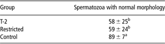
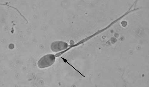
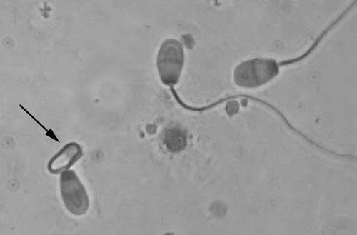
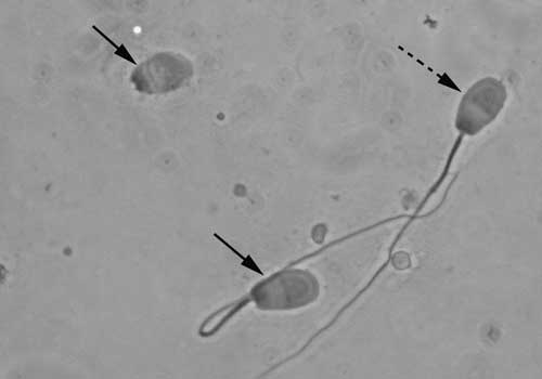
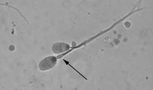
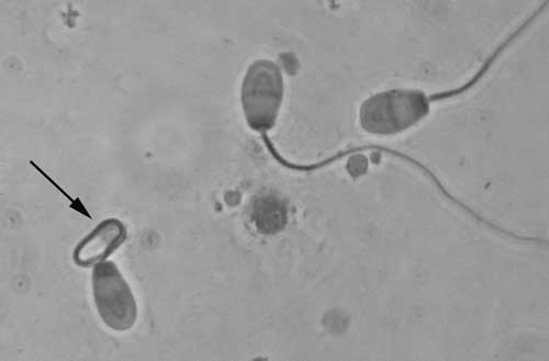
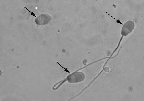
No effect of toxin treatment was detected on pH (ranging from 6.9 to 7.2 in each group), quantity of the semen (on average 1 ml in each group), and concentration of spermatozoa (ranging from 245 to 263 × 106/ml). The ratio of spermatozoa showing progressive forward motility decreased from 65% to 53% in the semen samples of toxin-treated animals compared to controls; however, this difference proved to be statistically not significant (P > 0.05). The ratio of spermatozoa with abnormal morphology increased, whereas that of the normal morphology decreased (P < 0.05) in the ejaculates collected from the toxin-treated and restricted animals (Table 3). The most frequent morphological abnormalities were: abnormality of the tail (60%), retention of cytoplasmic drop, absence of the acrosome and altered sperm head (Figures 1–3). A sperm cell was considered altered if at least one defect was present.
The ratio of spermatozoa with normal morphology in the semen 48 days after toxin treatment (%, mean ± s.d.)
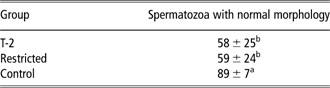
Numbers with different superscripts indicate significant differences (P < 0.05).
Rabbit spermatozoa with proximal retention of cytoplasmic drop.
Rabbit spermatozoa with twisted tail.
Rabbit spermatozoa with separated head, rolled tail (continuous arrow), and a normal spermatozoa (broken arrow).
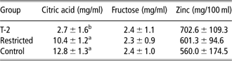
T-2 toxin applied in high doses decreased the concentration of citric acid in seminal plasma (P < 0.05). No effect of T-2 toxin on the concentrations of the other seminal plasma parameters (fructose and zinc) was observed (Table 4).
Concentration of citric acid, fructose and zinc in the seminal plasma 48 days after toxin treatment (mean ± s.d.)
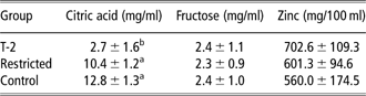
Numbers with different superscripts indicate significant differences (P < 0.05).
Testosterone production
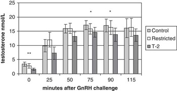
T-2 toxin decreased the basic testosterone level by 45% compared to control (P < 0.01) and resulted in lower (P < 0.05) GnRH-induced testosterone concentration (Figure 4). However, there was a ninefold increase in the hormone concentration 75 min after the GnRH injection in the toxin-treated animals compared to that of the controls, where a fivefold increase was observed.
Basic testosterone concentration and gonadotropin-releasing hormone (GnRH)-induced testosterone production (difference between control and T-2 groups, level of significance *P < 0.01, **P < 0.05).

Discussion
Acute effects of T-2 toxin occur after oral exposure to 0.06 to 10 mg/kg BW in various species. The effects observed include non-specific symptoms like weight loss, feed refusal, dermatitis, vomiting (cats, dogs, pigs and ducklings), diarrhoea, haemorrhage and necrosis of the epithelium of the stomach and intestine, bone marrow, spleen, testis and ovary (SCF, 2001). Rabbits can be considered to be rather sensitive to T-2 toxin, as reflected by the relatively low (1.1 mg/kg BW) LD50 values (Wannemacher and Wiener, Reference Wannemacher and Wiener1997), presumably due to the re-consumption of the toxin-containing caecal content by caecotrophy (Fekete et al., Reference Fekete, Tamas, Vanyi, Glávits and Bata1989b). The SCF (2002) has established a combined t-TDI (temporary tolerable daily intake) of 0.06 μg/kg BW per day for the sum of T-2 and HT-2. The T-2 toxin dose we applied was about 15 times higher compared to the TDI, near the LD50 value. No specific symptoms of the toxicosis were experienced, except feed refusal or the total lack of appetite (anorexia). The lack of dermatotoxic oral lesions on the palatinal mucosa, the tongue or the mouth – typical for T-2 effect – can be explained by the mode of toxin application, without any contact with the oral mucosa. The lesions and ulcers on the gastrointestinal epithelium found in the dead animals following acute exposure might be due to the direct cytotoxic effect (Fekete et al., Reference Fekete, Tamas, Vanyi, Glávits and Bata1989a). Pathological alterations in the liver, kidney, heart and lung, just like the feed-refusal effect, have been also described by several authors in rabbits (Niyo et al., Reference Niyo, Richard, Niyo and Tiffany1988; Glávits et al., Reference Glávits, Vanyi, Fekete and Tamas1989; Sándor and Ványi, Reference Sándor and Ványi1990) and/or in pigs and poultry (Sundstøl and Pettesson, Reference Sundstøl and Pettesson2004).
Trichothecenes are shown to alter the serotonin activity in the central nervous system. The inhibition of protein synthesis in the liver after T-2 exposure causes hyperaminoacidaemia, which leads to an increased brain uptake of tryptophan and serotonin synthesis, as a consequence. Serotonin receptors are thought to be important mediators in appetite regulation, and therefore an increased serotonin concentration is thought to be the reason for the decrease in or loss of appetite (Smith, Reference Smith1992). The lowest toxin concentration was 0.01 mg/kg BW in rabbits, which did not cause the feed-refusal phenomenon of T-2 (Fekete and Huszenicza, Reference Fekete and Huszenicza1993). However, the same concentration seemed to result in an impairment of the immune system, and an alteration in the liver, the kidney's glomerular function and the ovarian activity (Szilágyi et al., Reference Szilágyi, Fekete, Huszenicza and Albert1994).
Evaluation of sperm motility represents an important parameter because it is significantly correlated with the total number of offspring born, for example, fertility (Barnett et al., Reference Barnett, Tomlinson and Cooke1993; Krause, Reference Krause1995; Parkinson, Reference Parkinson2009). It is an accepted fact that normal ejaculates do contain a small percentage of spermatozoa that are abnormal in morphology. A semen sample containing a high percentage of abnormal sperm is indicative of impaired fertility (Kruger et al., Reference Kruger, Acosta, Simmons, Swanson, Matta and Oehninger1988; Gillan et al., Reference Gillan, Kroetsch, Maxwell and Evans2008; Parkinson, Reference Parkinson2009). According to our results, a high concentration of T-2 resulted in decreased motility and an increase in the ratio of morphologically abnormal cells, even 48 days after the exposure. A direct toxicity (cytotoxic effect) of T-2 toxin on the spermatogenic compartment of testis may be considered as one of the mechanisms of action of T-2 toxin in producing the abnormal spermatozoa, since trichothecenes are toxic for actively dividing cells. Cytotoxicity has been associated with either the impairment of protein synthesis by the binding of compounds to the ribosomes of eukaryotic cells, or the dysfunction of cellular membranes (WHO, 1990). The subsequent effect of the toxin (e.g. morphological abnormalities still on day 48 after toxin withdrawal) was probably attributable to the observations of Sprando et al. (Reference Sprando, Collins, Black, Olejnik, Rorie, Eppley and Ruggles2005), according to whom the cell types primarily affected by DON exposure were the pachytene spermatocytes, secondary spermatocytes, rounded spermatids and elongating spermatids, which, according to Ettlin et al. (Reference Ettlin, Bechter, Lee and Hodel1984), must have been exposed to the toxin 28 days prior to the time of tissue fixation and examination in rats.
In contrast with motility, sperm morphology was negatively influenced by feed restriction in our experiment. Severe feed restriction and weight reduction (50% to 60% of control values) may sharply impair rat reproduction. However, when adult Sprague–Dawley rats were fed a restricted diet and maintained at 90%, 80% and 70% of control BW for up to 17 weeks, fertility was not affected (Chapin et al., Reference Chapin, Gulati, Barnes and Teague1993). Feed restriction may result in reproductive disturbances in rabbits as well (Assane et al., Reference Assane, Katiellou, Sere, Gongnet and Missohou1995; Brecchia et al., Reference Brecchia, Bonanno, Galeati, Federici, Maranesi, Gobbetti, Zerani and Boiti2006). In our experiment, feed restriction caused only a slight (2% to 4%) decrease in BW, which can hardly explain the reduction in the ratio of cells with normal morphology. None of the other parameters examined confirmed the negative effect of the decreased nutrient intake.
Decreased motility in the toxin-treated group can be the result of the impaired epididymal function as well, that is, maturation of spermatozoa, leading to an impairment of sperm motility (Yeung, Reference Yeung1995; Parkinson, Reference Parkinson2009). As a general principle, a decrease in the concentration of the biochemical components of the seminal plasma produced by the accessory male organs indicates a dysfunction of the secreting organs (Cooper and Yeung, Reference Cooper and Yeung2000). The alteration of seminal plasma, namely the significantly lower concentration of citric acid found after toxin treatment, may also provide a sub-optimal environment for spermatozoa causing decreased motility (Cooper and Yeung, Reference Cooper and Yeung2000). All these effects could be detrimentally influenced by the lowered testosterone production, since androgens are known to play a pivotal role in the regulation of spermatogenesis (de Kretser and Kerr, Reference de Kretser and Kerr1994).
It is known that the blood-testis barrier is very selective and protects the organ from most of the toxic agents (Steinberger and Klinefelter, Reference Steinberger and Klinefelter1993). The trichothecenes are tetracyclic sesquiterpenoid compounds with a 12,13-epoxy group (SCF, 2002). Whether the toxin can pass through the blood-testis barrier or not has not been proven.
Nikodémusz and Mézes (Reference Nikodémusz and Mézes1992) observed that GnRH injection induced significantly lower testosterone production in ganders treated with T-2 toxin. Our result obtained in male rabbits is very similar since we found that T-2 toxin applied in high doses induced acute functional disturbances in Leydig cells, indicated by the decreased basic testosterone level, which resulted in a hypo-androgenic status. The possible explanation for this is that the T-2 toxin decreased testosterone production in Leydig cells, presumably by inhibiting early steps of the steroidogenic pathway, that is, the conversion of pregnenolone to progesterone (Fenske and Fink-Gremmels, Reference Fenske and Fink-Gremmels1990).
An other explanation can be that T-2 acts directly on the pituitary gland, or affects the ability of Sertolli cells to produce inhibin, as has been supposed in the case of DON (Sprando et al., Reference Sprando, Collins, Black, Olejnik, Rorie, Eppley and Ruggles2005). Disturbance in the function of Sertolli cells could be a reason for the increase in the ratio of cells with abnormal morphology as well. The Sertolli cell is an active participant in releasing sperms into the seminiferous tubule lumen and retaining defective spermatids. Failure of sperm release has been associated with a direct effect of various xenobiotics on Sertolli cells, a loss of stimulation of the testis and the selective removal of morphologically abnormal cells (Sprando et al., Reference Sprando, Collins, Black, Olejnik, Rorie, Eppley and Ruggles2005).
Since the male reproductive organs (testis, epididymis, accessory male organs, etc.) are strongly androgen-dependent organs in respect of structure as well as function and testosterone supports spermatogenesis, sperm maturation, seminal plasma production and sexual functions, the disruption of testosterone biosynthesis in Leydig cells can adversely affect male fertility (Weinbauer et al., Reference Weinbauer, Gromoll, Simoni and Nieschlag2000; Parkinson, Reference Parkinson2009).
In summary, the subsequent effect of T-2 toxin applied in high doses manifested in a decrease in sperm motility, an increase in the number of spermatozoa with abnormalities, an alteration of the composition of seminal plasma indicating a large drop in the concentration of citric acid, and a decrease in the basic testosterone level even after 48 days following a 3-day long acute toxicosis. All of these changes by themselves or in combination may result in decreased fertility (subfertility) or infertility in males. Further studies are needed to ascertain the specific mechanisms of action of T-2 toxin on function of male reproductive organs (spermatogenesis, maturation of spermatozoa, seminal plasma production, etc.) and semen quality.
Feed restriction, that is, less nutrient intake, resulted in more morphologically abnormal spermatozoa in the semen, but it did not cause significant loss in BW, motility of the spermatozoa, composition of the seminal plasma or testosterone concentration – its effect need further examination.
Acknowledgement
The research was funded by the Hungarian Academy of Sciences and the TéT foundation (project no. ZA-16/2008).