I. INTRODUCTION
Recent research efforts on the growth of diluted magnetic semiconductors (DMS) exhibiting Curie temperatures above 300 K have received a great attention due to their potential applications in spintronics devices.Reference Ohno1 Following the theoretical prediction of a possible room temperature ferromagnetism (RTFM) in Mn-doped ZnO,Reference Dietl2 studies on transition metal (TM)-doped ZnO system have intensified. RTFM has been reported in Co-,Reference Ramachandran, Tiwari and Narayan3 Mn-, Cr-, and NiReference Ueda, Tabata and Kawai4-doped ZnO systems. However, similar studies on Co- and Mn-doped polycrystalline ZnOReference Rao and Deepak5 and high quality ZnO:CoReference Kaspar, Droubay, Heald, Nachimuthu, Wang, Shutthanandan, Johnson, Gamelin and Chambers6 thin films grown by pulsed laser deposition (PLD) have not shown any ferromagnetism. Surprisingly, x-ray magnetic circular dichroism studies on Co-doped samples by Gacic et al.Reference Gacic, Jakob, Herbort, Adrian, Tietze, Brück and Goering7 showed that magnetism is not induced by the TM dopant atoms, either in solid solution or in the form of clusters or secondary phases. These conflicting results have generated controversy on the origin of observed ferromagnetism and raised questions about the role of dopants. Finally, the observation of RTFM in undoped HfO2Reference Venkatesan, Fitzgerald and Coey8 has challenged the understanding of magnetism and triggered researchers to revisit the role of dopants in controlling the magnetism of semiconducting and insulating oxides. Since then this so-called d 0 ferromagnetism has been observed in other systems as well, such as TiO2, In2O3,Reference Hong, Sakai, Poirot and Brize9 and nonstoichiometric CaB6.Reference Coey10 In case of TiO2 and HfO2 systems, the ferromagnetism has been attributed to the presence of oxygen vacancies (VO).Reference Kim, Hong, Park and Kim11 RTFM has also been reported in undoped ZnO powders, nanoparticles, nanorods, nanowires, and thin films.Reference Sudaresan, Bhargavi, Rangarajan, Siddesh and Rao12–Reference Kapilashrami, Xu, Strom, Rao and Belova15
An important but often overlooked aspect of the controversy regarding the observation of ferromagnetism in TM-doped ZnO is related to the role of intrinsic defects, metal precipitates, and TM–defect complexes.Reference Narayan, Nori, Pandya, Avasthi and Smirnov16 However, the ferromagnetism in undoped ZnO is linked to the poor crystalline quality, strain, grain boundaries, and intrinsic defects, namely, VO, zinc vacancies (VZn), and zinc interstitials (IZn). Recent ab initio calculations have suggested that the magnetic moment arises from the unpaired 2p electrons at the O sites surrounding the VZn with each nearest neighbor O atom carrying a magnetic moment ranging from 0.49 to 0.74 μB.Reference Wang, Sun, Chen, Kawazoe and Jena17 Recently, RTFM was induced in undoped ZnO powder by applying a mechanical forceReference Potzger, Zhou, Grenzer, Helm and Fassbender18 and then destroyed upon oxygen annealing. It was suggested in this case that the onset of FM had its origin in the unpaired spin states generated by mechanical strain and/or dislocations at the crystalline domain boundaries.
In our experience, point defects such as VO, VZn, and IZn are always present in the ZnO films grown by PLD and that these intrinsic defects play an important role in tailoring the physical properties of the films. We find that undoped ZnO films can be tailored to be ferromagnetic by invoking nonstoichiometric processing conditions, such as low oxygen partial pressure (pO2) growth, vacuum annealing, and laser irradiation, and further that the magnetization is destroyed upon high temperature oxygen annealing. These observations strongly suggest that the intrinsic point defects play an important role in introducing and controlling ferromagnetism in nominally pure ZnO. The primary focus of this study is to grow high-quality epitaxial single crystal films of pure ZnO on c-sapphire and to investigate their ferromagnetic and concomitant electrical and structural properties as a function of pulsed laser irradiation and thermal annealing in different environments. It is shown that these treatments can controllably vary the magnetic and electrical properties of ZnO films, which we infer to be associated with the introduction of defects, particularly Vo and VZn within the oxide matrix. Here, we report a detailed study of the electrical and magnetic properties of the films grown under different deposition conditions that were further subjected to different postgrowth annealing treatments. With the aid of Hall and electron paramagnetic resonance (EPR) data, the role of defects in modifying the magnetic properties of ZnO films has been illustrated and correlated with the electrical and other structural properties.
II. EXPERIMENTAL DETAILS
ZnO thin films have been deposited on single crystal c-plane sapphire substrates using PLD. A KrF excimer laser (λ = 248 nm, τ = 25 ns) with a pulse energy density of 3–4 J/cm2 at a repetition rate of 10 Hz was used to deposit thin films. The target–substrate distance was maintained at 4.5 cm during the film deposition. A high purity (5N) ZnO target was prepared using the conventional solid state reaction technique. Special care was taken to avoid any TM contamination, e.g., nonmagnetic plastic tweezers were used throughout the sample growth and characterization processes. A series of thin films were deposited for 15 min at substrate temperatures (T dep) ranging from room temperature to 750 °C and under different pO2 ranging from 10−5 to 1 Torr. The deposition chamber was evacuated to a base pressure of less than 1 × 10⁻⁶ Torr before the introduction of oxygen. Postdeposition annealing was carried out for 30 min in a high purity oxygen atmosphere at different annealing temperatures from 200 to 800 °C. Vacuum annealing was performed at 10−5 Torr for 3 h at 800 °C. Laser irradiation of films was carried out at room temperature in ambient air with a laser energy density of 0.2 J/cm2 and a frequency of 1 Hz for different numbers of pulses. The laser energy was kept below the threshold for melting, which was easily identified by the formation of cracks in the film. The structure of these films was characterized by x-ray diffraction (XRD) using a x-ray diffractometer (Rigaku Corporation, Tokyo, Japan) with Cu Kα radiation (λ = 0.154 nm) and a Ni filter. Microstructural characterization was carried out using a JEOL-2010 field emission transmission electron microscope (TEM; JEOL, Tokyo, Japan) equipped with a Gatan image filter tuning attachment (Gatan, Pleasanton, CA). The samples were examined by secondary ion mass spectrometer (SIMS) and an electron energy loss spectrometer (EELS) to determine their chemical composition and to assess for the presence of any unintended impurities. X-ray photoelectron spectroscopy (XPS) was performed using a LAS-3000 instrument (Riber, Rueil-Malmaison Cedex, France) with an Mg Kα x-ray source having an energy resolution of 0.9–1.0 eV. Room temperature electrical resistivity measurements were carried out using a four-point probe method, and Hall measurements were performed using an Ecopia Hall measurement system. Magnetic measurements were carried out using a vibrating sample magnetometer in conjunction with a physical property measurement system (Quantum Design Inc., San Diego, CA) and an alternating gradient magnetometer (Princeton Measurements Inc., Princeton, NJ). X-band (∼9.5 GHz) EPR spectroscopy measurements in the 4–300 K temperature range were carried out with an ER-200 spectrometer (Bruker, Santa Barbara, CA) equipped with a helium flow cryostat (Oxford Instruments, Concord, MA). The experimental spectra were least squares simulated using Lorentzian line shape models with the help of the EWVOIGT program.Reference Smirnov, Smirnova, Berliner and Bender19
III. RESULTS AND DISCUSSION
A. Structural characterization
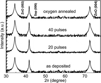
In these studies, the crystalline nature of the ZnO samples remained constant throughout the study. For example, Fig. 1 shows XRD characterization (2θ—intensity scan) of as-deposited (T dep = 750 °C, pO2 = 10−5 Torr) and laser-irradiated ZnO thin films with 20 and 40 pulses of laser output energy density of 200 mJ/cm2. The XRD pattern shows that the films are highly textured or epitaxial with a c-axis preferred orientation. No substantial change in (002) diffraction peak intensity and full width at half maximum (FWHM) was observed in the laser-irradiated ZnO thin films, indicating good crystalline quality of the films even after irradiation with 40 laser pulses. Within the detection limit of the XRD apparatus, there was no evidence of precipitation or impurity clustering in the films following laser irradiation.
Room temperature x-ray diffraction pattern of as-deposited, laser-irradiated, and oxygen-annealed films.

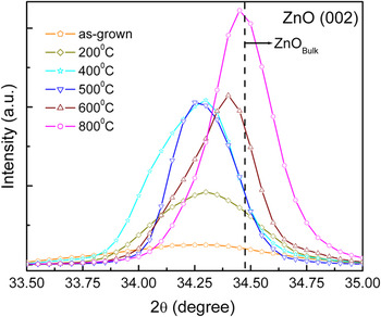
Figure 2 displays the XRD patterns of the ZnO (002) peak for the as-grown (T dep = 350 °C, pO2 = 10−6 Torr) and the oxygen-annealed (up to 800 °C for 30 min.) films. In the annealed sample, the angular position of ZnO (002) peak shifts toward 2θ = 34.44° (ZnObulk), indicating the relaxation of stress that occurs in high temperature oxygen-annealed samples. The FWHM decreases nearly linearly with increasing annealing temperature. Above 500 °C, the “c” lattice parameter of ZnO decreases with increasing temperature as revealed by the shift of ZnO (002) peak toward higher values. We believe that significant oxygen diffusion occurs in these samples above ∼500 °C.Reference Sudhakar, Kharel, Lawes, Suryanarayanan, Naik and Naik20 High temperature thermal annealing in oxygen environment leads to higher atomic mobility and oxygen diffusion into the film and hence to a reduction in the concentration of intrinsic point defects (VO and IZn) and compressive stress originating from them. To summarize, thermal annealing in oxygen atmosphere leads to (i) an increase in intensity of ZnO (002) reflection, (ii) decrease in FWHM, and (iii) relaxation of intrinsic residual stress.
ZnO (002) peak position of the films annealed in oxygen atmosphere at different temperatures.

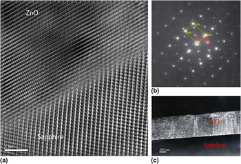
A detailed TEM study was performed to investigate the microstructure and verify the epitaxial relationship of the as-grown and laser-irradiated films. The low magnification bright-field TEM cross-section micrograph revealed the absence of any precipitates or clustering in the irradiated films (figure not shown). The high-resolution TEM micrograph [Fig. 3(a)], which is representative of the entire sample, reveals the highly epitaxial nature of the film and the atomically sharp interface with no evidence of any interfacial reaction or precipitation. The thickness of the film was determined to be around 650 nm. Both the fast Fourier transformation and the selected area diffraction pattern [Fig. 3(b)] show spots corresponding exclusively to the film and substrate. The detailed XRD and the TEM analyses suggest that laser irradiation with 40 pulses did not alter the crystalline quality substantially or result in precipitation and cluster formation in the ZnO films.
(a) Fourier-filtered high-resolution transmission electron microscope image of laser-irradiated ZnO (tilted to [ ] zone) showing highly epitaxial film on sapphire with an atomically sharp interface. (b) Indexed selected area diffraction pattern of film/substrate interface. (c) Bright-field image of the film showing the film thickness.

B. Magnetic and transport studies
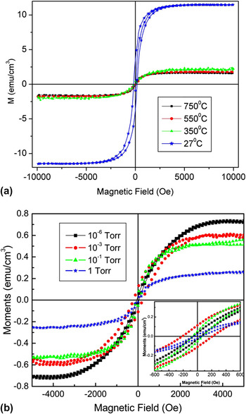
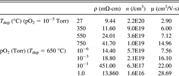
Magnetic and transport measurements were performed on as-grown ZnO thin film samples prepared under different growth conditions and annealed in different atmosphere. Figure 4 shows the room temperature magnetization data plotted as a function of the applied magnetic field for ZnO films grown (i) at different T dep and (ii) under different pO2. It can be seen from Fig. 4 that saturation magnetization (Ms) decreases with an increase in the T dep and pO2. ZnO films grown in different pO2 and at different T dep showed coercivity values in the range of 59–219 Oe; the highest coercivity (∼219 Oe) was observed in the films grown at 650 °C and 10−3 Torr pO2. Table I summarizes the important parameters, such as, ρ (mΩ-cm), n (/cm3), and μ (cm2/V·s) of the samples grown at different T dep and different pO2. Polycrystalline ZnO films grown at room temperature and pO2 of 10−6 Torr had the highest carrier concentration of ∼2.2 × 1020 cm−3. It is clear from the dependency of n on both T dep and pO2 that lowering the T dep is a more effective way of achieving higher n-type carrier concentrations. Resistivity gradually increases from 9.44 to 41.7 mΩ-cm as the T dep is increased from 27 to 750 °C. It also increases by three orders of magnitude (from 14.4 to 13,860 mΩ-cm) as the pO2 increases from 10−6 to 1 Torr, suggesting a strong dependence on pO2.
Room temperature M–H hysteresis curves of ZnO films grown at (a) different substrate temperatures under 10−5 Torr; (b) different oxygen partial pressures (pO2) at 650 °C.

Resistivity, carrier concentrations, and mobility of ZnO films grown at different T dep under 10−5 Torr pO2 and under different pO2 at 650 °C.

pO2, oxygen partial pressure.
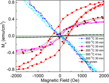
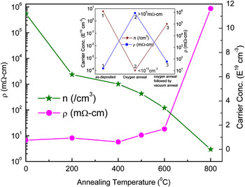
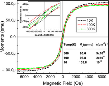
Figure 5 demonstrates the hysteresis (M–H) loops of the ZnO films annealed at different temperatures under oxygen atmosphere (760 Torr). It is clear from the plot that there is a gradual reduction in the magnetization value from ∼1 emu/cm3 (as-grown) as the annealing temperature is increased from 200 to 800 °C. As expected, longer anneals result in lower Ms values, e.g., the Ms for the sample annealed at 500 °C for 4 h is lower than the sample annealed for 30 min. Finally, when the films were annealed at temperatures 600 °C or higher they turn out to be diamagnetic. Figure 6 shows plots of carrier concentration and resistivity of ZnO films versus the annealing temperature. Annealing at up to 600 °C results in a modest change in the electrical properties (e.g., n decreased to ∼3.42 × 1019cm−3 and ρ increased to ∼18.3 mΩ-cm), whereas a dramatic change (n ∼1015 and ρ ∼870 Ω-cm) is observed after annealing at 800 °C. This change in electrical properties is consistent with the shift of the (002) ZnO peak toward a higher angle, as revealed in the XRD patterns (Fig. 2). It is important to note that films annealed at 600 °C for 30 min still exhibit a high carrier concentration of ∼3.4 × 1019 cm−3, although no ferromagnetism was observed in these films. Likewise, the temperature-dependent Hall measurementsReference Roro, Kassier, Dangbegnon, Sivaraya, Westraadt, Neethling, Leitch and Botha21 on an as-grown sample (T dep = 380 °C and pO2 = 200 Torr) show that the carrier concentration drops from ∼9 × 1017 cm−3 (300 K) to ∼1015 cm−3 (10 K), whereas Ms of undoped ZnO (T dep = 350 °C and pO2 = 10−6 Torr) increases only marginally with the decrease in temperature (Fig. 7), as is typical of ferromagnetic behavior. The lack of direct correlation between carrier concentration and magnetic moment leads us to conclude that carrier-mediated ferromagnetism does not appear to be the active exchange mechanism operating in the undoped ZnO system.
M–H plot of as-grown (T dep = 650 °C, pO2 = 10−5 Torr) and oxygen-annealed samples at different temperatures.

Carrier concentrations and resistivity of ZnO films annealed at different temperatures in 760 Torr oxygen atmosphere. Inset represents a schematic of the “on” and “off” states of the samples.

M–H hysteresis loops of as-grown sample (T dep = 350 °C, pO2 = 10−5 Torr) measured at different temperatures in the range of 300–10 K. [*data (n) has been taken from Ref. Reference Roro, Kassier, Dangbegnon, Sivaraya, Westraadt, Neethling, Leitch and Botha21]

The absence of any correlation between carrier concentration and magnetic moment was clearly demonstrated in some of our earlier studies where we had examined the RTFM of (Cu, Al):ZnOReference Chakraborti, Trichy, Narayan, Prater and Kumar22 systems. All the epitaxial-doped ZnO films showed ferromagnetic ordering at room temperature. The moment increased with increasing Cu concentration up to 5%. The 2% Cu sample had a total moment of 7 emu/cm3; 5% Cu sample had a total moment of 14 emu/cm3; and the 10% had a total moment of 10.5 emu/cm3. The drop in moment at highest Cu concentration could be due to an increased occurrence of antiferromagnetic coupling between Cu pairs occurring at shorter separation distances, which had been predicted in earlier theoretical studies.Reference Ye, Freeman and Delley23 The presence of Cu ions in ZnO increases the resistivity up to three orders. The resistivity of undoped ZnO is about 10–20 mΩ-cm and that of the Zn1−xCuxO films is of the order of 10 Ω-cm. Codoping the Cu:ZnO thin films with Al, an extrinsic donor, then leads to a large increase in the free carrier concentration. Al in +3 state behaves as a donor in ZnO and hence introduces carriers in the form of electrons. For example, for the 2% Cu-doped ZnO samples, the room temperature Hall measurements show that the electron concentration can be increased from 1.0 × 10¹⁷ to 1.5 × 10²⁰ by increasing Al concentration up to 5% Al. Further increase in Al content leads to a decrease in both sample conductivity and carrier density. Interestingly, an increase in carrier density of three orders of magnitude to 1020 cm−3 does not improve the ferromagnetism. Even though the role of free carriers cannot be entirely ruled out as the 2% Cu-doped ZnO still has a carrier concentration of 1017 cm−3, defects associated with oxygen-deficient compositions (e.g., VO) appear to be having a much greater effect in mediating ferromagnetic ordering in this ZnO-based DMS system. This further reinforces our belief that a free carrier-mediated mechanism (e.g., Ruderman-Kittel-Kasuya-Yosida (RKKY) or double exchange) is not a plausible explanation for ferromagnetism in ZnO-based DMS materials, and that a defect-mediated mechanism needs to be invoked.
C. Reversible ferromagnetism in ZnO
1. Annealing treatment
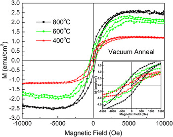
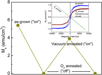
The effect of vacuum annealing on RTFM was further explored to extend our investigation to understand the role of defects in controlling the ferromagnetism in our undoped ZnO films. As-grown ZnO films were annealed in vacuum (10−5 Torr) at different temperatures for 3 h. As shown in Fig. 8, the Ms gradually increases when the annealing temperature is increased. Figure 9 shows a schematic of the reversible room temperature ferromagnetic ordering in the samples. The inset shows the room temperature hysteresis loops of the samples. As illustrated by this figure, RTFM can be controlled to switch between the “on” and “off” states reproducibly. For example, the RTFM found in as-grown films represents an “on” state that can be converted to the “off” (diamagnetic) state by oxygen annealing at 800 °C for 30 min. Finally, the “on” state can be restored by further vacuum annealing of the previously oxygen-annealed samples. It is important to point out here that the same sample has been used in the oxygen annealing and further vacuum annealing experiments and that the results have been reproduced multiple times. Vacuum annealing results in an increase in the carrier concentration from ∼1015 to ∼5 × 10¹⁸ and a decrease in ρ from ∼870 to ∼60 mΩ-cm. This is likely to be attributed to incorporation of the shallow donor level induced by VO and/or VO–Izn pair. It may be recalled here that the inset of Fig. 6 depicts the three states of samples. While “2” represents the “off” state (oxygen annealed at 800 °C and 760 Torr for 30 min), “1” and “3” represent the “on” state (as-grown at 550 °C and 10−5 Torr and oxygen annealing at 800 °C followed by vacuum annealing at 850 °C, respectively) of the ZnO films.
Room temperature magnetization curves of the vacuum (10−5 Torr)-annealed samples at different temperatures.

Schematic plot of the “on” and “off” magnetic states of as-grown, oxygen-annealed, and oxygen annealed followed by vacuum-annealed ZnO films. The inset shows the room temperature hysteresis loops of the same samples.

The oxygen content in the films has been characterized by XPS studies. An XPS survey spectra (5–1200 eV) and high-resolution scan for Zn and O were performed for the as-grown (T dep = 650 °C, pO2 = 10−5 Torr) and oxygen-annealed (800 °C, 760 Torr, 30 min) samples. The samples were etched by 5 kV Ar+ ion bombardment for 10 min before the survey scan. The C 1s peak at 285.0 eV was used for calibration. The binding energy of 2p3/2 (∼1022 eV) and 2p1/2 (∼1045 eV) levels of Zn corresponding to Zn–O bonding exhibited very similar features before and after oxygen annealing. Figure 10 shows the high-resolution scan of O 1s peak. The peak at ∼530 eV has been attributed previously to O2− ions in the stoichiometric wurtzite structure.Reference Cebulla, Weridt and Ellmer24, Reference Rao and Vinni25 The higher binding energy at ∼531.5 eV is associated with O2− ions in oxygen-deficient regions. An electron charge density in the region of a VO is reduced, which results in less screening of the O2−:1s electrons from their nucleus, thus raising the effective nuclear charge and hence binding energy of an O2−:1s electron.Reference Fan and Goodenough26 Therefore, the intensity of this component of O 1s peak is associated with the concentration of VO. It can be clearly seen from Fig. 10 that the intensity of 531.5 eV O 1s peak is significantly reduced in the oxygen-annealed films. Thus, oxygen annealing at a high temperature (800 °C, 30 min) promotes the diffusion of oxygen into the film and leads to a reduction in the concentration of VO, resulting in a more stoichiometric film.
X-ray photoelectron spectroscopy high-resolution scan of oxygen 1s peak for as-deposited (T dep = 650 °C, pO2 = 10−5 Torr) and oxygen-annealed (at 760 Torr, 30 min) films. The inset shows the room temperature magnetization curve of oxygen-annealed and oxygen annealed followed by laser-irradiated samples.

2. Laser irradiation treatment
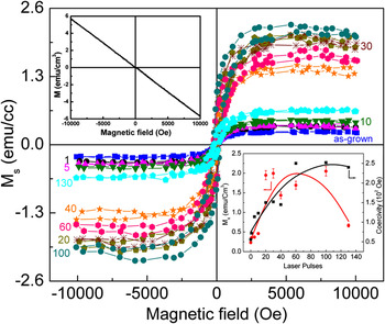
We have also investigated the role of low energy pulsed laser irradiation on magnetic properties. Magnetic measurements were performed for all the films in the temperature range 100–300 K and in magnetic fields up to 10 kOe. Figure 11 shows isothermal magnetization data plotted as a function of the applied magnetic field for both the as-deposited and the laser-irradiated films exposed to a series of laser pulses. The diamagnetic contribution from the sapphire substrate has been subtracted using the high-field slope between 5 and 10 kOe. Films deposited at high temperature (750 °C) and under high oxygen pressure of 10−2 Torr show a weak ferromagnetic coupling with a small coercive field, Hc (∼56 Oe), and a small Ms (0.2308 emu/cm3). As shown in the lower right inset of Fig. 11, the Ms of the films increases following laser irradiation due to the simultaneous formation of several defects, such as VZn, VO, and their interstitials. Both Ms and Hc increase with the increase in number of laser pulses up to ∼30 pulses where the Ms saturates while the coercivity continues to increase up to 130 pulses. A plateau or possible decrease in Ms was observed beyond 100 pulses, which could be attributed to the clustering of newly generated defects accompanied by a concomitant decrease in defect concentrations. Once the defects are in the form of clusters such as dislocation loop, they may lose their magnetic contribution. The Ms, however, increases only marginally with decreasing temperature down to 100 K, suggesting that the Curie temperature is well above 300 K. The magnetization for all the samples saturates at fields of ∼3000 Oe. These ferromagnetic ZnO films switched to a perfect diamagnetic state after annealing in oxygen ambient for 20 min at 800 °C, as shown in the top left inset of Fig. 11. Ferromagnetic ordering could again be restored in the oxygen-annealed samples, but only after laser irradiation with 40 pulses. The inset of Fig. 10 shows the room temperature magnetization curve for an oxygen-annealed sample and an oxygen-annealed sample that had then been laser irradiated. It should be pointed out that after irradiating the sample with 20 pulses the magnetization curve appeared very noisy and weak, but 40 laser pulses gave rise to a saturation moment of 0.42 emu/cm3, which is higher than the 0.3 emu/cm3 moment of the as-deposited film. The laser irradiation treatment also restored the carrier concentration in oxygen-annealed samples. After an O2 anneal at 800 °C for 20 min, the carrier concentration had dropped significantly to ∼1014/cm3, but then rises to ∼1019/cm3 after an exposure to 40 laser pulses.
Room temperature magnetization curves of pure ZnO films irradiated with different number of laser pulses, viz., as-grown, 1, 5, 10, 20, 30, 40, 60, 100, and 130 at 200 mJ/cm2. Inset (top left) shows diamagnetic behavior of laser-irradiated samples after annealing in O2 at 800 °C. Inset (bottom right) is the variation of room temperature saturation magnetic moment and coercivity with laser pulses. The solid (red) line represents the generic trend of the variation of magnetization as a function of laser pulses.

D. Defect characterization and phenomenological model
In our SIMS and EELS studies, we ruled out the possibility of ferromagnetic contamination as being responsible for the observed RTFM. Figure 12 shows the EELS spectra of the as-grown sample (T dep = 650 °C, pO2 = 10−5 Torr) in the range of 400–1300 eV. In the higher energy loss region, the only observed edges are that of the oxygen K-edge at ∼532 eV and the zinc L2,3-edges at ∼1020 and 1043 eV, respectively. No magnetic impurity was found within the detection limit of the EELS. The inset of Fig. 12 shows the depth profile of impurity concentration of as-grown ZnO films. Upon comparing with the standards, we found that the Fe concentration was about 1 ppm and that the Ni, Co, Mn concentrations were lower than that of Fe. It is important to note that EELS and SIMS studies revealed similar impurity levels in the oxygen-annealed and laser-irradiated samples. The lowest saturation moment of 0.2308 emu/cm3 was observed in the film grown at 750 °C in 10−2 Torr pO2. This moment would correspond to a concentration of 4.1 × 10¹⁸ atoms/cm3 of a ferromagnetic impurity having a moment of 6 μB/atom. SIMS study revealed that Fe contamination was at a concentration level of 1 ppm, which is two orders of magnitude below the above critical value. Oxygen-annealed samples also had the same amount of Fe contamination, but they did not show any ferromagnetism down to 10 K. CoeyReference Coey27 pointed out that iron could be diffusing from the substrate holder into the sapphire at high temperature (700 °C) and forming ferromagnetic nanoparticle precipitates, giving rise to RTFM. In our case, a very high saturation moment (11.5 emu/cm3) was observed in the polycrystalline samples grown at room temperature, where one would not expect any diffusion of iron into the sapphire. In addition, the x-ray and TEM studies provided no evidence for the presence of magnetic precipitates in the ZnO films. These results clearly rule out the possibility of any external ferromagnetic contamination and establish the fact that the observed ferromagnetism must be defect mediated rather than impurity induced.
Electron energy loss spectrometer spectra of as-grown (T dep = 650 °C, pO2 = 10−5 Torr) ZnO film. Inset shows the secondary ion mass spectrometer depth profile of Co, Si, Mn, Fe, and Ni impurities in as-grown (T dep = 650 °C, pO2 = 10−5 Torr) ZnO films.

The nature of the defects responsible for conductivity and ferromagnetism in our undoped ZnO films can be established as follows. The creation and destruction of point defects in semiconducting oxide films are governed by a thermodynamic equilibrium in the postgrowth annealing. Annealing in oxygen atmosphere definitely reduces the population of VO, whereas annealing in vacuum (10−5 Torr) may create both VZn and VO. As mentioned earlier, the very high formation energy of IZn in n-type ZnOReference Janotti and Van de Walle28 (∼4 eV) prevents their formation in the vacuum (10−5 Torr)-annealed samples. Further, it is well known that VZn are acceptors and give rise to the p-type conductivity. Since our samples continue to show the n-type conductivity after vacuum annealing, we are inclined to suggest that the vacuum annealing creates VO that act as a source of the n-type conductivity and ferromagnetism (assuming singly charged VO). The nature of defects which causes the intrinsic n-type characteristics has been widely debated in the literature recently.Reference Kim and Park29–Reference Lany and Zunger31 VO, Zni, and their complexes are theoretically considered to be the sources of n-type conductivity in ZnO29, although only the VO–IZn complex is predicted to be present as a shallow donor under oxygen-deficient conditions. The highly mobile nature of oxygen and IZnReference Janotti and Van de Walle32 also suggests that the oxygen interstitials will migrate rather quickly to the surface and be emitted. Since laser irradiation was performed in ambient atmosphere, the IZn will also be oxidized as soon as they reach out to the surface. All the above-presented arguments suggest that the n-type behavior in our studies is a result of a VO–IZn complex formation.
To further understand the local environment of the point defects present in the as-grown and annealed ZnO films, we used x-band (∼9.5 GHz) EPR spectroscopy on a set of samples grown and treated under conditions. The experiments were carried out by applying external magnetic field H both parallel and perpendicular to the film c-axis in the temperature range of 4–300 K.
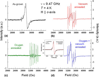
Figures 13(a)–13(d) show typical experimental EPR spectra (T = 4 K) of as-grown and annealed ZnO films in various environments for the H ‖ c-axis orientation. The observed spectral components for both H parallel and perpendicular to the c-axis are as follows. (i) A prominent broad peak (ΔH pp ∼80 G, g ∼2) was observed for all the samples except for the oxygen-annealed ZnO. This signal is attributed to the point defects in the ZnO films and here onwards it is referred to as the “ZnO signal.” (ii) Six sharp lines, overlapping the ZnO signal, correspond to the Mn2+ (with electronic spin S = 5/2 and nuclear spin I = 5/2) impurities. While the six lines correspond to the central |MS = −1/2〉→ |MS = +1/2〉 (ΔMI = 0) hyperfine transition, the less intense intermediary sets of the lines are due to the forbidden nuclear spin transitions, i.e., ΔMI = ±1. (iii) The hyperfine coupling constant A = 80 ± 2 Oe is same for the both film orientations studied, i.e., H ‖ and ⊥ to c-axis, indicating that the Mn2+ ions are in a high symmetry environment. (iv) The additional clusters of six lines flanking the intense central six lines correspond to the |MS = −3/2〉 → |MS = −1/2〉 and |MS = +1/2〉 → |MS = +3/2〉 (ΔMI = 0) transitions. The zero-field splitting parameter |D| can then be estimated as ∼225 ± 10 Oe. The observed D and A values for Mn2+ in our ZnO films are in agreement with other systems containing Mn2+ as either an impurity or a dopant.Reference Abragam and Bleaney33, Reference Jain and Lehmann34
Experimental x-band electron paramagnetic resonance spectra of ZnO films grown and treated under the conditions (a) As-grown, (b) annealed in vacuum, (c) annealed in the presence of oxygen, and (d) oxygen followed by vacuum annealing. The inset shows the experimental (black) and least square fit (red) spectra of as-grown ZnO shown in (a). The fit is done using EWVOIGT programReference Smirnov, Smirnova, Berliner and Bender19 with the parameters: Lorentzian line shape, g = 1.997 and ΔH pp = 78 G.

The surprising prominent varying intensities of the Mn2+ spectrum with respect to the ZnO signal in our samples could be due to a combination of various factors such as EPR cavity quality (Q) factor that was varied due to different dielectric losses in the partially conductive sample, microwave power at the sample site, and/or other changes in the experimental configuration. However, despite its prominence, the integrated area under the Mn2+ EPR peak is significantly smaller (by a factor of ∼103) than that of the ZnO signal in all the samples. The Mn2+ represents a minor impurity and the rest of the discussion will focus only on the ZnO signal.
Annealing of the as-grown films in presence of oxygen resulted in a complete disappearance of the ZnO EPR signal [Fig. 13(c)]. However, the ZnO signal reappears upon subsequent vacuum annealing of the films [Fig. 13(d)]. These observations are fully consistent with the “on” and “off” behavior observed in the magnetization and the changes in conductivity of the ZnO films. This indicates that the spins that are responsible for the magnetism in these ZnO films could be the same as those being directly observed by EPR.
The electronic g-factors and peak-to-peak line widths (ΔH pp) of the ZnO signal in all the samples studied were obtained by performing a nonlinear least squares fitting of the experimental spectra (see inset of Fig. 13) using the EWVOIGT programReference Smirnov, Smirnova, Berliner and Bender19 and a Lorentzian line shape model that includes a variable dispersion contribution. The admixture of dispersion was found to be essential for accounting for the observed line shape, which is attributed to a shift of the microwave phase within the conductive ZnO film (i.e., skin depth effects are responsible for the signal asymmetry that is known as a Dysonian line shapeReference Dyson35).
Comparison of the literature data on EPR of the single crystal ZnO with our EPR results is useful. But it must be emphasized that most of the reported EPR studies in the literature used single crystal ZnO samples either pristineReference Son, Ivanov, Kuznetsov, Svensson, Zhao, Willander, Morishita, Ohshima, Itoh, Isoya, Janzén and Yakimova36, Reference Wang, Vlasenko, Pearton, Chen and Buyanova37 or irradiated by high energy electrons.Reference Son, Ivanov, Kuznetsov, Svensson, Zhao, Willander, Morishita, Ohshima, Itoh, Isoya, Janzén and Yakimova36–Reference Taylor, Filipovich and Lindeberg43 In contrast, our samples were thin films (∼700-nm thick) and not subjected to any electron irradiation. Also, EPR spectra from our samples were observed without any in situ illumination by 325–515-nm light used in the earlier studies.Reference Son, Ivanov, Kuznetsov, Svensson, Zhao, Willander, Morishita, Ohshima, Itoh, Isoya, Janzén and Yakimova36–Reference Taylor, Filipovich and Lindeberg43 Even though the nature of our ZnO samples and the EPR experimental conditions were quite different than those of the previous studies,Reference Son, Ivanov, Kuznetsov, Svensson, Zhao, Willander, Morishita, Ohshima, Itoh, Isoya, Janzén and Yakimova36–Reference Taylor, Filipovich and Lindeberg43 it is worthwhile to highlight and compare the major features of the observed signals. First, for the ZnO films studied in this study both the dominating ZnO and the minor Mn2+ impurity signals vanished beyond the EPR detection limit at temperatures above 40 K. In contrast, the EPR signals from ZnO crystals have been reported at temperatures as high as 90 K.Reference Son, Ivanov, Kuznetsov, Svensson, Zhao, Willander, Morishita, Ohshima, Itoh, Isoya, Janzén and Yakimova36–Reference Taylor, Filipovich and Lindeberg43 Although this could be attributed to a higher spin count in larger crystals versus thin films, the differences in electronic relaxation mechanisms would be another possible explanation. Second, the ZnO EPR signals reported in the literatureReference Son, Ivanov, Kuznetsov, Svensson, Zhao, Willander, Morishita, Ohshima, Itoh, Isoya, Janzén and Yakimova36–Reference Taylor, Filipovich and Lindeberg43 were as narrow as 2–6 G, while ΔH pp of the EPR lines in our samples were 70–95 G. We speculate that the larger line width in our samples could be due to a combination of inherent strains (e.g., lattice mismatch with respect to the substrate) present in the film, a faster relaxation of the unpaired spins due to the interaction with the Mn2+ impurity, and finally additional magnetic interactions (both dipolar and spin exchange) with the different point defects.
The observed g values of the ZnO line observed in our studies of this film lie in between the reported g-values for VO (g iso ∼1.996)Reference Locker and Meese39–Reference Kappers, Gilliam, Evans, Halliburton and Giles42 and VZn (g iso ∼2.013)Reference Galland and Herve38, Reference Kappers, Gilliam, Evans, Halliburton and Giles42, Reference Taylor, Filipovich and Lindeberg43 in the ZnO crystals and, therefore, the exact nature of the point defects in the ZnO thin films cannot be ascertained by the EPR data alone. However, combining EPR results with the magnetization and conductivity data, we tentatively assign the observed ZnO peak, with g iso = 2.00–2.01 to the VO.
IV. CONCLUSIONS
In summary, we have systematically studied the structural, chemical, electrical, and magnetic properties of undoped ZnO thin films grown on c-sapphire by PLD under different growth conditions and annealed in different atmosphere. The magnetic properties of the nominally pure ZnO samples have been found to depend strongly on processing conditions and postgrowth annealing treatments. TEM studies have ruled out the presence of any magnetic precipitates and second phases as the source of the magnetism. SIMS and EELS analyses indicated that TM contamination (Fe and Mn with lesser amounts of Ni and Co) in the pure ZnO samples were below 10 ppm. We have found that ferromagnetism is always correlated with conditions that results in films that are substoichiometric in oxygen, while stoichiometric ZnO films always tend toward diamagnetism. Here, we have successfully demonstrated reproducible switching between “off” and “on” ferromagnetic states using oxygen annealing in combination with either laser irradiation or vacuum annealing treatments. High temperature annealing in oxygen brings the films to near-stoichiometric conditions and appears to relax the intrinsic residual stress resulting from the presence of point defects. A suspected decrease of VO upon oxygen annealing has been confirmed in our own XPS studies. Detailed room temperature Hall measurements reveal that the resistivity and carrier concentration of undoped films also vary with growth conditions and can be switched “off” and “on” by using high temperature oxygen annealing and further vacuum annealing, respectively. But, studies on the TM-doped samples have shown that magnetism does not correlate with neither resistivity nor carrier concentration. Three orders of magnitude variations in both resistivity and carrier concentration can be realized without significantly altering the magnetic properties of the sample. As already indicated, we have discovered that pulsed laser irradiation at low laser energy densities, just below the level where melting occurs, can be an effective way of introducing defects into ZnO that lead to RTFM. This would appear to provide a means of patterning ZnO surfaces with magnetic and diamagnetic features. The magnetic moment increases with the number of laser pulses up to about 40 hits and then saturates and/or begins to tail off. The laser-irradiated samples also show a significant increase in n-type electrical conductivity and an increase in free carrier concentration. The induced ferromagnetism can be reversed upon thermal annealing in pure oxygen. We believe that nanosecond laser irradiation introduces a large population of vacancies (particularly VO) and dangling bonds. These in turn introduce donor states that explain the increase in free carrier densities and unpaired electrons (e.g., electron spins in oxygen atoms in tetrahedron sites around VZn) that introduce localized magnetic moments.
EPR measurements indicate the presence of a broad peak at g = 2.01. This would be most consistent with the magnetic moment arising from the VO (g = 1.996), although the possible contribution from VZn (g = 2.013) cannot be entirely ruled out. We believe that the long-range magnetic exchange interaction that leads to the ferromagnetism is actually mediated by the VO, which form bound magnetic polarons that effectively couple nearby moments. The occurrence of ferromagnetism in some other transparent oxides has been explained by the above-mentioned mechanism.
All these results have clearly established that for ZnO there is a strong correlation between RTFM and presence of trapped nonequilibrium concentrations of defects (vacancies, dangling bonds, and other radiation-induced defects). It is concluded that there exist defect-assisted mechanisms that both introduce magnetic moments and lead to the occurrence of long-range ferromagnetic ordering in ZnO films at room temperature even in the absence of other 3d TM ion dopants. We have forwarded what would seem to be a plausible explanation for this observed behavior. But, we have not been able to conclusively identify the exact defect states that are producing the magnetism in ZnO. However, we have clearly captured the phenomenology and hope that this will help others in the future to finally identify the exact mechanisms that lead to magnetism in both pure and TM-doped ZnO films.
ACKNOWLEDGMENTS
The authors thank Prof. A.I. Smirnov and Dr. S. Nellutla for the EPR measurements. The authors are pleased to acknowledge the support of the Army Research Office under Grant No. W911NF-04-D-0003.