A 6-year-old boy recovering from an upper respiratory infection had an oxygen saturation of 85%, mild nail clubbing, and cyanosis. An echocardiogram showed mild hypoplasia of the tricuspid valve and right ventricle and an atrial septal defect with bidirectional flow.
A 36-year-old man presented with severe dizzy spells and palpitations. He had undergone brain surgery for a brain abscess 5 years ago and was previously diagnosed with “polycythemia”. He had moderate nail clubbing and saturations in the mid 80s. An echocardiogram showed an atrial septal defect with bidirectional flow.
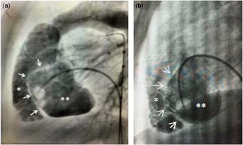
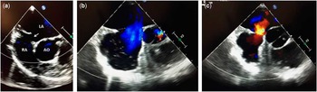
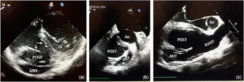
At catheterisation, in both patients, the right atrial and pulmonary arterial pressures were normal. A right ventriculogram showed a muscle bundle coursing from near apical towards the outflow tract, seen best on the lateral view (Fig 1). The posterior “chamber” was smooth-walled, and the anterior “chamber” was trabeculated. There was no gradient between the two “chambers”. Transoesophageal echocardiography revealed an atrial septal defect with bidirectional flow (Fig 2) and two right ventricular “chambers” (Fig 3). Temporary balloon occlusion of the atrial septal defect caused no increase in right atrial pressure. The atrial septal defect in each patient was closed with an Amplatzer septal occluder. Both patients were asymptomatic with saturations in the high 90s 6 months and 1 month later, respectively.
Lateral view right ventriculogram of patient 1 ( a ) and patient 2 ( b ) showing a double-chambered right ventricle (arrows show the dividing muscle bundle) with a smooth-walled posterior chamber (**) and a more trabechulated anterior chamber (*). There was no gradient between the chambers, no pulmonary valvar stenosis, no pulmonary hypertension, and no ventricular septal defect.

Transoesophageal images showing the atrial septal defect (arrow) in patient 1 ( a ) and bidirectional shunting (left-to-right, b , right-to-left, c ) in patient 2. AO=aortic valve in cross-section; LA=left atrium; RA=right atrium.

Transoesophageal images in the four-chamber view showing the double-chambered right ventricle with the tricuspid valve, which was mildly hypoplastic (arrows) and the two chambers in patient 1 ( a ). In patient 2, the two right ventricular chambers are shown in a short-axis view ( b ) and a right ventricular inflow-outflow view ( c ). ANT=anterior; AO=aorta; LV=left ventricle; POST=posterior; RVOT=right ventricular outflow tract.

Restivo et alReference Restivo, Cameron, Anderson and Allwork 1 described seven different morphological divisions of the right ventricle in double-chambered right ventricle. We describe an unusual anatomical variant and speculate that the muscle bundle altered the compliance of the right ventricle, resulting in right-to-left shunting at the atrial level.
Acknowledgements
None.
Financial Support
This research received no specific grant from any funding agency or from commercial or not-for-profit sectors.
Conflicts of Interest
None.
Ethical Standards
This study does not involve human and/or animal experimentation.