Implications
The Japanese quail (Coturnix japonica) belongs to the order Galliformes and the family Phasianidae. They have been used for meat and egg production since 1900. After World War II, the Japanese quail has become an important experimental animal, similar to the mouse and rat, for scientific research in universities. Recently, they have been accepted as a model animal for studying the relationship between the endocrine status and the reproductive behaviors. Not enough data are available about the relationship between serum estradiol-17β (E2) concentration and egg-laying performance of the Japanese quail. A few studies were conducted to measure the effect of intramuscular E2 benzoate injection on egg-laying performance, whereas the majority of other studies mainly focused on the effect of phytoestrogen (daidzein and genistein)-containing diet on the egg-laying performance of the Japanese quail, female chicken and ducks. Thus, this study will supply some data for the physiological knowledge about the effect of E2 on reproductive physiology of the Japanese quail.
Introduction
Estradiol (E2) is a main reproductive hormone affecting growth, development, maturation and functioning of reproductive tract, as well as the sexual differentiation and the behavior (Lien et al., Reference Lien, Cain and Forrest1985; Laugier et al., Reference Laugier, Courion, Pageaux, Fanidi, Dumas, Sandoz, Nemoz, Prigent and Pacheco1988; Balthazart et al., Reference Balthazart, Cornil, Charlier, Taziaux and Ball2009). It is an intraovarian factor affecting the function of hypothalamus, pituitary, liver, skeleton and calcium (Ca) homeostasis (Connolly and Callard, Reference Connolly and Callard1987; Williams et al., Reference Williams, Paul and Herring1991; Turner et al., Reference Turner, Riggs and Spelsberg1994).
Blood E2 concentration of Japanese quail varies with age and the nuclear E2 receptors have been shown in hypothalamus, pituitary, ovary, shell gland, isthmus, magnum, vagina and liver (Griffin et al., Reference Griffin, Flouriot, Sharp, Grene and Gannon2001; Hrabia et al., Reference Hrabia, Ha and Shimada2004; Brunström et al., Reference Brunström, Axelsson, Mattsson and Halldin2009). Treatment of immature Japanese quail with E2 increases BW and enhances growth of the oviduct, formation of tubular secretory glands, epithelial cell differentiation and causes early sexual maturation (Lien et al., Reference Lien, Cain and Forrest1985; Forgó et al., Reference Forgó, Péczely, Dong Xuan and Hargitai1996). Estradiol-17β (E2) initiates mRNA expression and synthesis of albumen proteins from the oviduct (Pennequin et al., Reference Pennequin, Robins and Schimke1978; Stevens, Reference Stevens1991; Salomaa et al., Reference Salomaa, Joensuu, Sannisto, Ylikomi, Kulomaa and Tuohimaa1992). It has an indirect effect on liver IGF-1 synthesis. Liver-derived IGF-1 has stimulatory effect on ovalbumin synthesis in the presence of E2. In Japanese quail oviduct cell culture, the stimulatory effect of IGF-1 on ovalbumin synthesis has been reported to be depending on its concentration in the medium in the presence of E2 (Kida et al., Reference Kida, Miura, Takenaka, Takahashi and Noguchi1995).
E2 affects liver function and stimulates egg yolk precursors vitellogenin (VTG) and very-low-density lipoprotein (VLDL) by increasing capacity for de novo fatty acid synthesis (Gruber et al., Reference Gruber, Bos and Ab1976; Dashti et al., Reference Dashti, Kelley, Thayer and Ontko1983). According to a study, VTG level was increased in Japanese quail injected intramuscularly with E2 (Gibbins and Robinson, Reference Gibbins and Robinson1982). In another study, in zebra finches, injection of E2 to non-breeding females elevated plasma levels of VTG and VLDL, whereas no significant changes were observed in plasma VTG or VLDL levels in breeding females as compared with controls (Willams, Reference Willams1999).
E2 indirectly affects gonadotropin secretion by modifying the secretion of gonadotropin-releasing hormone (GnRH) from the hypothalamus, or a direct effect on gonadotropin-secreting cells in the anterior pituitary gland. Low physiologic dose of E2 decreases the secretion of gonadotropins. It is effective in inhibiting secretion of both luteinizing hormone (LH) and follicle-stimulating hormone (FSH) in young photo-stimulated quail by decreasing the activity of mRNA encoding the alpha and beta subunits of LH and FSH (Davies et al., Reference Davies, Massa and James1980). However, higher physiologic doses of E2 stimulates the pituitary secretion of LH and FSH, which in turn stimulate gonadal function. However, addition of E2 to pituitary cell culture inhibited or stimulated spontaneous secretion of FSH, depending on the species (Miller and Wu, Reference Miller and Wu1981).
In many studies, male and female quail embryos have been treated with E2, E2 esters and synthetic estrogen analogues to study their effect on development of reproductive tract, sexual maturation, behavior and egg-laying performance (Aste et al., Reference Aste, Panzica, Viglietti-Panzica and Balthazart1991; Halldin et al., Reference Halldin, Berg, Brandt and Brunström1999; Berg et al., Reference Berg, Holm, Brandt and Brunström2001). There is a limited number of studies concerning the effect of E2 on Japanese quail egg production. A few studies were conducted to measure the effect of E2 benzoate injection on egg-laying performance, whereas the majority of the studies mainly focused on the effect of phytoestrogen (daidzein and genistein)-containing diet on the egg-laying performance of the Japanese quail (Akdemir and Sahin, Reference Akdemir and Sahin2009), female chicken (Liu and Zhang, Reference Liu and Zhang2008) and female ducks (Zhao et al., Reference Zhao, Zhou, Ni, Lu, Tao, Chen and Chen2005; Ni et al., Reference Ni, Zhu, Zhou, Grossmann, Chen and Zhao2007). In Japanese quail, the effect of external administration of E2 on performance requires more study. Therefore, the main aim of this study was to measure the effect of E2 on the egg-laying performance of Japanese quail.
Material and methods
Animal, feeding and grouping
Female Japanese quail (Coturnix japonica), 13 weeks old, reared from the same hatch at the Selçuk University animal farm. Birds were housed in cages (25 × 35 × 48 cm) under 16L : 8D light–dark cycle. During the course of the study, birds were fed ad libitum with a diet supplying 20% CP, 2901 kcal/kg metabolic energy (ME), 2.50% Ca, 0.35% phosphorus (P), 1.02% lysine, 1.02% methionine and cysteine mixture. After a 7-day adaptation period, the birds were randomly assigned to three groups, that is, one control group (n = 10) and two test groups (n = 11, n = 12).
E2 injection and data collection
E2 (E1024, Sigma-Aldrich, St Louis, MO 63103, Missouri, USA) was dissolved in 1 ml sesame oil (0108SSM01, Ulusoy Drag and Cosmetic Manufacturing Company, Konya, Turkey) containing 9% ethanol. The birds were weighed before the first injection and then before every following injection by using a balance (DS-30, 99 Reagent Lane, Fair Lawn, NJ 07410, USA). The control group was subcutaneously injected 0.2 ml sesame oil–ethanol mixture, whereas the test groups were injected with 0.2 ml sesame oil–ethanol mixture containing 0.1 or 0.2 mg E2, twice in a week (Tuesday and Friday at 1000 h) for 3 weeks. One day after the first injection (at 1000 h), egg number, egg weight and food conception were recorded daily.
Blood sampling and serum extracting
On the last day of the experiment, the birds were injected at 1000 h, and 3 h later (at 0100 h) seven birds from each group were randomly selected for bleeding. Blood samples (2 ml/bird) were collected from the jugular vein by using 5 ml syringes (8696569000227, Hayat Medical Equipment Manufacturing Company, Istanbul, Turkey) provided together with 20 G needles. The needles were taken off from the syringes and the blood gently poured into 8.5 ml tubes (6248500202, Improve Medical Instruments Co., Ltd, Guangdong, China) containing gel and clot activator at room temperature. The tubes were Arabic numbered and brought to Academy Hospital Biochemistry Lab (Furkandede Mah. No: 18, Meram, Konya, Turkey). To separate the serum, clotted blood samples were centrifuged at room temperature at 4000 r.p.m. for 10 min.
Measurement of serum E2 concentration
Serum E2 concentrations were measured by using VITROS estradiol kit reagent pack (Cat: 855 2630) and VITROS immunodiagnostic products estradiol calibrators on the VITROS ECi immunodiagnostic system (Ortho-Clinical Diagnostics, Inc. 2003–2006, 100 Indigo Creek Drive Rochester, NY 14626-5101, USA). According to the supplier's recommended protocol, serum concentration of E2 was measured.
Measurement of serum FSH concentration
Serum FSH concentrations were measured according to the procedure provided by a kit (Novatein Biosciences, Cat: E60046, Novatein Biosciences, Cambridge, MA 02138, Massachusetts, USA) using double-antibody sandwich ELISA.
Measurement of serum Ca and P concentrations
Serum Ca content was colorimetrically measured by VITROS chemistry products Ca slide assay by using the VITROS chemistry products calibrator kit I (Ortho-Clinical Diagnostics, Inc. 2003–2006). According to the supplier's recommended procedure, serum Ca content was measured by using arsenazo III dye method. Serum P content was colorimetrically measured by VITROS chemistry products phos slides assay. It was performed using the VITROS chemistry products calibrator kit 1 on VITROS chemistry systems. According to the supplier's procedure, serum concentration of P was measured by ammonium molybdate technique. The test results were reported in milligrams per deciliter (mg/dl).
Measurement of egg weight and eggshell resistance
Eggs laid per group were weighed daily. Eggshell strength was measured by using egg force reader (06-UM-001 Version B, Orka Technology Ltd, Ramat HaSharon, Israel).
Statistical analysis
Data were analyzed by one-way ANOVA and unpaired t-test by using Minitap 10.5 for windows.
Results
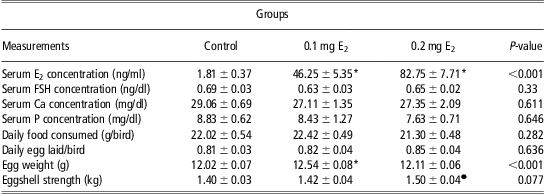
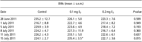
Injection of 0.1 or 0.2 mg E2 significantly increased serum concentrations of E2, whereas serum FSH, Ca and P concentrations were not significantly changed (Table 1). Injection did not cause any significant changes in daily food conceptions and egg production/bird. Injection of 0.1 mg E2 caused a significant increase in egg weight over the other two groups (Table 1). Eggshell strength was significantly high (T = 2.17, P = 0.032) in the 0.2 mg E2-injected group as compared with others (Table 1). Injection of 0.1 mg E2 significantly increased BW (P < 0.05) as compared with other two groups (Table 2).
Serum concentrations of E2, FSH, Ca, P, daily food conceptions/bird, daily egg production/bird, egg weight and eggshell strength in control and test groups
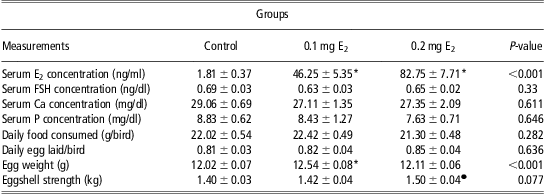
E2 = estradiol-17β; FSH = follicle-stimulating hormone; Ca = calcium; P = phosphorus.
Data were displayed as mean ± s.e.m.
*Indicates that the difference is significant as compared with others.
•Indicates that it is only significant over the control group when t-test applied.
Differences in BWs (g) along the study, measured before the injections
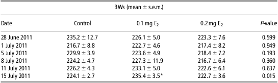
BWs were measured twice, weekly.
*Indicates that the difference is significantly high as compared with control and 0.2 mg E2-injected groups.
Discussion
In this study, birds were kept in the same room; the lighting, food and water availability were all the same. For each treatment, 10, 11 and 12 birds were used. Similar numbers of animals per treatment were also used by Wilhelms et al., (Reference Wilhelms, Cutler, Proudman, Anderson and Scanes2006) to test the effects of atrazine on sexual maturation of female Japanese quail. In this study, E2 was subcutaneously injected, twice in a week. The same, E2 injection procedure, was also applied in rat to study its effect on serum Ca level and bone development (Yeh et al., Reference Yeh, Chen and Aloia1997; Ohmori et al., Reference Ohmori, Kanda, Kawano, Kambe and Seo2000). At present, injection of E2 dose dependently increased serum E2 concentrations. Similar trends of increases in plasma E2 concentration, after the administration of E2, were reported by other researchers. In a study, force-molted laying white leghorn hens were implanted compudose pellets and plasma E2 concentration increased in parallel to the dose of E2 (Qin and Klandorf, Reference Qin and Klandorf1995). In another study, ovariectomized (OVX) rats were implanted subcutaneously with different amounts of E2-containing implants and it was reported that the relationship between graded doses of E2 and serum concentration of E2 was linear (Mannino et al., Reference Mannino, South, Inturrisi and Quinones-Janab2005).
At present, subcutaneous E2 injection caused slight reductions in serum FSH concentrations. According to the result of some in vitro studies, addition of E2 inhibited spontaneous secretion of FSH in sheep, pig and cow pituitary cell cultures. The reason for the decrease in FSH secretion, due to the E2 treatment, had been linked to the suppression of the expression of the gene encoding activin B (Nett et al., Reference Nett, Turzillo, Barata and Rispoli2002). A parallel decrease in concentrations of mRNA for FSH-β subunit and activin-βB was reported. However, the observed decrease in expression of the activin-βB gene after treatment with E2 was less than that for FSH-β. Therefore, it was concluded that a relatively smaller reduction in activin concentrations may be sufficient to inactivate the second messenger cascade and thereby greatly reduce FSH synthesis and secretion (Nett et al., Reference Nett, Turzillo, Barata and Rispoli2002). E2 treatment has no effect on rabbit pituitary cell FSH secretion, whereas it stimulated spontaneous secretion of FSH in rat pituitary cell cultures (Miller and Wu, Reference Miller and Wu1981). Therefore, different breeds of animal and birds respond differently to E2 administration (Takahashi and Jensen, Reference Takahashi and Jensen1985). In addition, different estrogens at different doses administered by different routes do not have the same pharmacodynamic effects (de Lignières and Silberstein, Reference de Lignières and Silberstein2000).
In this work, injection of 0.1 mg E2 significantly increased BW over the other two groups. Daily food consumptions per bird in the 0.1 mg E2-injected group were slightly higher than the food consumption of birds in other groups. BWs in the 0.2 mg E2-injected group were even reduced as compared with the control group. One bird in the 0.2 mg E2-injected group was dead, probably as a result of individual health problems, and this was accepted as experimental loss. It cannot be said that the birds injected with 0.2 mg E2 get stressed as BWs were only slightly decreased as compared with the BWs at the beginning of experiment. Food consumption in the 0.2 mg E2-injected group was also slightly decreased as compared with the control and 0.1 mg E2-injected groups, whereas egg-laying performance was slightly high. Injections caused some stress to all animals, as BWs decreased in all groups, on 1 July 2011 (Table 2). Subsequently, slight decreases in BW were observed in the control and 0.2 mg E2-injected groups along the course of the study. However, there were regular increases in BWs in the 0.1 mg E2-injected group as compared with others.
In an experiment, 10-week-old female Japanese quails (C. japonica) were fed with a diet containing different amount of phytoestrogen. It was reported that BWs and food consumptions were significantly increased, but there were no significant changes in serum vitellogenin concentration in the group fed with the diet containing 10 ppm E2. However, food consumptions and BWs decreased and serum VTG concentration increased in females fed with the diet containing 1000 ppm E2 (Shibuya et al., Reference Shibuya, Wada, Mizutani, Sato, Itabashi and Sakamoto2005). Briefly, lower doses of E2 stimulate body growth, but decrease the serum VTG concentration, whereas higher doses have inhibitory effect on growth and food consumption. However, high doses have stimulatory effect on serum VTG concentration.
In poultry, the two major hormones required for the full expression of growth are growth hormone (GH) and triiodothyronine (T3) and both require the anterior pituitary gland. Studies in women have shown a concomitant rise in serum E2 concentration and in GH secretion, in the late follicular phase of the normal menstrual cycle (Faria et al., Reference Faria, Bekenstein, Booth, Vaccaro, Asplin, Veldhuis, Thorner and Evans1992; Ovesen et al., Reference Ovesen, Vahl, Fisker, Veldhuis, Christiansen and Jorgensen1998). Similar results were also obtained in sheep (Landefeld and Suttie, Reference Landefeld and Suttie1989; Malven et al., Reference Malven, Haglof and Jiang1995; Scanlan and Skinner, Reference Scanlan and Skinner2002), mice (Yan et al., Reference Yan, Jones, Hernandez, Liu, Simpson and Chen2004) and rats (Childs et al., Reference Childs, Iruthayanathan, Akhter, Unabia and Whitehead- Johnson2005). In a study, female rat pituitary cells were cultured for 24 h in defined media containing vehicle or E2 in different concentrations. Addition of low-dose (0.01 to 1 nM or 0.01 to 10 nM) E2 increased the percentage of GH mRNA-bearing cells, whereas high dose of E2 (100 to 250 nM) either had no effect or inhibitory effects on the percentage of GH mRNA-bearing cells (Childs et al., Reference Childs, Iruthayanathan, Akhter, Unabia and Whitehead- Johnson2005). Therefore, E2 dose dependently increases GH secretion. According to another report, GH-releasing hormone (GHRH) mRNA expression was lower in aromatase knockout female mice (Yan et al., Reference Yan, Jones, Hernandez, Liu, Simpson and Chen2004). E2 replacement in aromatase knockout female mice increased the mRNA expression of GHRH, GH and and pituitary transcription factor-1 (Pit-1; Yan et al., Reference Yan, Jones, Hernandez, Liu, Simpson and Chen2004). In contrast, Lam et al. (Reference Lam, Lee, Tam and Srivastava1996) reported that injections of 25 μg/kg day estrogen given to OVX rats for 5 days decreased both GH mRNA and GHRH receptor mRNA. The increase in mRNA expression of GHRH, GH and Pit-1 may depend on the physiological state of the animal, as well as the concentration of E2 in their blood.
There is strong evidence that the effects of GH and thyroid hormones are mediated by hepatic production of IGF-1. Circulating concentrations of IGF-1 are markedly decreased in hypophysectomized young chickens, with GH partially reversing this effect (Huybrechts et al., Reference Huybrechts, King, Lauterio, Marsh and Scanes1985; Lazarus and Scanes, Reference Lazarus and Scanes1988). GH also elevates plasma IGF-1 level in intact adult chickens (Scanes et al., Reference Scanes, Proudman and Radecki1999). It was reported that IGF-I release from chicken hepatocytes in vitro was elevated in the presence of GH and synergistically with GH and insulin (Houston and O'Neill, Reference Houston and O'Neill1991). These studies indicate that the positive effect of E2 on GH synthesis depend on the physiological state of the animal, as well as the blood concentration of estrogen.
Here, injection of 0.1 mg E2 caused significant increase in egg weight as compared with others (Table 1). This was because of the significant increase in BW. In the 0.1 mg E2 group, egg weight was increased in parallel to the increase in BW. It has been reported that BW in Japanese quail is positively correlated with egg weight (Arora and Samples, Reference Arora and Samples2011; Ojo et al., Reference Ojo, Ayorinde and Fatoki2011). Egg number was not significantly altered by E2 injections in this study, but it was slightly increased in the E2-injected group. In a study, intramuscular injection of 3- to 5-week-old female Japanese quails with 100 μg/bird per day E2 benzoate (Elnagar and Abd-Elhady, Reference Elnagar and Abd-Elhady2009) increased egg number and egg weight, whereas in broiler hens, except peak E2 levels, no correlation between plasma concentration of E2 and egg-laying performance was reported (Onagbesan et al., Reference Onagbesan, Metayer, Williams, Decuypere and Bruggeman2006). Our results are slightly different, because the effect of E2 depends on the species, age, reproductive status, dose applied and the route of the administration.
At present, it was expected that E2 treatment will increase serum concentration of Ca and P, as E2 increases Ca absorption from the gut (Hansen et al., Reference Hansen, Beck, Scheideler and Blankenship2004). However, there were even slight reductions in serum Ca and P concentrations after E2 injection. Eggshell strength was slightly increased in the 0.1 mg E2-injected group, whereas it was significantly increased by the injection of 0.2 mg E2 as compared with other two groups. These reductions in serum Ca and P concentrations were probably caused by increased Ca and P utilization for eggshell formation and quality.
In conclusion, the function of E2 is dependent on the dose applied. Injection of 0.1 mg E2 increased BW probably by increasing GH or IGF-1 secretion or both. Injection of 0.2 mg E2 causes a reduction in BW but increases Ca and P utilization.
Acknowledgment
The authors thank Selçuk University, Department of Animal Science, for providing the study material and environment.