Systemic lupus erythematosus (SLE) is a disease characterised by loss of B-cell tolerance to autoantigens, particularly nucleic acids and their binding proteins. Although autoantibodies to nuclear antigens are elicited in healthy individuals during protective immune responses (Ref. Reference Wardemann1) and are present in the serum of up to 20% of the population, they are not sufficient to cause autoimmune disease. Clinical onset of SLE is often preceded by epitope spreading with development of antibodies to multiple nuclear antigens (Ref. Reference Arbuckle2). In addition, recruitment of inflammatory cells and mediators to susceptible target organs is required. Multiple genetic pathways predispose to development of SLE, some of which are shared by other autoimmune diseases; usually there are contributions from three or more susceptibility alleles in individual patients (Ref. Reference Fairhurst, Wandstrat and Wakeland3). The disease may be triggered in genetically susceptible individuals by environmental factors, including microbial antigens, drugs, toxins and hormones. Because a single cause for SLE has not been identified, therapy has relied on global immunosuppression; however, this causes significant morbidity and mortality from unwanted side effects including infections, osteoporosis, infertility and premature atherosclerosis.
More than a hundred single genetic defects affecting the immune system cause SLE-like disease in mice (Refs Reference Fairhurst, Wandstrat and Wakeland3, Reference Theofilopoulos and Kono4, Reference Lauwerys and Wakeland5), suggesting that there are many pathways that can be targeted therapeutically. The major challenge to assembling a new therapeutic armamentarium for SLE is the difficulty in conducting large-scale clinical trials in this highly complex disease. In addition, because of the heterogeneity of the disease, targeting a particular immune pathway may not be equally successful for each patient. Furthermore, restoration of homeostasis may be difficult to achieve because insufficient or excessive cell activation can predispose to autoimmunity. For example, insufficient B-cell signalling can result in selection of autoreactive B cells into the naive B-cell repertoire (Ref. Reference Grimaldi6), whereas excessive B-cell signalling can result in the escape of autoreactive cells during antigenic stimulation (Ref. Reference Cornall and Goodnow7). In addition, many inflammatory mediators have pleiotropic effects that are dependent on the microenvironment and the cell activation state. Finally, synergistic combinations of drugs that allow smaller doses and therefore less toxicity need to be identified.
The innate immune system as a target for SLE therapy
The innate immune system comprises cells and soluble molecules that are the first responders to an immune insult. Receptors on cells of the innate immune system recognise microbial components and induce cellular activation and cytokine release that induce activation and proliferation of T and B cells. The innate immune system is also crucial for noninflammatory clearance of apoptotic material generated from normal cell turnover. Circulating natural antibodies, complement and other acute-phase proteins opsonise and help specialised cells to clear foreign material. A defect in these functions can give rise to autoimmunity.
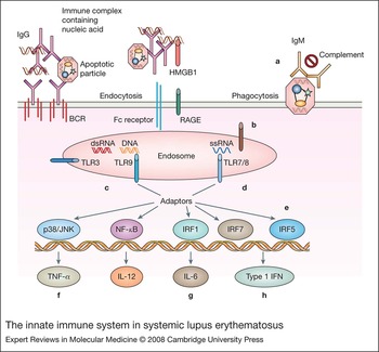
An emerging concept in the pathogenesis of SLE is that an excessive load of apoptotic particles containing nuclear antigens or of immune complexes containing autoantigens can overcome self-tolerance mechanisms and trigger autoimmunity (Refs Reference Mahoney and Rosen8, Reference Navratil, Liu and Ahearn9). Loss of tolerance may be due to excessive generation of such material, deficiencies of circulating ‘natural’ IgM antibodies, complement and other proteins that are involved in opsonisation and clearance, or alterations in thresholds for signalling of the innate immune response (Refs Reference Bickerstaff10, Reference Manson, Mauri and Ehrenstein11, Reference Manderson, Botto and Walport12, Reference Lee-Kirsch13). One mechanism for the pathogenicity of increased antigenic or apoptotic load is through activation of Toll-like receptors (TLRs) that recognise foreign and endogenous nucleic acids (Ref. Reference Christensen and Shlomchik14). Four TLRs – TLR3, TLR7, TLR8 and TLR9 – expressed predominantly by antigen-presenting cells (APCs) and B cells belong in this category (Ref. Reference Lenert15). There has been much focus recently on TLR9 (which recognises bacterial CpG-rich DNA) and TLR7 (which recognises viral single-stranded RNA), which are normally sequestered inside endosomes away from circulating endogenous nucleic acids (Refs Reference Lenert15, Reference Wagner and Bauer16). However, these endosomes are situated adjacent to phagosomes that take up apoptotic material. In addition, nucleic acids within immune complexes can be delivered to TLR-containing endosomes after cellular uptake via either cell-surface B-cell receptors (BCRs) or Fc receptors (Refs Reference Lau17, Reference Wagner18). Blockade of this process underlies the therapeutic effect of antimalarial drugs in SLE patients (Ref. Reference Rutz19).
Stimulation of TLR7 and TLR9 leads to transcription of genes encoding interleukin 6 (IL-6), IL-12, tumour necrosis factor α (TNF-α), type I interferons (IFNs) and other innate immune effectors (Refs Reference Akira, Uematsu and Takeuchi20, Reference Honda, Takaoka and Taniguchi21, Reference Pandey and Agrawal22) (Fig. 1). Polymorphisms of the TLR-induced transcription factor gene Irf5 are associated with SLE in humans, indicating the importance of this pathway in disease (Ref. Reference Sigurdsson23). One important mechanism by which TLR ligation might contribute to SLE pathogenesis is by induction of type I IFNs. IFN-α is overexpressed in human SLE patients and accelerates SLE in humans and in some genetically predisposed strains of mice (Refs Reference Ronnblom and Alm24, Reference Mathian25) (see below). Other functions reported as a result of TLR engagement include blockade of suppressor cell function (Ref. Reference Pasare and Medzhitov26), maturation of monocytes into macrophages and dendritic cells (Ref. Reference Krutzik27), and inhibition of shedding of inducible costimulator (ICOS) ligand from B cells, resulting in increased T-cell help through ICOS (Ref. Reference Logue28).
The innate immune system in systemic lupus erythematosus. Immune complexes containing nucleic acid antigens and apoptotic bodies are internalised by B cells via the BCR and Fc receptor, by dendritic cells via the Fc receptor and by macrophages via Fc receptors and endocytosis. Complexes are directed to endosomes containing nucleic-acid-binding TLRs. Via recruitment of adaptor molecules, this leads to activation of transcription factors (such as p38, JNK, NF-κB, IRFs) that drive the production of inflammatory cytokines. Alterations in many different components of this pathway may induce or offer protection from SLE. (a) Immune complexes and apoptotic particles are cleared by various molecules including natural IgM, complement, DNases and serum amyloid P. Deficiencies of these molecules can cause SLE. (b) An extra copy of TLR7/8 (part of the Yaa locus) accelerates murine SLE. (c) TLR3 deficiency has no effect on murine SLE and TLR9 deficiency decreases anti-DNA antibodies but does not improve disease outcome. (d) TLR7 deficiency decreases anti-RNA antibodies and improves disease outcome in mice. (e) Polymorphisms of IRFs are associated with SLE. (f) TNF-α blockade can induce SLE but has also been shown to induce remission of established kidney disease. Thus its effects are stage specific. (g) IL-6 blockade delays disease in murine models. (h) Type I IFN signature is observed in active SLE patients. Deficiency of type I IFN receptor prevents disease in some SLE models but administration of type I IFN is protective in others. Polymorphism of IFN-receptor-associated signalling molecule Tyk2 is associated with SLE in humans. Abbreviations: BCR, B-cell receptor; HMGB, high-mobility group box 1; IFN, interferon; IRF, interferon regulatory factor; JNK, Jun kinase; RAGE, receptor for advanced glycation end product; TLR, Toll-like receptor.

TLR9 ligands can induce either inflammation or tolerance, depending on the mode and timing of delivery (Ref. Reference Ehlers and Ravetch29). The recent in vitro finding that release of type I IFNs following activation of TLR9 ligands is dependent on the presence of the inflammatory mediator HMGB1 suggests a mechanism by which inflammation controls the outcome of TLR9 ligation (Ref. Reference Tian30). The effect of TLR9 deficiency in murine models of SLE is strain dependent. Although the titres of anti-double-stranded DNA and nucleosome antibodies are decreased in autoimmune-prone TLR9-deficient mice (Ref. Reference Christensen31), protection has been observed in some mice (Ref. Reference Ehlers32) but increased autoantibodies to RNA antigens and increased mortality have been observed in others (Ref. Reference Wu and Peng33). By contrast, TLR7 clearly contributes to SLE pathogenesis. TLR7-deficient SLE-prone mice fail to mount antibodies to ribonuclear antigens and have a more moderate disease course than their wild-type counterparts (Refs Reference Christensen31, Reference Berland34). Given this emerging knowledge, it has therefore been of much interest that the Y-linked Yaa gene – an accelerator of SLE in the BXSB male mouse – was found to be a reduplication of part of the X chromosome containing Tlr7 and Tlr8 (Ref. Reference Pisitkun35). NZW/BXSB mice express high titres of antibodies to RNA and have a marked expansion of the B-cell, monocyte and dendritic cell lineages that express TLR7 (Ref. Reference Akkerman36). Immune complexes containing RNA are potent stimulators of TLR7 and TLR8, especially in the presence of IFN-α and can therefore amplify and perpetuate disease. Further complexity is added by the differential expression of TLR7, TLR8 and TLR9 in different cell types and their ability to crossregulate each other (Ref. Reference Christensen and Shlomchik14).
Oligodeoxynucleotide inhibitors of TLR signalling (ODNs) containing methylated DNA or DNA containing CCGG or TTAGGG motifs have recently been developed and inhibit IFN-α production in response to immune complexes or viruses (Ref. Reference Lenert15). Synthesis of inhibitors with dsDNA-like structure may be selective for autoreactive cells because these are preferentially taken up into anti-dsDNA-secreting B cells via the BCR (Ref. Reference Kanzler37). Administration of ODNs to SLE-prone mice results in delay in both the onset and progression of SLE nephritis (Ref. Reference Dong38). Interestingly, in a mouse model of SLE, an inhibitor of both TLR7 and TLR9 did not add clinical benefit to the effects of specific TLR7 or TLR9 inhibitors and had less effect on anti-dsDNA titres than TLR9 inhibition alone (Ref. Reference Anders and Schlondorff39). Since much of the effect of TLR9 ligation is mediated by IFN-α, inhibition of this cytokine is also currently being tested in human clinical trials.
Another important component of the innate immune system is the Fc receptor family, which regulates responses to immune complexes (Ref. Reference Nimmerjahn and Ravetch40). Four different classes of human IgG Fc receptors have been identified, with differing affinities for IgG and for specific IgG isotypes and with differential expression on particular lymphoid cell types. FcRI, FcRIIA and FcRIIIA are activating receptors that are broadly expressed on cells of the haematopoietic lineage. FcRIIB is an inhibitory receptor expressed on all immune cells and is the only Fc receptor expressed on B cells. FcRIIB inhibits B-cell responses to immune complexes and helps terminate humoral immune responses once there is antibody excess over antigen. Expression of FcRIIB is constitutively low in several mouse models of SLE and the disease can be reversed in these mice by inducing only 40% more FcRIIB expression on B cells (Ref. Reference McGaha, Sorrentino and Ravetch41). Polymorphisms of FcRIIB are associated with human SLE, but also with protection from infectious diseases (Ref. Reference Clatworthy42). In addition, humans with SLE fail to upregulate FcRIIB on memory and plasma cells, a defect that is partly genetically determined and partly acquired (Refs Reference Mackay43, Reference Su44). Complete absence of FcRIIB in mice leads to autoimmunity and SLE-like disease in a strain-specific manner (Ref. Reference Bolland and Ravetch45). Analysis of these mice has indicated that FcRIIB regulates B-cell tolerance in the periphery, acting as a brake on the differentiation of autoantibody-producing plasma cells (Ref. Reference Fukuyama, Nimmerjahn and Ravetch46).
FcRIIIA and its newly described mouse homologue FcRIV are intermediate-affinity receptors that recognise immune complexes but not monomeric IgG. These receptors are thought to be the activating receptors for immune complexes in vivo, because high-affinity Fc receptors are already occupied with monomeric Ig (Ref. Reference Nimmerjahn and Ravetch40). Polymorphisms of FcRIIIA that lower antibody-binding affinity are associated with susceptibility to SLE nephritis and with poorer responses to therapy with B-cell-depleting agents. Low-affinity binding FcRIIA polymorphisms have also been associated with SLE and with antiphospholipid syndrome (Refs Reference Harley, Kelly and Kaufman47, Reference Karassa, Trikalinos and Ioannidis48). These polymorphisms of activating receptors may result in decreased immune complex clearance.
The relative expression of activating and inhibitory Fc receptors influences the outcome of immune-complex stimulation of cells, as does the isotype of the antibodies in the complexes. During inflammation, FcRIIB is downregulated whereas activating Fc receptors are upregulated, resulting in activation of immune cells (Ref. Reference Nimmerjahn and Ravetch40). This may contribute to the failure of upregulation of FcRIIB on B cells in patients with active SLE. One proposed mechanism of action of intravenous immunoglobulin is that it upregulates FcRIIB, thus altering the balance of activating to inhibitory receptors (Ref. Reference Nimmerjahn and Ravetch49). This effect appears to be dependent on a sialic-acid-bearing IgG glycoform that acts through an as yet unidentified receptor (Ref. Reference Nimmerjahn and Ravetch49). Further understanding of this pathway may lead to novel therapies for SLE based on modulating FcR function.
Targeting activation of the acquired immune response in SLE
Triggering of the innate immune system by exposure to exogenous antigen is crucial for the activation of antigen-specific T and B cells and their subsequent clonal proliferation and differentiation into memory cells. Pathogenic autoantibodies in SLE are isotype switched and somatically mutated – functions dependent on T-cell help (Ref. Reference Diamond50). Self-tolerance of T cells is maintained by many mechanisms, including sequestration from self-antigen, deletion of highly self-reactive cells in the thymus, and peripheral regulation either by APCs that have not been activated by innate mechanisms or by other regulatory cell subsets (Refs Reference Abbas51, Reference Miyara and Sakaguchi52). Very little is known about the antigenic specificity of T cells in SLE. Therefore, T-cell-directed therapies for SLE have focused on non-antigen-specific pathways. Although the specificity of the T-cell response is provided by the interaction of the major histocompatibility complex (MHC)–peptide complex with the T-cell receptor (TCR), T-cell activation requires a second set of costimulatory signals between T cells and APCs. The last decade has seen the identification of many costimulatory receptor–ligand pairs that are potential therapeutic targets.
CD28–B7 family members
CD28 is constitutively expressed on T cells and is upregulated upon T-cell activation. CD28 engagement by its ligands CD80 and CD86 (B7-1 and B7-2) amplifies signals through the TCR and stabilises the immunological synapse, thus lowering the threshold for naive T-cell activation, and facilitating entry into the cell cycle and secretion of IL-2 (Refs Reference Mondino and Mueller53, Reference Vincenti and Luggen54). Engagement of the TCR in the absence of CD28 costimulation may lead to cell death or induction of anergy (Refs Reference Lohr55). This maintains tolerance to self-antigens that are usually chronically encountered in the absence of costimulatory signals. The CD28-homologous molecule CTLA4 has much higher affinity for CD80 and CD86 and serves as a negative regulator of T-cell function. CTLA4, unlike CD28, signals through CD80 and CD86 to induce several regulatory molecules including indoleamine 2,3-dioxygenase (IDO), an enzyme that helps maintain tolerance through alterations in tryptophan metabolism (Refs Reference Mellor56, Reference Mellor57). A recently identified polymorphism of CTLA4 is associated with autoimmunity in humans (Ref. Reference Ueda58).
Blockade of CD28 is achieved by administration of the extracellular domain of CTLA4 fused to immunoglobulin heavy chain (CTLA4Ig, Abatacept) (Ref. Reference Vincenti and Luggen54). CTLA4Ig acts both as a competitive antagonist of B7–CD28 interactions and as an inducer of regulatory pathways in APCs. CTLA4Ig may also alter expression of adhesion molecules and chemokine receptors, resulting in inhibition of traffic of inflammatory cells to target organs (Ref. Reference Mirenda59), and prevents the upregulation of ICOS (Ref. Reference Akkerman36). However, under some circumstances, administration of CTLA4Ig might be detrimental. For example, although CTLA4Ig can induce T-cell deletion and anergy, it may prevent tolerance induction in circumstances where signalling through CTLA4 is necessary to establish active tolerance (Ref. Reference Davidson60).
CTLA4Ig and its higher-affinity mutant LEA29Y (Belatacept) have been successfully used in patients with psoriasis, rheumatoid arthritis and transplantation (Refs Reference Vincenti and Luggen54, Reference Moreland61). In mice, CTLA4Ig prevents the onset of SLE (Ref. Reference Finck, Linsley and Wofsy62) but does not reverse active inflammation (Ref. Reference Daikh63). For this reason, combination therapies have been tested. Administration of CTLA4Ig with concomitant CD40 or CD40L blockade to SLE-prone mice is a highly synergistic regimen that prevents the onset of SLE for many months after a short course of treatment. This is due to long-lasting blockade of T-cell help for autoreactive B cells (Ref. Reference Wang64). Dual treatment with a single dose of the alkylating agent cyclophosphamide and continuous CTLA4Ig is highly synergistic for the treatment of active SLE nephritis in mice and induces complete remission of histological changes in the kidney. The mechanism for this effect is not completely understood, but includes depletion of activated T and B cells, together with altered renal chemokine and chemokine receptor expression, and consequent dampening of the inflammatory response to glomerular immune-complex deposition (Ref. Reference Schiffer65). Clinical trials of Abatacept in active SLE and in combination with cyclophosphamide in SLE nephritis have been initiated.
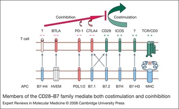
B7–CD28 interactions are essential for activation of naive T cells, but have a lesser role in regulating effector responses and responses outside secondary lymphoid organs. Expression of other B7 family members allows regulation of effector T cells at peripheral sites (Ref. Reference Greenwald, Latchman and Sharpe66) (Fig. 2). ICOS is a CD28-like molecule that is expressed on activated and memory T cells and preferentially induces IL-4 and IL-10 production (Ref. Reference Hutloff67). ICOS is highly expressed on IL-10-expressing effector cells (Ref. Reference Lohning68) and on IL-21-secreting CXCR5-positive follicular T-helper (Th) cells located in the apical zone of germinal centres where somatic mutation occurs (Ref. Reference Rasheed69). Ligation of ICOS results in upregulation of CD40L on T cells, which costimulates immunoglobulin synthesis and differentiation of B cells to memory and plasma cells (Ref. Reference McAdam70). Dramatic defects of the humoral immune response occur in ICOS-deficient mice and humans as well as defects in IL-10 and IL-17 production (Refs Reference Dong71, Reference Shilling, Bandukwala and Sperling72). Antagonism of ICOS during the effector phase of Th2-mediated disease attenuates inflammation in murine models (Ref. Reference Shilling, Bandukwala and Sperling72). The ICOS ligand B7RP-1 is expressed on B cells and macrophages and in inflamed tissues. ICOS expression is increased on T cells from SLE-prone mouse strains, particularly mice that bear a Yaa mutation (Ref. Reference Akkerman36) and on T cells in SLE patients (Ref. Reference Hutloff73). The pathogenic role of ICOS has recently been highlighted by the development of B-cell autoimmunity in mice deficient in roquin, a repressor of ICOS expression (Ref. Reference Vinuesa74), and by studies showing that blockade of ICOS ligand prevents and treats disease in NZB/W SLE-prone mice (Ref. Reference Iwai75). Clinical trials of ICOS blockade in humans with SLE are now under way.
Members of the CD28–B7 family mediate both costimulation and coinhibition. Two signals are required for T-cell activation. Signal 1 is mediated through interaction of the T-cell receptor (TCR) with MHC/peptide complex shown in blue. Signal 2 is mediated by non-antigen-specific interactions such as that of B7 with CD28. CD28-like receptors are shown on the surface of the T cell and B7-like ligands (blue) are shown on the surface of the antigen-presenting cell (APC). Receptors shown in green transduce a positive (costimulatory) signal to the T cell. Receptors shown in red transduce a negative (coinhibitory) signal to the T cell. Not all the receptors have been identified (indicated by ?). Abbreviations: BTLA, B and T lymphocyte attenuator; HVEM, herpesvirus entry mediator; ICOS, inducible costimulator; MHC, major histocompatibility complex.

The receptor programmed death 1 (PD-1), which is expressed on both B and T cells, binds to two ligands: PDL1, which is widely expressed on many tissues and PDL2, which is expressed mainly on activated monocytes. Like CTLA4, PD-1 transduces a negative signal into the cell and helps to maintain peripheral tolerance (Ref. Reference Sharpe76). Studies in mouse models of multiple sclerosis and diabetes have shown that this effect is mediated via the interaction of PD-1 with PDL1 but not with PDL2 (Refs Reference Ansari77, Reference Fife78). PD-1-deficient mice develop spontaneous strain-specific autoimmunity including SLE (Ref. Reference Okazaki, Iwai and Honjo79) and recently a PDCD1 polymorphism associated with human SLE has been identified (Ref. Reference Prokunina80).
Several other members of the CD28–B7 family have been characterised. B and T lymphocyte attenuator (BTLA), like CTLA4 and PD-1, is a negative regulator of T-cell responses and its deficiency results in increased susceptibility to induced autoimmunity. BTLA is unusual because it binds herpesvirus entry mediator (HVEM), a member of the tumour necrosis factor receptor family (Refs Reference Watanabe81, Reference Sedy82). B7-H3 and B7-H4 bind to unidentified receptors (Ref. Reference Greenwald, Freeman and Sharpe83). Little is currently known about the role of these molecules in autoimmune disease pathogenesis; however, these molecules appear to have their effects only at low antigen doses.
The TNF–TNFR family
Multiple members of this family mediate costimulation of T cells and B cells, with 4-1BB, CD40L and B-cell activating factor (BAFF) being most relevant to SLE therapies.
4-1BB
The engagement of the T-cell costimulatory receptor 4-1BB (CD137) paradoxically prevents germinal centre formation in a T-cell-dependent fashion. In SLE-prone mice, three doses of an agonistic anti-4-1BB antibody confers prolonged inhibitory effects on autoantibody production starting 1–2 weeks after antibody administration (Ref. Reference Foell84). In the chronic graft-versus-host (CGVH) model of SLE, administration of the anti-4-1BB antibody resulted in rapid depletion of donor T cells by activation-induced death (Ref. Reference Kim85). By contrast, in the NZB/W model, CD4+ T cells were not depleted, but cytokine secretion by these cells was markedly diminished (Ref. Reference Foell84). Transfer of either activated T cells or APCs from untreated mice overcame the T-cell anergy and precipitated disease. Another hypothesis for the activity of anti-4-1BB is that T-regulatory cells (T regs) are generated (Refs Reference Foell84, Reference Choi86). Although a regulatory population of CD8+ cells has been described, it is clear that the antibody still mediates tolerance in CD8-deficient mice (Ref. Reference Foell84). Finally, production of IFN-γ has been shown to enhance tolerance in the Murphy Roths Large (MRL)/lpr model (Ref. Reference Sun87) but not in the CGVH model, suggesting that IFN-γ is not essential for tolerance induction (Ref. Reference Kim85). This system is interesting because it shows that long-lasting restoration of tolerance can be achieved in mice with spontaneous SLE in which B and T cells are continuously being activated. Further studies in this system may reveal the mechanisms for maintenance, loss and restoration of tolerance in autoimmunity.
CD40L
CD40L is an important costimulatory molecule, whose expression on activated T cells is regulated by CD28 and by ICOS (Ref. Reference Greenwald, Freeman and Sharpe83). Engagement of CD40 on APCs by CD40L synergises with TLR signalling and results in increased expression of MHC and B7 molecules and secretion of IL-12, thus enhancing their antigen-presenting function (Ref. Reference Salomon and Bluestone88). Engagement of CD40 on B cells delivers a survival signal and is essential for formation of the germinal centre and for reactivation of memory B cells (Refs Reference Castigli89, Reference Grewal and Flavell90). Absence of CD40 or CD40L in humans results in a profound humoral immunodeficiency (Ref. Reference Vogel and Noelle91). Antibodies to CD40L prevent onset and delay progression of SLE in mouse models (Ref. Reference Wang92), but two Phase II human clinical trials failed because of the unexpected development of thrombotic events in some of the treated patients (Ref. Reference Sidiropoulos and Boumpas93). Although one of the anti-CD40L trials failed to show clinical efficacy at the dose used (Ref. Reference Kalunian94), some of the SLE patients treated with a different anti-CD40L antibody showed clinical improvement (Ref. Reference Boumpas95). Mechanistic studies in these patients revealed that despite unaltered total B-cell numbers, treatment diminished the number of autoantibody-producing B cells (Refs Reference Boumpas95, Reference Huang96, Reference Grammer97). It has been suggested that the effect of anti-CD40L is due to specific B-cell unresponsiveness rather than T-cell anergy, because tolerance cannot be broken in mice by transfer of pathogenic T cells (Ref. Reference Mohan98). However, in humans with SLE, B cells producing anti-DNA antibody reappeared after cessation of anti-CD40L, indicating that tolerance had not been established (Ref. Reference Huang96). A striking therapeutic effect can be observed in mouse models when anti-CD40L is combined with CTLA4Ig. This combination, given for two weeks to SLE-prone mice, induces a prolonged effect on disease when given early in the course (Ref. Reference Daikh63), and, more importantly, maintains B-cell tolerance to autoantigens without preventing immune responses to exogenous antigens (Ref. Reference Wang64). However, this combination is not effective in the setting of active nephritis and requires the addition of a single dose of cyclophosphamide, which kills activated effector cells (Ref. Reference Schiffer65). Although anti-CD40L antibodies are not safe for use in humans, antibodies to CD40 have been recently developed for human use and appear to be safe for the treatment of B-cell tumours (Ref. Reference Drachman99). This may allow for a renewed attempt at CD40 blockade in human SLE.
BAFF
The TNF family member BAFF is a B-cell survival factor expressed by many different cell types that binds to three different BAFF receptors (TACI, BCMA and BAFF-R), which are variably expressed on the B-cell surface during development (Refs Reference Sutherland, Mackay and Mackay100, Reference Ramanujam and Davidson101). BAFF-R and TACI are the predominant receptors on most mature B cells, except for plasma cells, which express BCMA (Refs Reference Mackay102, Reference O'Connor103). APRIL, a closely associated ligand of BAFF, binds to TACI and BCMA and to proteoglycans such as CD138 on the plasma cell surface; CD138 serves as a coreceptor to enhance binding of APRIL to BCMA (Ref. Reference Ingold104). Maintenance of peritoneal cavity B1 B cells is BAFF independent (Refs Reference Schiemann105, Reference Ramanujam106) and germinal centre B cells are only partially dependent on BAFF. Thus BAFF-deficient mice can still develop autoimmunity, although they do so in a much delayed fashion (Refs Reference Vora107, Reference Jacob108). BAFF transgenic mice on a nonautoimmune genetic background develop a form of SLE that is due to production of autoantibodies by marginal zone and B1 cells and this depends on B-cell signalling through TLRs but does not require T cells (Ref. Reference Groom109). BAFF levels may control B-cell selection because competition for BAFF limits selection of autoreactive B cells into the mature compartment. Depletion of B cells results in excess production of BAFF, which may then decrease the stringency for B-cell selection (Refs Reference Lesley110, Reference Thien111). It remains unclear whether the increased sensitivity of autoreactive B cells to survival signals mediated through BAFF and APRIL is applicable to memory cells, plasmablasts or plasma cells (Ref. Reference Martin and Chan112). Patients with SLE and other autoimmune diseases have increased serum levels of BAFF (Refs Reference Zhang113, Reference Cheema114) and in murine SLE nephritis, BAFF is also found locally at the site of inflammation where it is made by activated macrophages and dendritic cells (Ref. Reference Schiffer207). Activated macrophages express BAFF receptors and thus may mediate their own survival in an autocrine fashion (Ref. Reference Chang115).
BAFF blockade has been achieved either with a monoclonal anti-BAFF antibody or with soluble BAFF receptors. BAFF-R-Ig selectively blocks BAFF, whereas TACI-Ig blocks both BAFF and APRIL, and therefore is able to deplete plasma cells that are dependent on APRIL–BCMA interactions. Despite these differences, both selective and nonselective BAFF blockers prevent SLE in mouse models (Ref. Reference Ramanujam106), indicating that BAFF is more important than APRIL in SLE pathogenesis. BAFF blockade depletes B cells by 50% but does not significantly decrease the titres or affinity of autoantibodies, nor does it prevent T-cell activation. By significantly decreasing B-cell numbers and spleen size, BAFF blockade results in a decrease in the total number of activated T cells and monocytes, thus decreasing the total inflammatory load and the capacity of inflammatory cells to migrate to the kidney. The combination of BAFF blockade and CTLA4Ig, but not either drug alone, can induce remission of SLE in NZB/W mice by mechanisms that are not fully delineated (Refs Reference Ramanujam106, Reference Ramanujam116). A human clinical trial of the anti-BAFF antibody Belimumab in moderately active SLE showed only modest clinical effects, with failure to meet initial efficacy end points. However, long-term follow up and post-hoc analysis of patients that were serologically active at initiation has shown a greater sustained improvement in treated patients than in placebo-treated controls (Ref. Reference Ginzler117). More extensive trials of this agent are in progress. TACI-Ig is also entering clinical trials in SLE.
B-cell modulation and depletion
SLE is characterised by the production by B cells of high-affinity pathogenic autoantibodies that mediate tissue injury. In addition, B cells have other important functions that can contribute to disease pathogenesis. Activated autoreactive B cells that take up autoantigen through their antigen receptor can function as APCs and serve to diversify the epitopes of self-antigen presented to autoreactive T cells. B cells also produce cytokines involved both in lymphoid regulation and in inflammatory processes (Ref. Reference Grammer and Lipsky118). Because autoreactive B cells play a role in both the inductive and the effector arms of autoimmune disease, there has been much interest in B-cell depletion or modulation as a treatment strategy for autoimmune disease. One such strategy has been the use of anti-CD20 antibodies.
CD20, a transmembrane protein with four subunits, is upregulated on most B cells past the pre-B-cell stage but is absent on plasma cells (Refs Reference Riley and Sliwkowski119, Reference Uchida120). Anti-CD20 monoclonal antibodies successfully deplete peripheral human B cells for periods ranging from three months to more than one year through mechanisms involving Fc-receptor dependent killing, complement-dependent killing and antibody-dependent apoptosis (Refs Reference Clynes121, Reference Uchida122). Studies in mice show that the kinetics of B-cell depletion varies between different B-cell compartments with 90% follicular B cells in the spleen being depleted within two days of anti-CD20 monoclonal antibody administration but less effect on marginal zone B cells. The peritoneal cavity (particularly B1) and the germinal centre B-cell compartments demonstrate the greatest relative resistance to anti-CD20 monoclonal antibody treatment although marginal zone, germinal centre and peritoneal B1 B cells express comparable levels of CD20 and appear to have access to the antibody. These differential sensitivities depend partly on the number and localisation of mononuclear phagocytes within tissues that mediate antibody-dependent cell-mediated cytotoxicity (Refs Reference Uchida122, Reference Gong123, Reference Hamaguchi124). Hence, location and microenvironment influence the extent of B-cell depletion (Ref. Reference Gong123). Because B-cell depletion causes a compensatory increase in BAFF levels, blockade of BAFF synergises with anti-CD20 monoclonal antibody to enhance B-cell depletion (Ref. Reference Gong123). A new approach that may avoid the need for two drugs is the use of a depleting anti-BAFF-R antibody. This antibody blocks the effects of BAFF and also depletes BAFF-R-bearing cells. However, like anti-CD20, this antibody does not deplete long-lived plasma cells that no longer express BAFF-R (Ref. Reference Lin125).
Preliminary clinical experience with a mouse–human chimaeric anti-CD20 antibody (Rituximab) in patients with SLE (Ref. Reference Sfikakis, Boletis and Tsokos126) and other autoimmune diseases has been encouraging (Refs Reference Looney127, Reference Leandro128, Reference van Vollenhoven129), with rapid resolution of symptoms in some patients that depends on B-cell depletion but not on depletion of autoantibodies. Autoantibodies continue to be produced because plasma cells do not express CD20. These studies confirm the role of B cells as important effector cells independently of their ability to produce antibodies. The degree of B-cell depletion is affected by FcRIIIA genotype; there is less depletion in patients bearing the low-affinity allele, which is also a SLE-susceptibility allele (Ref. Reference Anolik130). In addition, the rapid induction of human antichimaeric antibodies (HACA) has prevented B-cell depletion in some SLE patients. A fully human anti-CD20 is now available and may help solve this problem. In some patients, remissions are long-lived and returning B cells are predominantly of the transitional phenotype with low numbers of memory B cells (Ref. Reference Anolik131). The reappearance of memory B cells is also associated with earlier relapse in rheumatoid arthritis patients treated with anti-CD20 (Ref. Reference Leandro132). Long-term remissions have been associated with the absence of antibodies to extractable nuclear antigens (Ref. Reference Ng133). Progressive multifocal leukoencephalopathy has been observed in two SLE patients that received more than one course of anti-CD20 together with other immunosuppressive therapy. Most of the reports of the efficacy of anti-CD20 in SLE have been uncontrolled case series in small numbers of treatment-resistant patients in which the drug has been combined with other therapies including high doses of corticosteroids and cyclophosphamide. A placebo-controlled Phase III trial of anti-CD20 in SLE patients is now in progress.
CD22 is a member of the sialoadhesin (siglec) family of adhesion molecules that is coexpressed with the B-cell receptor on mature B cells and up to the late germinal centre stage (Refs Reference van Vollenhoven129, Reference Tedder134, Reference Crocker, Paulson and Varki135). CD22 has both immunoreceptor tyrosine activation motif (ITAM) and immunoreceptor tyrosine inhibitory motif (ITIM) domains in its intracellular domain and can act both as a negative regulator of BCR signalling and as a costimulatory molecule, depending on the context in which it is activated (Ref. Reference Otipoby136). Because of this complexity, the precise effect of CD22 upon BCR signalling in B cells at the various developmental stages is not fully elucidated. B cells from CD22-deficient mice have a shortened lifespan and the mice have reduced circulating B-cell numbers; however, these mice also have hyper-reactive B cells and may develop autoantibodies with age (Ref. Reference O'Keefe137). Epratuzumab, a humanised monoclonal anti-CD22 antibody, can mediate antibody-dependent cellular cytotoxicity and has been used to treat human B-cell malignancies (Ref. Reference Leonard138). The rationale for using anti-CD22 in human autoimmunity is based on the notion that modulating B-cell function may be safer than therapies that deplete B cells. However, given the complexity of CD22 signalling in B cells and the ability of this molecule to mediate functions both in a ligand-dependent and a ligand-independent fashion, it is difficult to predict what the effect of an anti-CD22 antibody will be in humans with autoimmune disease. The first clinical trial of epratuzumab in 14 SLE patients with mild to moderately active SLE has just been reported (Ref. Reference Dorner139). Treatment with anti-CD22 appeared to be safe and resulted in modest B-cell depletion in some of the patients. Serum levels of IgG and C3 were not altered, and autoantibody levels either increased or stayed the same. Although clinical efficacy could not be assessed in this study, improvements in BILAG (British Isles lupus assessment group) scores were reported. Mechanistic studies of patients in this clinical trial showed preferential targeting of CD27-negative naive or transitional B cells that have high levels of cell surface CD22 but no decrease in memory B cells or plasma cells (Ref. Reference Jacobi140). Cell-surface CD22 levels were downregulated by epratuzumab. Cell culture experiments further showed that epratuzumab modulated the abnormal proliferative response of SLE B cells to TLR or CD40 ligation. An antiproliferative effect was also observed in vitro when human B-cell lines were immobilised and stimulated with anti-IgM (Ref. Reference Carnahan141). Further studies will be needed to determine whether these modest B-cell effects result in a measurable clinical effect in controlled trials.
Depletion of autoantibodies or specifically of autoantibody-producing B cells is another approach that is currently in development. Abetimus sodium is a compound consisting of four double-stranded 20-mer ODNs that binds to anti-dsDNA antibodies and clears them from the serum. In mice, this drug has been shown to decrease splenic autoantibody-producing B cells, but it is not clear whether this also occurs in humans. Although the drug depletes anti-DNA antibodies in SLE patients by 30%, two Phase III clinical trials have failed to reach their primary endpoints of decreased time to renal flare and clinical benefit was demonstrated only using post-hoc analysis of a patient subset with high-affinity abetimus-binding antibodies at trial initiation (Ref. Reference Alarcon-Segovia142). Further studies of this agent are in progress. Other compounds directed specifically at autoantibody-producing B cells have been developed and are described in a recent review (Ref. Reference Wiesendanger143).
Cytokines in SLE – pleiotropic activities may complicate treatment
Cytokine inhibition has been used successfully to treat several autoimmune and inflammatory diseases but has not yet been applied to the treatment of SLE. One reason is that a number of pro-inflammatory cytokines are also required for the active maintenance of tolerance and their blockade may exacerbate the loss of tolerance to ubiquitous antigens. For example, TNF-α blockade enhances the production of SLE-related autoantibodies. Crosstalk between innate immune system signals and cytokines can be dysregulated in a systemic inflammatory environment and this may result in proinflammatory functions of cytokines that are usually anti-inflammatory. For example, IL-10 usually signals through STAT-3 but in the presence of type I IFNs it switches to the proinflammatory STAT-1 (Refs Reference Herrero144, Reference Hu145). In addition, upregulation of costimulatory receptors and integrins, and alteration in the ratio of activating to inhibitory Fc receptors, can alter the cellular response to cytokines.
IL-6
IL-6 is a multifunctional cytokine that is critical for the development and functioning of many different cells and plays a crucial role in immune responses and inflammation (Refs Reference Liang146, Reference Kishimoto147). In the setting of activation of the innate immune system, IL-6 dampens T reg function (Ref. Reference Pasare and Medzhitov26). Conversely, IL-6 has a regulatory role in the resting immune system as it has been shown to maintain anergy of resting B cells chronically exposed to self-antigens (Ref. Reference Wiesendanger208). Excessive secretion of IL-6 is found in many autoimmune diseases (Ref. Reference Mihara, Nishimoto and Ohsugi148) including SLE (Ref. Reference Linker-Israeli149), where it induces plasma cell and effector T-cell differentiation (Refs Reference Pasare and Medzhitov26, Reference Tackey, Lipsky and Illei150). A critical role for IL-6 in the pathogenesis of SLE was demonstrated by the beneficial effects of IL-6 receptor blockade and the exacerbating effect of IL-6 in NZB/W F1 mice (Refs Reference Liang146, Reference Mihara151, Reference Finck, Chan and Wofsy152). A Phase I clinical study using an anti-IL6R monoclonal antibody (MRA) in SLE has been completed but the development of dose-dependent neutropaenia may complicate the use of this agent in human SLE (Ref. Reference Illei153).
TNF-α
TNF-α is a pleiotropic cytokine that can mediate both cell survival and cell death (Ref. Reference Hill and Lunec154). The importance of this cytokine in maintaining the balance of the immune system is highlighted by acceleration of autoimmunity in a TNF-α-deficient background (Refs Reference Jacob and McDevitt155, Reference Kontoyiannis and Kollias156) and the triggering or maintenance of autoimmunity in the setting of excess TNF-α (Refs Reference Mountz157, Reference Romas, Gillespie and Martin158, Reference Feldmann and Maini159). TNF-α antagonists have a remarkable therapeutic effect in a number of autoimmune diseases including rheumatoid arthritis and Crohn disease (Refs Reference Sandborn and Hanauer160, Reference Feldmann and Maini161, Reference Tobin and Kirby162). However, TNF-α blockade appears to exacerbate certain autoimmune diseases (Ref. Reference Kollias163) and has resulted in the emergence of new autoimmunity in up to 50% of treated patients with the development of antinuclear antibodies and even clinical SLE (Ref. Reference Swale164). By contrast, TNF-α is known to be pathogenic in mouse models of nephritis and is produced by intrinsic renal cells as a result of exposure to inflammatory cytokines including IL-1 (Ref. Reference Tipping and Holdsworth165). Based on these findings, successful anecdotal use of TNF-α blockade has been reported in a small number of patients with refractory SLE nephritis (Ref. Reference Aringer166). Not surprisingly, autoantibody titres increased in the treated SLE patients, confirming that some features of autoimmunity are exacerbated by this approach. One hypothesis for this effect is that TNF-α blockade increases the load of apoptotic cells (Ref. Reference Aringer167). One group found increased expression of TNFRII on CD4+ CD25+ T regs of SLE patients. Signalling through this receptor resulted in loss of suppressive function of T regs that could be reversed by IL-2 supplementation (Ref. Reference Valencia168). Other hypotheses include the induction of IFN-α following TNF blockade and the requirement for TNF-α in negative regulation of activated T and B cells. Since TNF-α is made as a soluble and a membrane protein and can bind to two different receptors with different functions (Refs Reference Chan and Lenardo169, Reference Locksley, Killeen and Lenardo170, Reference Fu and Chaplin171), there has been recent interest in selective TNF-α blockade as a strategy for a safer and more specific therapeutic response (Refs Reference Kollias163, Reference Kassiotis and Kollias172).
Type I interferons
Type I IFNs made by plasmacytoid dendritic cells are implicated in the pathogenesis of SLE (Refs Reference Pascual, Farkas and Banchereau173, Reference Ronnblom and Alm174). Their therapeutic use can induce SLE-like syndromes, and peripheral blood mononuclear cells from a subset of patients with SLE display increased expression of IFN-α-regulated genes (Refs Reference Bennett175, Reference Baechler176). IFN-α promotes the differentiation of monocytes into dendritic cells, induces differentiation of CD8+ T cells and, together with IL-6, induces differentiation of B cells into plasma cells. However, the role of IFN-α in autoimmunity is pleiotropic and absence of IFN-α or its receptor has had opposite effects in different mouse models of SLE (Ref. Reference Koutouzov, Mathian and Dalloul177). The explanation for this might lie in genetic differences and/or in the different effects of IFN-α on resting and activated dendritic cells. In vitro experiments have shown that triggering of TLR9 in naive peripheral blood mononuclear cell cultures results in an IFN-α-dependent inhibition of inflammatory Th1 cytokine secretion upon subsequent activation (Ref. Reference Meyers178). For this reason, type I IFN therapy has been used to treat autoimmune diseases such as multiple sclerosis. However, if type I interferons are administered to activated dendritic cells, proinflammatory cytokines are generated (Ref. Reference Nagai179). This suggests that the outcome of treatment with IFN-α antagonists in patients may be stage dependent and may not always be predictable. In addition, the recent finding that the interferon signature can be elicited in human monocytes in the absence of IFN-α by ligation of activating Fc receptors, together with blockade of inhibitory Fc receptors, suggests that there are multiple pathways for induction of inflammation by immune complexes (Ref. Reference Dhodapkar180). Early clinical trials of anti-IFN antibodies are in process and should more definitively define the pathogenic role of this group of cytokines in SLE.
IL-10
IL-10 is increased in patients with active SLE and correlates with disease activity (Refs Reference Park181, Reference Hagiwara182). Although IL-10 is classically considered an anti-inflammatory cytokine, its role in SLE appears to be an inflammatory one. In the presence of IFN-γ and immune complexes, there is attenuation of the suppressive functions of IL-10 mediated by a decrease in expression of the IL-10 receptor and decreased activation of JAK-1 (Ref. Reference Ji183). In the presence of IFN-α there is a gain of proinflammatory function of IL-10 leading to STAT-1-dependent expression of genes that are normally induced by IFN-γ. In NZB/W mice, continuous administration of IL-10 accelerated onset of renal disease and treatment with an anti-IL-10 monoclonal antibody delayed disease onset (Ref. Reference Ishida184). By contrast, in NZM2410 mice, IL-10 was protective when administered early in disease (Ref. Reference Blenman185). Similarly, in MRL/lpr mice, IL-10 deficiency resulted in a more severe disease (Ref. Reference Yin186). The differences between these models may be related to the amount of concomitant inflammation at the time of IL-10 administration or depletion. In a pilot open-label short-term study of a mouse anti-IL-10 monoclonal antibody in a small number of active SLE patients, disease improvement was evident at 2 months, with continued responses over 3–6 months (Ref. Reference Llorente187).
Interferon γ
IFN-γ accelerates disease in SLE-prone NZB/W mice whereas treatment with anti-IFN-γ is protective (Refs Reference Jacob, van der Meide and McDevitt188, Reference Ozmen189). IFN-γ–/– MRL/lpr mice were also protected from early death with a reduction in the severity of glomerulonephritis. These studies highlight the importance of IFN-γ in accelerating SLE development, presumably by increasing MHC expression and autoantigen presentation to otherwise quiescent nontolerant anti-self T cells, and also by promoting local immune and inflammatory processes (Ref. Reference Balomenos, Rumold and Theofilopoulos190). By contrast, results in STAT-4- and STAT-6-deficient SLE-prone mice showed surprisingly that a decrease in IFN-γ and an increase in IL-4 accelerated SLE nephritis even though autoantibody titres were diminished (Ref. Reference Singh191). This may be due to early regulatory effects of IFN-γ on the development of Th17 effector cells. IL-17-secreting cells appear to be critical effector cells in rheumatoid arthritis and multiple sclerosis (Ref. Reference Weaver192), but very little is known as yet about the role of this cytokine in SLE.
The inflammatory process in SLE nephritis
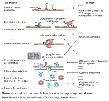
Proliferative glomerulonephritis is the most severe form of SLE nephritis and is characterised by mesangial proliferation and infiltration of the kidney parenchyma by inflammatory cells. This process is initiated by deposition of antibodies in the renal glomeruli but it is increasingly recognised that this is not sufficient for renal inflammation to occur. In NZB/W SLE-prone mice deficient in activating Fc receptors, autoantibody and complement deposition in glomeruli do not result in renal damage (Ref. Reference Clynes, Dumitru and Ravetch193). Subsequently it was shown that the important Fc-receptor-bearing cells are of haematopoietic origin and that circulating monocytes bearing the activating receptors are sufficient to restore disease (Ref. Reference Bergtold194). In SLE patients, polymorphisms of Fc receptors have been associated with the susceptibility to renal disease (Ref. Reference Karassa, Trikalinos and Ioannidis48). Other downstream effector pathways including chemokines and cell death molecules are also required to mediate kidney damage. By contrast, in the MRL/lpr model, interstitial inflammation can occur in the kidney without immune-complex deposition and in the absence of Fc receptors, perhaps indicating that endothelial activation is sufficient to initiate inflammatory cell migration into target organs in this mouse (Refs Reference Chan195, Reference Matsumoto196). A model for the effector pathways involved in renal inflammation is shown in Figure 3. Microarray analysis of human SLE biopsies has shown considerable heterogeneity in gene expression between patients. Infiltration by B cells and myeloid cells was observed in some patients, as was evidence of fibrosis. As observed in peripheral blood specimens, some patients also had a type I IFN signature in the kidney (Ref. Reference Peterson197).
The events that lead to renal failure in systemic lupus erythematosus. Disease is initiated by deposition of immunoglobulin and complement on the glomerular basement membrane. This is followed by an inflammatory cascade that involves engagement of activating Fc receptors by circulating monocytes, endothelial activation, chemokine secretion, recruitment of activated lymphocytes and finally release of proapoptotic factors that result in irreversible renal cell death. Potential therapies (shown in red) may target this cascade in a stage-specific manner. Abbreviations: B, B cell; T, T cell; MØ, macrophage; NO, nitrous oxide; TGF-β, transforming growth factor β; TNF-α, tumour necrosis factor α.

Invasion of the glomerulus and the interstitium by inflammatory cells involves different chemokines and receptors. In particular, CCR2 and CCR5 mediate glomerular cell invasion whereas CCR1 mediates interstitial invasion (Refs Reference Anders198, Reference Anders, Ninichuk and Schlondorff199). Studies of chemokine expression in the kidneys of NZB/W mice have shown that a limited panel of chemokines is expressed in the early stages of nephritis. The onset of proteinuria coincides with expression of CCR2 and CCR5 ligands and with infiltration and activation of interstitial macrophages that secrete IL-1 and other inflammatory molecules. With progression of disease, there is extensive spreading of the inflammatory response to include multiple chemokines and cytokines (Ref. Reference Schiffer207). These data suggest that it may be difficult to induce remission of active nephritis with single chemokine or cytokine inhibitors but that such reagents may be useful once most activated cells have been purged from the kidneys. Furthermore, these studies predict stage-specific therapies for SLE nephritis. For example, CCR2 and CCR5 blockade may be useful for treating the early stages of glomerular disease whereas IL-1 or TNF-α blockade may be useful for antagonising the proinflammatory effects of activated infiltrating macrophages. Since upregulation of CCR1 occurs only in mice with established proteinuria, CCR1 blockade may be effective for treatment of late- rather than early-stage disease. Consistent with this result, a CCR1 antagonist does not affect systemic immune activation or glomerular damage in SLE-prone MRL/lpr mice but slows progression of interstitial renal disease and fibrosis (Ref. Reference Anders198). As more is learned about the inflammatory process in the SLE kidney, application of therapies to prevent renal cell infiltration are likely to be tested.
Clinical implications and research in progress
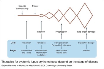
Maintenance of immune homeostasis requires a finely tuned immune system that needs the flexibility to respond quickly to pathogens but does not mount immune responses to self-antigens. Both immunodeficiency and exaggerated immune responses can predispose to autoimmunity. In addition, cytokines and cell receptors that are balanced to maintain tolerance in the setting of a resting immune system can mediate inflammatory responses when the immune system is activated. Multiple defects of immune tolerance can lead to SLE and predisposing causes can include both immunodeficiency in the resting immune system and excessive immune responses in the activated immune system. For this reason, restoration of immune homeostasis in SLE patients poses many challenges. It is increasingly recognised that therapies for active disease, characterised by lymphoid cell proliferation and production of multiple inflammatory mediators, may need to be different from therapies that prevent disease flares or those that might eventually prevent disease onset in genetically predisposed individuals (Fig. 4). Despite these difficulties, many new drugs are being developed for the treatment of SLE and clinical trials are in progress.
Therapies for systemic lupus erythematosus depend on the stage of disease. The various stages of SLE consisting of a pre-disease period followed by flares and remissions, are associated with differences in the state of immune activation which may require different therapeutic approaches.

Preclinical testing of novel therapies in murine models
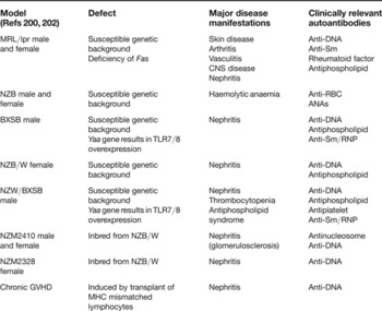
As discussed above, immune activation pathways involved in initiation of SLE may not always be the same as those that are pathogenic during the effector phase. Furthermore, inflammatory environments can alter signalling cascades or provide redundant survival signals. Because the clinical effects of new therapeutics may not always be predictable from in vitro studies or studies in non-autoimmune mice, it is important to test these drugs in preclinical models. Many different mouse models of SLE now exist (Refs Reference Fairhurst, Wandstrat and Wakeland3, Reference Theofilopoulos and Kono4, Reference Lauwerys and Wakeland5) and several of these have been extensively characterised and used for therapeutic studies (Ref. Reference Hahn, Wallace and Hahn200) (Table 1). Most have been used for studies of prevention or treatment of SLE nephritis or of the effects of treatment on autoantibody production and immune cell activation. In addition, the MRL/lpr mouse can be used for study of vasculitis, arthritis and skin disease, whereas the NZW/BXSB mouse can be used to study the effects of therapy on antiphospholipid syndrome (Ref. Reference Akkerman201). It is important to recognise that murine SLE models have a number of limitations. First, mouse immune pathways are not always the same as human pathways. Second, not all clinical SLE manifestations are present in each mouse strain. In particular, there are no good mouse models for the treatment of the central nervous system manifestations of SLE. Third, responses to some therapies are strain dependent and therapies that improve disease in one model may worsen it in another. In particular, a number of differences have been observed in responses to therapy of MRL/lpr mice and NZB/W mice. Fourth, most mouse models do not undergo spontaneous disease remissions, although it is now possible to study disease relapses after remissions have been induced with biological therapies. Nevertheless, animal models allow analysis of basic immune mechanisms of disease and therapy that are not possible in human studies and can help direct appropriate clinical use of new therapeutics.
Common mouse models of systemic lupus erythematosus used for therapeutic studies

Abbreviations: ANAs, antinuclear antibodies; CNS, central nervous system; GVHD, graft-versus-host disease; MHC, major histocompatability complex; TLR, Toll-like receptor; RBC, red blood cell; RNP, ribonucleoprotein; Sm, Smith antigen.
Clinical trials in human SLE
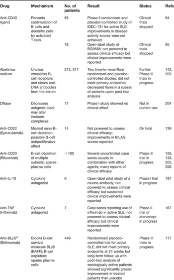
New agents for which the results of clinical trials have been published are shown in Table 2. Most of these trials were Phase II studies and had insufficient statistical power to assess clinical efficacy. However, several large Phase III studies are now in progress and results can be expected in the next few years. The design of SLE clinical trials will be of crucial importance in determining optimal therapies for induction of disease remission and for maintenance of disease remission. Much thought is being given to recruitment of patient populations, disease activity measurements, disease response measurements, primary outcome measures and mechanistic analyses of immunological responses in treated patients. Most clinical trials now employ the BILAG, a scoring system that measures the severity of disease in individual organ systems. Some drugs are being tested in remission induction studies whereas others are being tested in ‘time-to-flare’ studies. The choice of design will depend on the expected mechanism of action of each drug. For example, blockade of inflammatory cytokines or depletion of whole cell populations would be expected to induce remission of active disease whereas therapies that prevent naive cell activation or modulate B-cell selection would be expected to have a preventive rather than a remission induction effect. Testing in preclinical models may help direct trial design for each new agent.
Therapeutic trials of novel agents for systemic lupus erythematosus

aPublished in abstract form only but included here because it is a large Phase III study. Abbreviations: BAFF, B-cell-activating factor belonging to the TNF family (also known as BLyS, B lymphocyte activator); BILAG, British Isles lupus assessment; SLE, systemic lupus erythematosus; TNF, tumour necrosis factor.
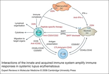
The range of new drugs being developed reflects the global nature of the immune defects in SLE (Fig. 5). Approaches include measures to decrease antigen load (DNase), measures to dampen the innate immune system (ODN and IFN-α blockade), drugs that antagonise activation of the acquired immune system (costimulatory blockade, modulation of BCR signals), blockade of the soluble mediators derived from the effector arm of the immune response (cytokine antagonists), protection of target organs from damage (chemokine inhibitors, complement inhibitors) and even depletion of whole cell populations (B-cell depletion, stem cell transplant). In addition, there are agents designed to specifically target autoreactive B cells through induction of anti-idiotypic antibodies. The scope of this article does not allow discussion of all the new drugs being developed, but for a comprehensive review see Ref. 143. The challenge will now be to determine at which stage of disease each reagent is most likely to be effective and which reagents will work together. Optimal therapy will also require continued research that will eventually allow identification of the underlying immune defect in each patient. The ultimate goal is to selectively correct deficits that contribute to autoimmunity without impacting protective immunity.
Interactions of the innate and acquired immune system amplify immune responses in systemic lupus erythematosus. Therapeutic interventions for SLE (shown in red) target multiple arms of the immune system (see Ref. 143 for review). Abbreviations: APRIL, A proliferation inducing ligand; BAFF, B-cell-activating factor belonging to the TNF family; DC, dendritic cell; ICOS, inducible costimulator; IFN-α, interferon α; ODN, oligodeoxynucleotide; TLR, Toll-like receptor.

Acknowledgements and funding
M.R. and A.D. are supported by grants from the NY SLE Foundation, NY Arthritis Foundation, Lupus Research Institute, Alliance for Lupus Research and the NIH. M.R. is an Arthritis Foundation Young Scholar. A.D. is a Kirkland Lupus Scholar. We thank our panel of peer reviewers for helpful suggestions in improving this manuscript.